
Apparatus for strengthening vertebral bodies

a technology for strengthening vertebrae and vertebrae, applied in the field of vertebroplasty and a method and apparatus for strengthening vertebrae, can solve the problems of reducing the chances of maintaining sterility, wasting unused biomaterials and equipment, and reducing so as to reduce the risk of spinal cord compression

Active Publication Date: 2009-07-21
IZI MEDICAL PROD
View PDF71 Cites 12 Cited by
  • Summary
  • Abstract
  • Description
  • Claims
  • Application Information

AI Technical Summary

Benefits of technology

[0009]It is therefore an object of the present invention to provide a novel method and apparatus for strengthening vertebral bodies which obviates or mitigates at least one of the disadvantages of the prior art.
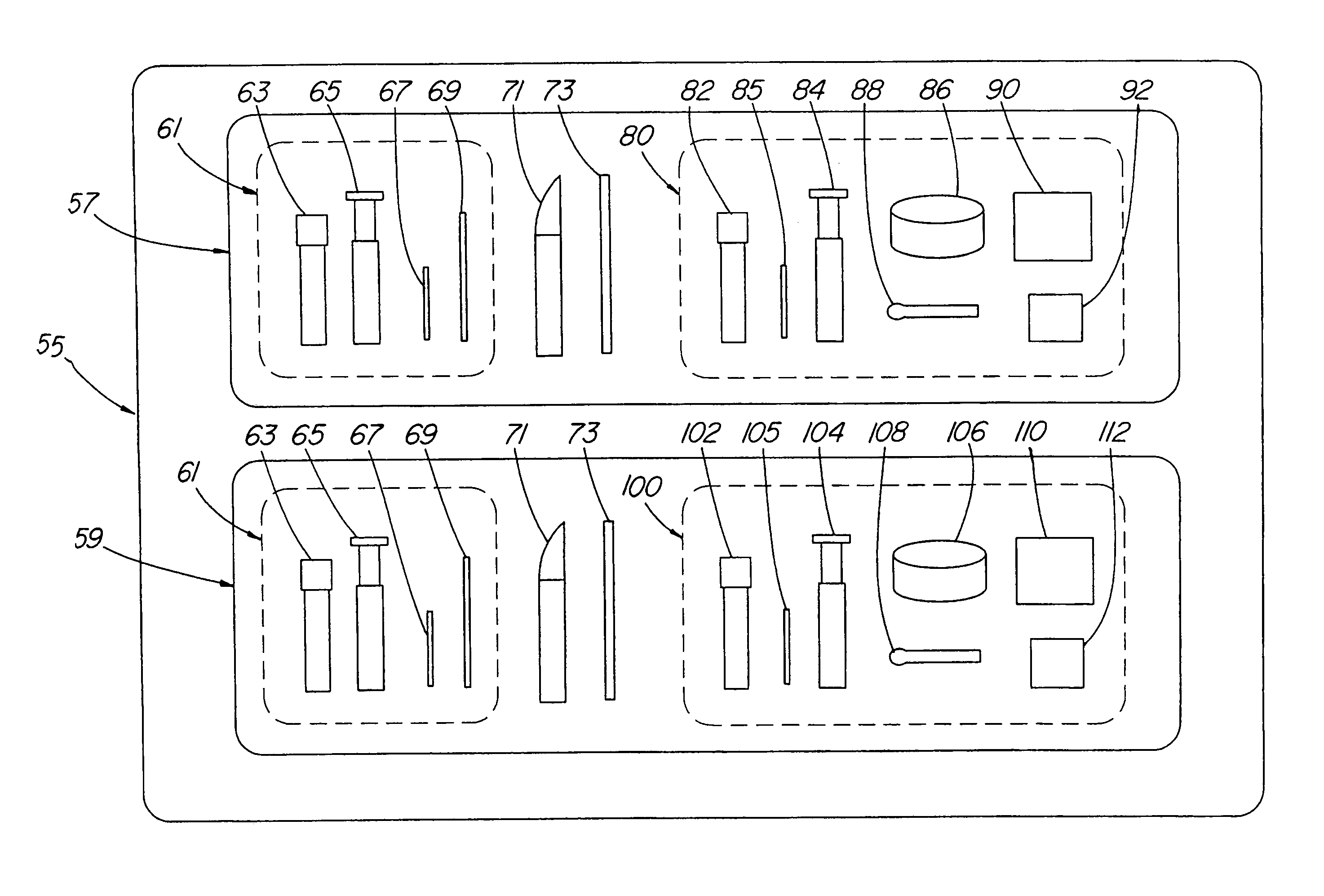
[0011]An apparatus and method for performing vertebroplasty are provided. In one embodiment of the invention, there is provided a kit for performing vertebroplasty having two packs, each pack having the necessary components for performing vertebroplasty via each pedicle of the damaged vertebrae. Each pack is sterilized, so that if the vertebroplasty performed via one pedicle is sufficient, then the second pack can be saved for later use. In another embodiment, each pack within the kit has two cements, each cement having different imaging properties, such that each cement will appear different when viewed with an imaging device in the lateral plane and / or will be viewable when overlapping. A presently preferred embodiment involves two packs having methylmethacrylate powder but each pack has a different amount of opacifier to be added to the powder, such that when each cement is mixed and injected, each cement is visible when exposed to X-ray lateral fluoroscopy. A method is also provided that utilizes the kit, and which allows a medical professional to monitor a second injection of cement via the second pedicle and thus reduce the risk of spinal cord compression due to unwanted flow of cement into the spinal cord, or nerve compression.

Problems solved by technology

However, should the nature of the vertebral defect only require injections from one side of the vertebrae, then the unused biomaterial and equipment is wasted.
Furthermore, after the equipment has been provided and arranged for use, it must be kept clean and sterile.
If the equipment is laid out too far in advance of a procedure, the chances of maintaining sterility decreases.
Contaminated or unused equipment is either replaced or discarded, thus making for a very time consuming and costly exercise.
However, this system is designed for any bone filling procedure and thus does not include all of the necessary components for percutaneous vertebroplasty.
In sum, the present commercially available kits are not directed towards percutaneous vertebroplasty procedures, thus resulting in equipment being wasted.

Method used


Image

  • Apparatus for strengthening vertebral bodies
  • Apparatus for strengthening vertebral bodies
  • Apparatus for strengthening vertebral bodies

Examples


Embodiment Construction

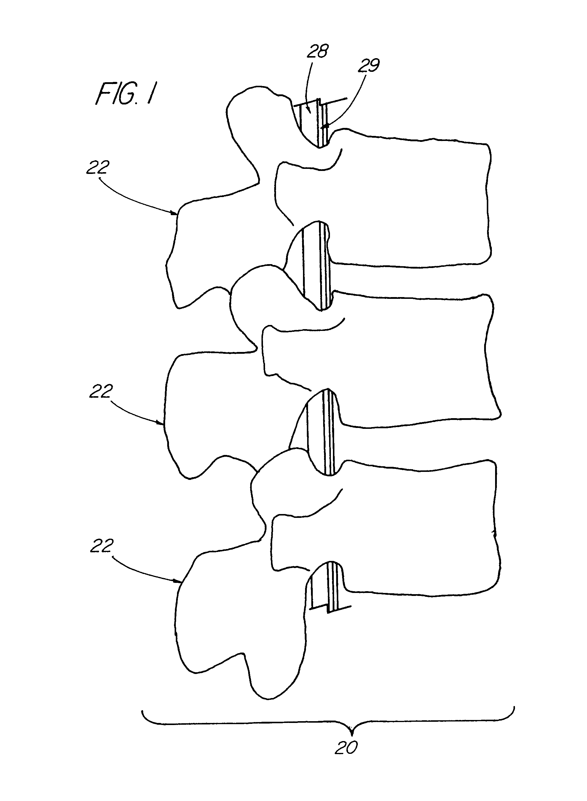
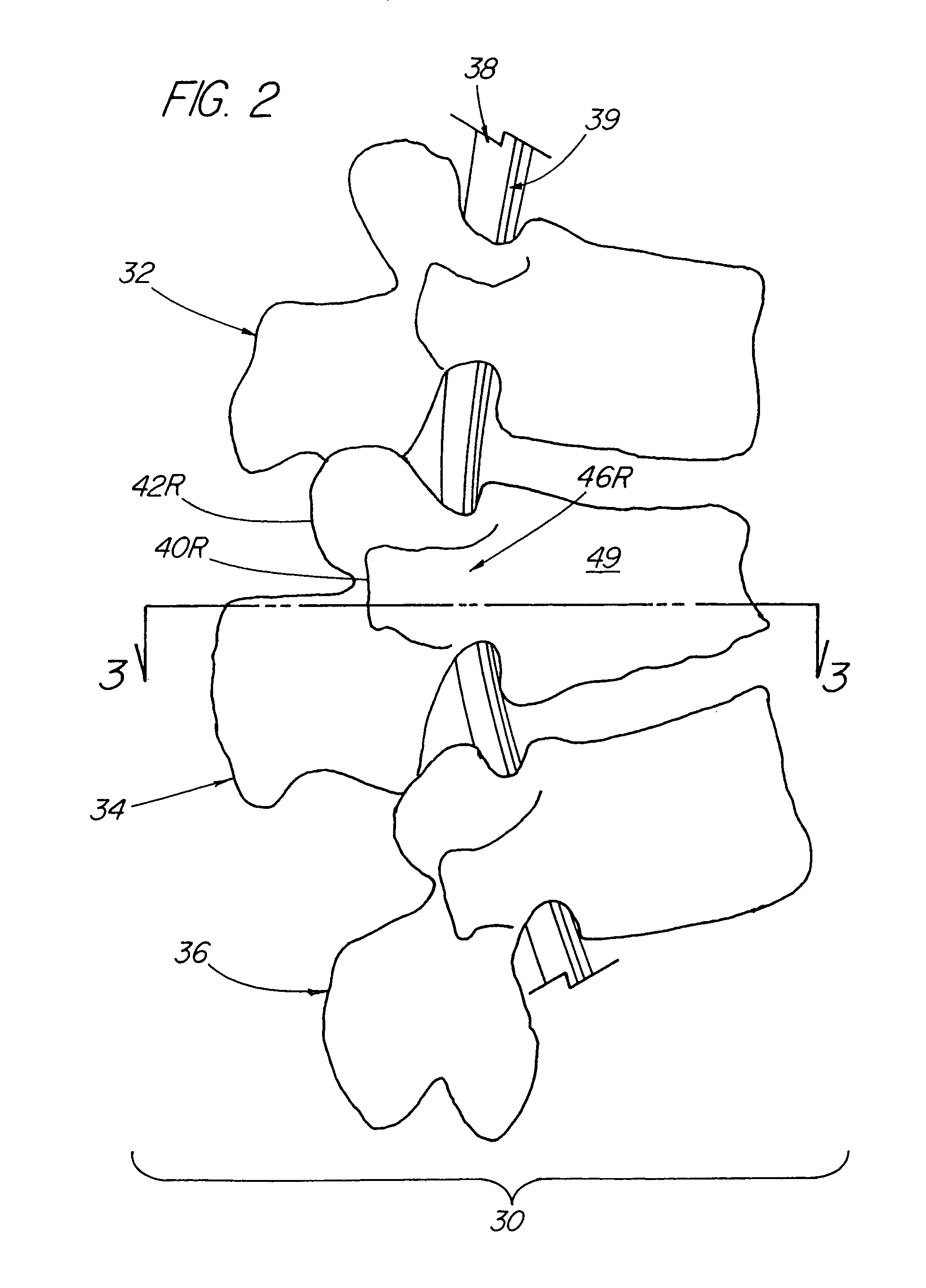
[0026]Before discussing embodiments of the present invention, various components of vertebrae and the spine will be discussed. FIG. 1 is a right lateral view of a segment 20 of a normal spine. Segment 20 includes three vertebrae 22. The spinal cord 28 and epidural veins 29 run through the spinal canal of each vertebrae 22. In contrast to segment 20 of FIG. 1, FIG. 2 shows a right lateral view of a segment 30 of a spine wherein at least one of the vertebra has a condition suitable for treatment by vertebroplasty. Segment 30 includes a first vertebra 32, a compressed middle vertebra 34 and a third vertebra 36. Spinal cord 38 and epidural veins 39 run through the spinal canal of each vertebrae 32, 34 and 36.

[0027]As shown in FIGS. 2 and 3, vertebra 34 has a right and left transverse process 40R, 40L, a right and left superior articular process 42R, 42L, and a spinous process 44 at the posterior of vertebra 34. Right and left lamina 45R, 45L lie intermediate spinous process 44 and super...


PUM


Abstract

An apparatus and method for performing vertebroplasty are provided. In one embodiment of the invention, there is provided a kit for performing vertebroplasty having two trays, each tray having the necessary components for performing vertebroplasty via each pedicle of the damaged vertebrae. Each tray is sterilized, so that if the vertebroplasty performed via one pedicle is sufficient, then the second tray can be saved for later use. In another embodiment, each tray within the kit has two cements, each cement having different imaging properties, such that each cement will appear different when viewed with an imaging device in the lateral plane and / or will be viewable when overlapping. A presently preferred embodiment involves two trays having methylmethacrylate powder but each tray has a different amount of opacifier, either as supplied or added by the vertebroplasty professional, such that when each cement is mixed and injected, each cement is visible when exposed to X-ray lateral fluoroscopy. A method is also provided that utilizes the kit, and which allows a medical professional to monitor a second injection of cement via the second pedicle and thus reduce the risk of spinal cord compression or venous filling due to unwanted flow of cement into the spinal canal.

Description

RELATED APPLICATION INFORMATION[0001]This is a continuation-in-part application of U.S. patent application Ser. No. 09 / 425,480 filed Oct. 22, 1999, now U.S. Pat. No. 6,273,916.BACKGROUND OF THE INVENTION[0002]1. Field of the Invention[0003]The present invention relates to vertebroplasty and a method and apparatus for strengthening vertebral bodies.[0004]2. Description of the Prior Art[0005]Percutaneous vertebroplasty is a technique involving the injection of a biomaterial into a vertebral body in order to treat the defects therein. It is performed using a set of surgical equipment assembled to complement such a procedure. For example, it is necessary to have equipment for prepping the patient's skin in order to provide antimicrobial effectiveness to the skin overlying the vertebrae. Equipment is also required to maintain the surgical area as clean and sterile as possible to help reduce the risk of infection. Equipment is further required to anaesthetize the patient to produce a reve...

Claims


Application Information

Patent Timeline
IPC IPC(8): A61B17/58A61B17/56A61B17/70A61B17/88A61B19/02A61F2/00A61F2/44A61F2/46A61L24/00A61L27/50
CPCA61B17/70A61B17/8802A61B19/026A61L24/001A61L27/50A61B19/0271A61B19/54A61L2430/38A61F2/441A61F2/4601A61F2310/00353A61B2017/564A61B50/30A61B50/33A61B90/39
Inventor MURPHY, KIERAN P. J.
Owner IZI MEDICAL PROD
Learn More