Flexible electrosurgical electrode with manipulator

Active Publication Date: 2009-03-24
ELLIQUENCE
View PDF5 Cites 23 Cited by
  • Summary
  • Abstract
  • Description
  • Claims
  • Application Information

AI Technical Summary

Benefits of technology

[0012]In accordance with a feature of the invention, an electrosurgical probe comprises a very long, thin, flexible, insulated wire electrode, so thin and flexible that it can be used with a miniature or micro-sized endoscope combining imaging optics and an instrument channel with an overall diameter below about 3 mm, hereinafter referred to as a micro-fibre. In accordance with a principal feature of the present invention, the micro-fibre electrode is removably mounted in a novel handpiece or handle for controlling the micro-fibre electrode while also being able to manipulate and guide the micro-fibre electrode in a relatively stationary, tissue touching technique, improving surgical outcomes in procedures such as endolaryngeal surgery. The combination of the micro-fibre electrode with the novel handpiece results in an innovative endoscopic instrument allowing precise, accurate cutting and coagulation of laryngeal lesions, for example, when hand manipulating the micro-fibre tip and thus being able to navigate and guide the micro-fibre tip more easily to the targeted tissue. The targeted tissue can be removed, ablated, vaporized, coagulated, incised, or excised where histological examination is required. The movement range of the preferred instrument allows the micro-fibre electrode tip to be moved from a neutral position aligned with the longitudinal axis of the handpiece in an upward direction about 45° or down about 15°, allowing access to a wide range of targeted lesions in a wide range of locations on a patient's anatomy.
[0013]The micro-fibre electrode handpiece that serves as the manipulator device offers many significant clinical and technical advantages with video endoscopic assisted instrumentations. The advantages include safety, tactile feedback, wide range of accessibility, bloodless field, precise and accurate removal of targeted tissue, much less scarring, faster, high quality healing, excellent tactile feedback, and less pain and swelling. Pressureless cuts minimize the risk of injury to laryngeal structures. Economically it combines a shorter operating time with a cost effective unit and instrument. A further advantage is the ability to excise tissue for histologic evaluation if desired.
[0015]The micro-fiber electrode used in the invention is simple to construct, and uses a minimal amount of precious material. It therefore provides a safe, sterile, surgical, disposable single patient use tool available for each patient in a cost effective manner. The manipulator of the invention in which it is used, while relatively less expensive to construct than re-useable surgical tools, has the big advantage that the re-usable manipulator is only the navigating guide and holder for the disposable RF micro-fiber electrode which actually contacts the tissue surgically. Thus sterilization of the manipulator is much less of a problem for the surgeon and hospital and contributes to patient safety.
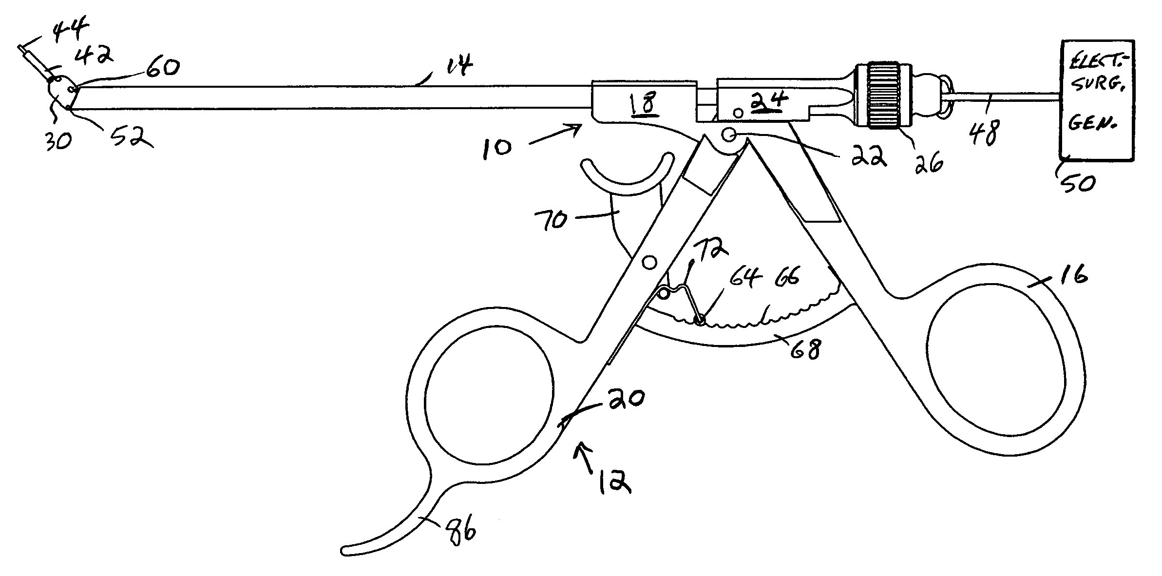
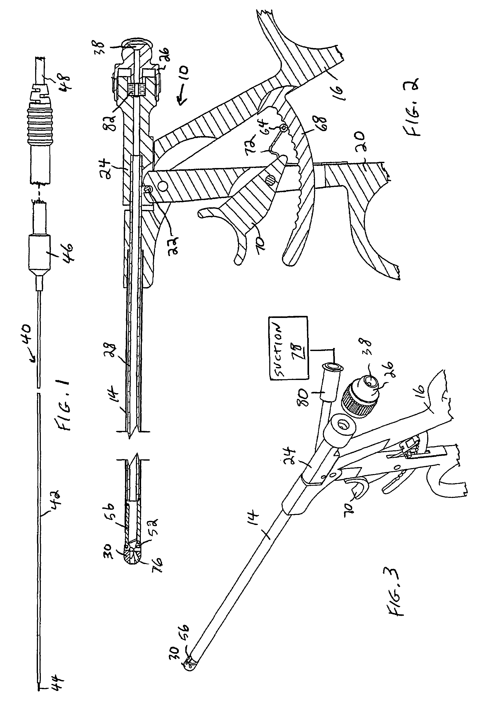
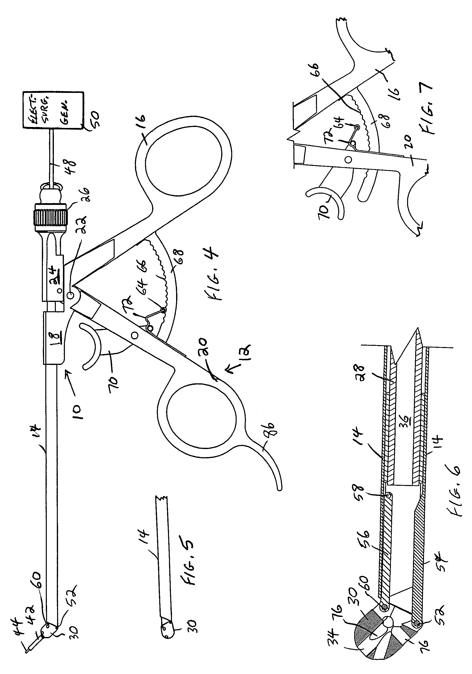
[0016]In a preferred embodiment, the handpiece is constructed such that, not only can the working end be deflected after insertion in the patient, but also the deflected position can be clamped by the surgeon, with its position easily changed by the surgeon with a finger of the same hand upon releasing the clamp whenever desired. The length that the fibre extends from the end of the deflector can also be easily controlled in the handpiece of the invention.
[0019]In still another preferred embodiment, the manipulator is configured to provide a channel for suctioning off separated tissue or plume or other fluids created at the surgical site. The reduced plume / fluids at the site improves visibility for the surgeon.
[0022]The construction of the invention will provide important benefits for all MIS arthroscopic or endoscopic procedures and in many cases enables the efficient delivery of radiofrequency (RF) energy technology for controlled precise tissue cutting, absorption and other tissue effects and in a safe manner. It is cost effective and considerably less expensive than other surgical modalities such as lasers where the novel electrode configuration may be of importance, as well as for general electrosurgical procedures where the volumetric reduction of tissue or ablation of tissue that is hard to reach with the known electrodes is desirable. Examples of particular procedures for which the electrosurgical electrode of the invention is particularly suitable is spinal disc reduction and endonasal procedures.

Problems solved by technology

However, their disadvantage is that they require expert manual skills and rigorous practice, and in addition frequently result in diffuse bleeding at the surgical site, obscuring the operative field and making it difficult to access the progress of the surgery.
Powered instruments (micro-debriders, shavers) have been used, in which tissue is excised by a rotating blade and aspirated out of the sheath, but there is a high risk of tissue damage and tissue may be chewed up rapidly and cannot be evaluated histologically.
Their disadvantages include combustion of ventilation tube materials and anesthetic gas mixtures during laser use in larynx tissue destruction and alteration, complicated safety precautions for doctors, staff and patients (ie. protective glasses, bouncing of the laser beam off metal instruments causing inadvertent burns) and possible electrical hazards, eye and skin injuries, as well a steep learning curve for doctor and staff, and their high price.

Method used


Image

  • Flexible electrosurgical electrode with manipulator
  • Flexible electrosurgical electrode with manipulator
  • Flexible electrosurgical electrode with manipulator

Examples


Embodiment Construction

[0033]The reader is directed to the referenced prior patents for a more detailed description of electrosurgical procedures and principles of operation which will assist in understanding the invention described in the present application.

[0034]As described in the co-pending application, a very thin electrosurgical electrode in the form of a fibre, referred to herein as a micro-fibre, is provided for use with a miniature micro endoscope. The present invention describes a novel manipulator which receives the micro-fibre and in turn is sufficiently thin to fit in a channel of a miniature micro endoscope in order to allow the operating surgeon to control the position of the active end of the fibre while inside the patient or at the surgical site.

[0035]In a preferred embodiment illustrated in the figures, the manipulator 10 in accordance with the invention comprises a proximal handle end 12 to which is mounted an elongated tubular-shaped outer tube 14. The right handle half 16 is connecte...


PUM


Abstract

An electrosurgical instrument that is configured for use in MIS electrosurgical procedures, comprises a handpiece with a separable electrosurgical electrode in the form of a micro-fiber, comprising a long, thin, flexible, insulated wire. The combination is configured to cooperate with the cannula of an endoscope to reach interior tissue. The handpiece is constructed to allow the tip with the micro-fiber active end to be deflected to navigate the fiber end in diverse directions under control of the user, and also allows the deflected position to be releasably clamped with the same hand that manipulates the micro-fiber active end.

Description

RELATED APPLICATION[0001]This application is a CONTINUATION-IN-PART of a commonly owned patent application, Ser. No. 10 / 914,740, filed in the U.S. Patent And Trademark Office on Aug. 9, 2004 and entitled FLEXIBLE ELECTROSURGICAL ELECTRODE FOR TREATING TISSUE, now U.S. Pat. No. 7,160,295.[0002]This invention relates to an electrosurgical probe for treating ailments or diseases by minimally invasive surgery (MIS) or similar endoscopy procedures.BACKGROUND OF THE INVENTION[0003]Our prior U.S. Pat. No. 5,505,728, whose contents are incorporated herein by reference, describes a novel electrosurgical electrode for ablating or shrinking throat tissue in a surgical procedure. This is accomplished by an electrosurgical electrode activated by electrosurgical currents that is applied by the surgeon to the patient.[0004]Our prior U.S. Pat. No. 6,447,510, whose contents are incorporated herein by reference, describes a novel electrosurgical electrode for the treatment of benign and malignant les...

Claims


Application Information

Patent Timeline
IPC IPC(8): A61B18/18A61B18/14
CPCA61B18/148A61B18/1492A61B18/22A61B19/22A61B2218/007A61B34/70
Inventor GARITO, JON C.ELLMAN, ALAN G.
Owner ELLIQUENCE
Why Eureka
Try Eureka