
Apparatus and method for registration, guidance and targeting of external beam radiation therapy

Active Publication Date: 2008-10-21
IMPAC MEDICAL SYST
View PDF187 Cites 116 Cited by
  • Summary
  • Abstract
  • Description
  • Claims
  • Application Information

AI Technical Summary

Benefits of technology

[0021]It is yet another object of the invention to provide an improved imaging system for a patient threatment facility and which includes a therapeutic radiation beam's eye views and is also capable of controlling a field size.
[0023]It is yet another object of the invention to provide an improved imaging system that permits an exporting of images to therapeutic treatment planning systems for fusion with either CT or standard simulation images for the localization of internal soft tissue treatment targets.
[0024]It is another object of the invention to provide an improved imaging system that provides for rapid patient alignment using ultrasound imaging in accordance with a treatment plan.
[0025]It is still another object of the invention to provide an improved treatment system that provides for the accurate localization and targeting of therapy for a soft tissue treatment volume.
[0027]It is yet another object of the invention to provide an improved and integrated treatment system that allows for continuous monitoring during every aspect of a therapeutic treatment procedure.
[0028]In accordance with the above objects, the present invention accurately and definitively localizes and fixates the position of a prostate gland (and other portions of the human body) during all phases of the radiation therapy, thus permitting accurate targeting and delivery of escalated dose to the patient without increased morbidity and damage to the surrounding healthy tissues.

Problems solved by technology

Treatment for this stage is problematic with significantly low control rates using traditional doses of radiation, which is the main-line therapy.
Treatment of prostate cancer is difficult because of the extreme proximal position of tissues that are sensitive to radiation, such as the bladder and rectum.
Radiation treatment, which is typically delivered in daily fractionated doses over the course of several weeks, is further complicated by prostate motion relative to the radiation field on a daily basis.
However, these dose-escalated treatments have met with problems due to increased dose delivered to normal tissues that are in the radiation field, producing many unacceptable complications such as rectal fistulas and bladder perforation and / or sloughing.
Therefore, dose-escalated, conformal treatments cannot be delivered without significantly increased morbidity unless the exact position of the prostate can be visualized and registered, and this field localization maintained during the course of the treatment.
One of the primary problems associated with radiation treatment of prostate cancer is the location of the prostate during treatment planning.
The prostate is not visible on simulation X-rays and is difficult to define in simulation CT data.
The resulting beam definitions are not optimally conformal with respect to the prostate, resulting in potential under-dosage of the cancerous tissue and / or overdosage of nearby healthy tissue.
Another significant problem during radiation therapy is caused by prostatic movement between treatment sessions.
Since numerous treatments are delivered over a period of weeks or months, this migration can result in significant loss of targeting accuracy due to prostatic movement with respect to the isocenter.
Likewise, there is also an issue of prostatic movement during the actual treatment session.
Any movement by the patient may move the prostate with respect to the treatment isocenter, reducing beam / prostate conformance and impairing the effectiveness of the treatment.
Another significant issue is the unwanted radiation exposure to the rectum and bladder.
BAT does not offer a treatment planning component.
The BAT cannot be used during treatment, because it would interfere with the therapeutic beams and because it would be difficult to ensure continued ultrasound-transparent contact with the patient throughout the treatment.
Furthermore, the BAT is not integrated with the treatment plan and is only used to visually position the center of the prostate with respect to the isocenter.

Method used


Image

  • Apparatus and method for registration, guidance and targeting of external beam radiation therapy
  • Apparatus and method for registration, guidance and targeting of external beam radiation therapy
  • Apparatus and method for registration, guidance and targeting of external beam radiation therapy

Examples


Embodiment Construction

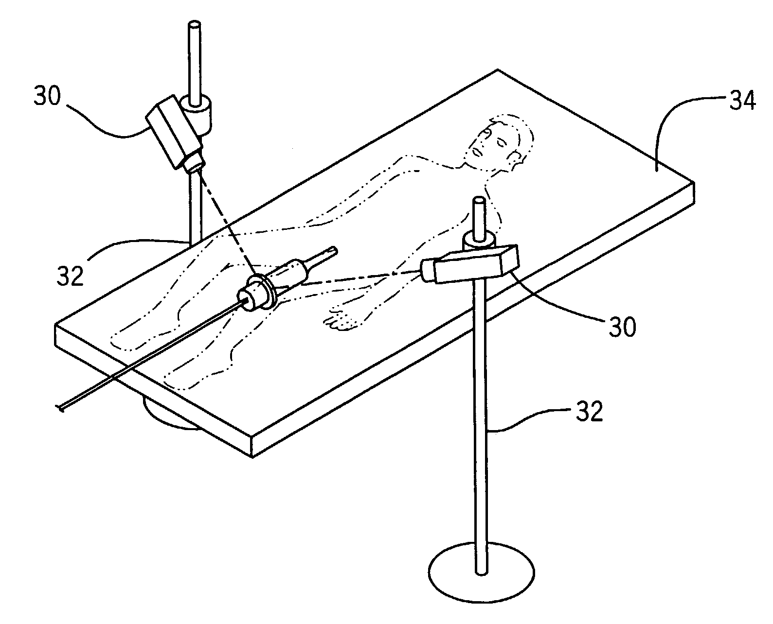
[0044]The present invention comprises a technique and integrated hardware and software system to provide improved planning, registration, targeting, and delivery of conformal, external beam radiation therapy of prostate cancer and other soft-tissue diseases. Real time ultrasound imaging during planning and treatment is used for localization of soft tissue treatment targets and fused with radiographic or CT data for conformal treatment optimization. The fusion technique of the present invention provides accurate localization of the prostate (or other tissue) volume in real time. In particular for treatment of prostate cancer, visualization of the prostate gland is achieved using transrectal ultrasonography and the fusion of that image in the precise location of the prostate within the pelvic region. This makes possible accurate determination of the location of the prostate target by transformation of the ultrasound image data on both the ultrasound and x-ray / CT images. With unambiguo...


PUM


Abstract

An improved system and method for optimizing the planning, registration, targeting, and delivery of conformal, external beam radiation therapy of prostate cancer and other soft-tissue diseases. Real-time ultrasound imaging during planning and treatment is used forlocalization of soft tissue treatment targets and fused with radiographic or CT data for conformal treatment optimization. The fusion technique provides accurate localization of the prostate volume in real time. For treatment of prostate cancer, visualization of the prostate gland is achieved using transrectal ultrasonography and the fusion of that image in the precise location of the prostate within the pelvic region, accurately determining the location of the prostate target by transformation of the ultrasound image data on both the ultrasound and X-ray / CT images. The radiation field may be optimized to significantly reduce the volume of irradiated normal tissue, minimizing the exposure of the surrounding healthy tissues and increasing dosage delivered to the prostate treatment target.

Description

FIELD OF THE INVENTION[0001]The present invention relates generally to systems for radiation therapy. More particularly, the present invention relates to a system for targeting soft tissue for external beam radiation therapy.BACKGROUND OF THE INVENTION[0002]Prostate adenocarcinoma is the most commonly diagnosed cancer in the U.S. male population (excluding skin cancer). Over 20% of these cases are locally-advanced non-metastatic cancers. Treatment for this stage is problematic with significantly low control rates using traditional doses of radiation, which is the main-line therapy. Treatment of prostate cancer is difficult because of the extreme proximal position of tissues that are sensitive to radiation, such as the bladder and rectum. Radiation treatment, which is typically delivered in daily fractionated doses over the course of several weeks, is further complicated by prostate motion relative to the radiation field on a daily basis. More aggressive radiation treatment technique...

Claims


Application Information

Patent Timeline
IPC IPC(8): A61B8/00A61B6/00A61B6/03A61B8/08A61B8/12A61B17/00A61B17/34A61B19/00A61N5/10
CPCA61B6/5247A61B8/08A61B8/463A61B8/5238A61B19/5244A61N5/103A61B6/00A61B6/03A61B8/12A61B19/50A61B2017/00274A61B2017/3413A61B2018/00547A61B2019/207A61B2019/5236A61B2019/5238A61B2019/5255A61B2019/5276A61B2019/5291A61B2019/5295A61B2019/5483A61N2005/1058A61B8/4245A61B8/4263A61B2090/3983A61B2034/2055A61B2090/367A61B34/20A61B2090/376A61B2090/101A61B34/10A61B2090/374A61B2090/378A61B2090/365
Inventor BURDETTE, EVERETTE C.DEARDORFF, DANA L.
Owner IMPAC MEDICAL SYST
Learn More