
Method and apparatus for determining symmetry in 2d and 3d images

a technology of symmetry and image, applied in image data processing, instruments, character and pattern recognition, etc., can solve the problems of hardly applicable algorithms in clinical environments, prone to noise and skull appearance, and human brains almost never perfectly symmetric, so as to maximize the modified local symmetry index and accurate and fast determination of fissure line segments

Inactive Publication Date: 2008-08-05
KENT RIDGE DIGITAL LABS
View PDF9 Cites 16 Cited by
  • Summary
  • Abstract
  • Description
  • Claims
  • Application Information

AI Technical Summary

Benefits of technology

[0011]It is also an object of this invention to provide a clinically applicable solution to automatically determine the MSP of 3D radiological images. Ideally the solution should be specially tailored in terms of reasonably acceptable time for processing for a common hardware setup, robustness to variations in clinical data, user-friendliness, being fully automatic and stable.SUMMARY OF THE INVENTION
[0013]In essence the present invention stems from the realization that a localized characteristic measurement of a line segment in an image at points adjacent the line segment provides information at relatively low cost that can contribute to a relatively accurate determination of the symmetry of the entire image. A further realization of the present invention applies this localized characteristic measurement with histogram analysis to reject image anomalies that detract from a relatively accurate determination of the symmetry in an iterative manner that omits these anomalies from further processing, which would otherwise provide relatively low efficiency in image determination and analysis. In preferred embodiments, a further realization of the present invention combines the localized characteristic measurement with a further localised characteristic measurement of points along a given line segment in an image which is particularly useful in 3D image determination and processing.
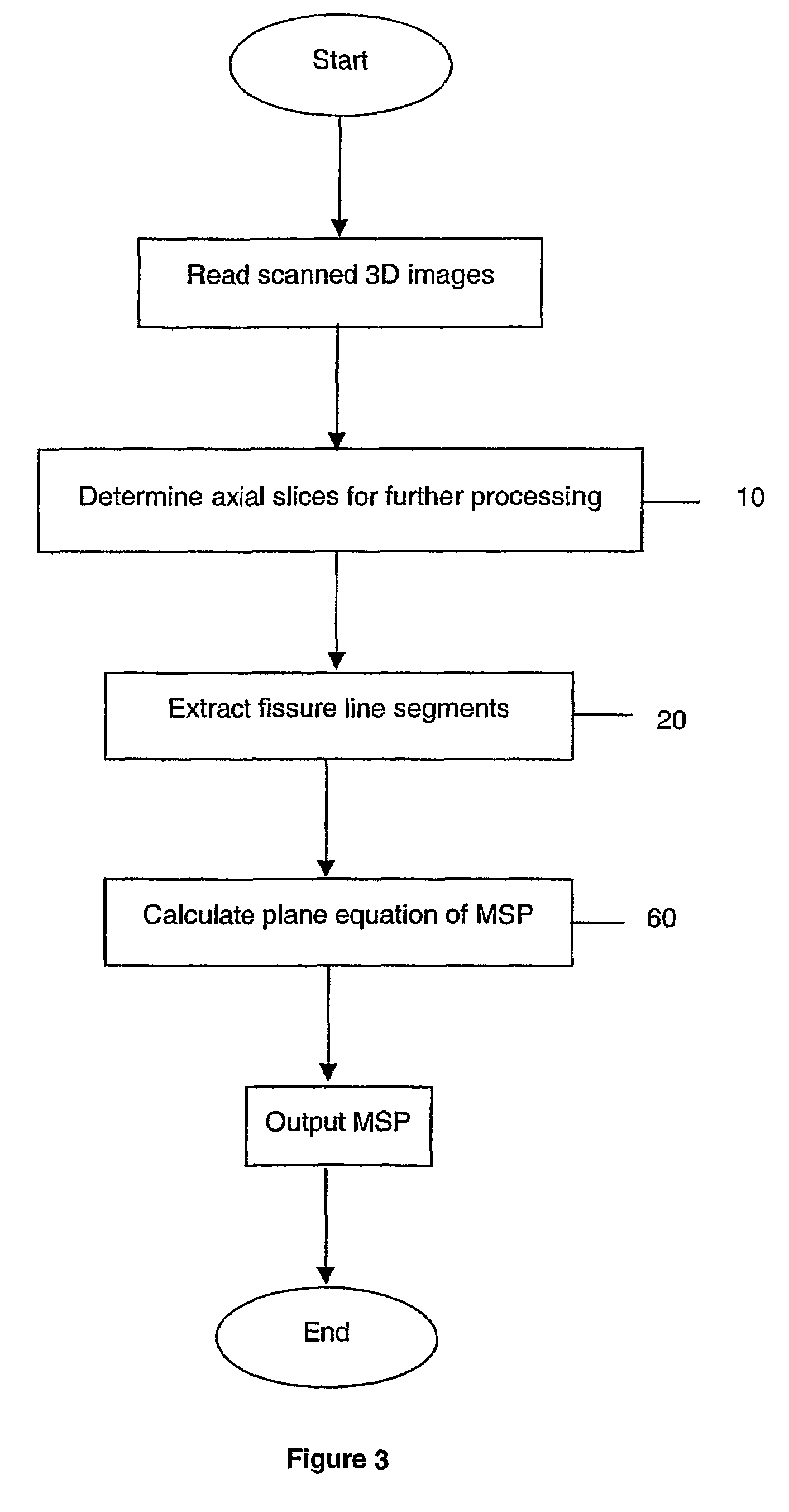
[0067]Embodiments of the present invention are made possible using the novel concepts of local symmetry index, fissure pattern measure, and a novel way to calculate plane equation based on histogram analysis. The present invention makes use of the geometric property (approximate symmetry measured by local symmetry index) and may also make use of grey level (fissure pattern measure) properties of fissure line segments on axial slices for accurate and fast determination of fissure line segments. The robust approximation method to calculate the plane equation of MSP from extracted fissure line segments via histogram analysis allows the present invention to deal with pathological images correctly where either tumor(s) or mass effects dominate some of the axial slices, since the extracted fissure line segments in these abnormal slices will be regarded as outliers and are excluded from plane equation determination of MSP.
[0068]According to another aspect, the present invention provides a method of extracting symmetry line from radiation images via maximizing the modified local symmetry index.

Problems solved by technology

However, human brains are almost never perfectly symmetric (R. J. Davidson, K. Hugdahl, Eds., Brain Asymmetry.
One of the limitations of the algorithm is its dependence on the edge map, which is quite prone to noise and skull appearances.
Furthermore, the algorithm is hardly applicable to clinical environments since it is too time consuming (as long as 7 minutes for a typical volume).
The algorithm was very sensitive to asymmetry in the 3D images, and could not deal with pathological images.
The algorithm failed in those axial slices where the lateral ventricle was present.
The algorithm also failed if any of the fissure lines were not straight or too broad, as in the case of excessive CSF.
The algorithm needed pre-processing to get appropriate gradient modulus that should be fissure line dominant, which was difficult if not impossible due to noise, skull and skin appearances.
In addition, an incorrect detection of a fissure line in one slice would contribute to the final extraction, because there was no way to correct automatically.
If the subjects being imaged were pathological such as containing excessive amount of CSF or having tumors, the algorithm also failed.
Whilst the first method noted above by Liu et. al may deal with both normal and pathological images, none of the suggested algorithms are suitable for clinical application either due to excessively time intensive computing or due to an inability to process the real clinical data with pathology or ubiquitous asymmetry presented in axial slices.
None of the existing methods meet the current clinical requirements in terms of speed, accuracy, and robustness to clinical data.

Method used


Image

  • Method and apparatus for determining symmetry in 2d and 3d images
  • Method and apparatus for determining symmetry in 2d and 3d images
  • Method and apparatus for determining symmetry in 2d and 3d images

Examples


Embodiment Construction

Head Coordinate System

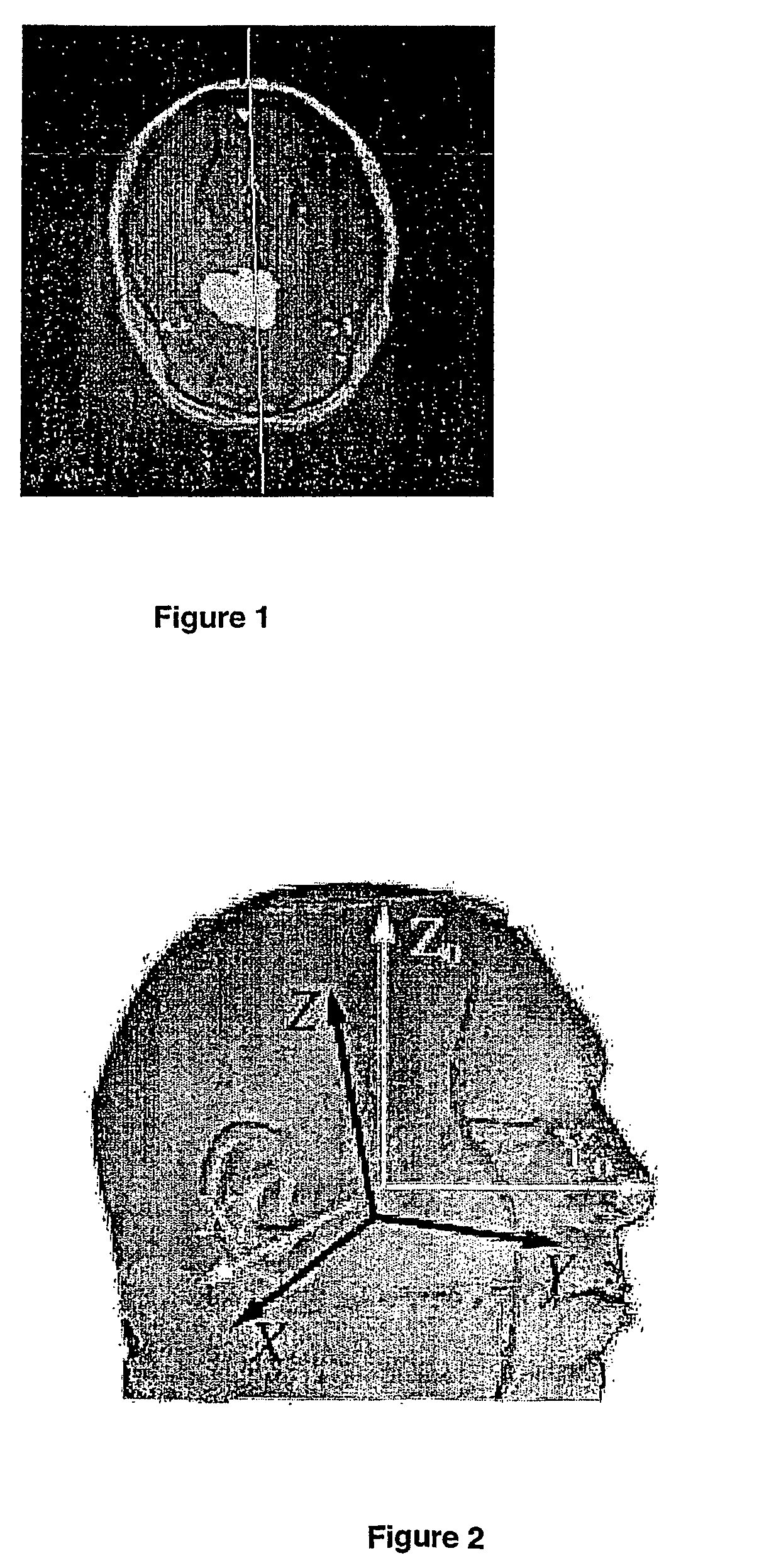
[0084]Referring to FIG. 2, the ideal head coordinate system is centered in the brain with positive X0, Y0, Z0 axes pointing to the right, anterior, and superior directions, respectively. In clinical practice, the imaging coordinate system XYZ (FIG. 2, black coordinate axis) differs from the ideal coordinate system due to positioning offsets (translations) and rotation of the head when imaged. With respect to this coordinate system, the plane equation X0=0 is the MSP of the brain, and it is an objective of this invention to find the plane equation of MSP in the XYZ coordinate system.

3D Images and Axial Slices

[0085]Neuroradiology scans are 3D volumetric data expressed as a stack of 2D slices.

[0086]The 3D images can be obtained in 3 different ways: slices that are scanned along X, Y, and Z directions are called sagittal, coronal, and axial scans respectively. The scanned 3D images are linearly interpolated in space so that the voxel sizes are 1×1×1 mm3. Since from...


PUM


Abstract

A method of determining symmetry in an image, a method of determining a symmetry plane line segment of a 3D image, a method of determining a symmetry line of a 2D image, and a computer program product. The method includes a) determining at least one searching line segment within a predefined search area of an image portion, the at least one searching line segment including a reference point (x, y) at its center and an angle θ with respect to a predetermined axis of the image portion; b) for each searching line segment, determining a first local characteristic in accordance with a measurement at points adjacent the searching line segment; c) determining the symmetry in the image in accordance with a calculation based on the first local characteristic.

Description

[0001]This application is a 371 of PCT / SG02 / 00006 filed on Jan. 18, 2002.FIELD OF THE INVENTION[0002]The present invention relates to the processing of images. More particularly, the present invention relates to the processing of two-dimensional (2D) images or the processing of three-dimensional (3D) images, where such 3D images comprise a plurality of 2D image slices. In one aspect, the present invention is suitable for use in the determination of a plane of symmetry in a 3D medical image. For example, the present invention is suitable for use in determining the midsagittal plane (MSP) of the human brain from radiological images, and it will be convenient to hereinafter describe the invention in relation to that exemplary application. It should be appreciated, however, that the invention is not limited to that application, only.BACKGROUND OF THE INVENTION[0003]Human brains exhibit an approximate bilateral symmetry with respect to the inter-hemispheric (longitudinal) fissure bisecti...

Claims


Application Information

Patent Timeline
Patent Type & Authority Patents(United States)
IPC IPC(8): G06K9/00G06K9/32G06K9/36G06T7/40G06T7/60
CPCG06T7/608G06T2207/30016G06T7/68
Inventor HU, QINGMAONOWINSKI, WIESLAW LUCJAN
Owner KENT RIDGE DIGITAL LABS
Learn More