
Minimally invasive surgical access device

a minimally invasive, access device technology, applied in the field of minimally invasive access devices, can solve the problems of affecting the long term health of the spine, affecting the function and strength of the spine, and longer hospital stays and recovery, so as to reduce the risk of damaging anatomical structures

Inactive Publication Date: 2008-08-05
THOMPSON MIS
View PDF6 Cites 37 Cited by
  • Summary
  • Abstract
  • Description
  • Claims
  • Application Information

AI Technical Summary

Benefits of technology

The device allows safe and controlled access to spinal pathology with reduced risk of anatomical damage, minimizing muscle and ligament resection and stabilizing the surgical site, thereby reducing post-surgical pain and complications.

Problems solved by technology

These supporting structures are not causing the patient any discomfort or pain, however, because the surgeon must be able to visualize the surgical operation, they need to be removed or detached.
As a result, these tissues never return to their normal anatomical position, which is disrupted in the surgical process, weakening their function and strength.
Also, the patient may experience significant pain and discomfort resulting in longer hospital stays and recoveries.
The long term health of the spine can also be affected because these supporting structures are not able to perform their function normally.
This can result in further pain and discomfort, and can even lead to additional surgeries.
A frequently encountered problem is transitional syndrome whereby the nerves adjacent to an open fusion and instrumentation become compressed.
This may in-part be due to the initial fusion procedure dissecting supporting muscles and ligaments creating an iatrogenic instability that leads to adjacent level stenosis.
In addition, large open procedures often result in extensive scar formation that can lead to conditions such as arachnoiditis and failed back syndrome.
These patients suffer significant and debilitating pain which is often refractory to additional surgery.
Many can no longer work or conduct normal activities of daily living.
Unfortunately, a number of complications have occurred using the previously described system.
Additionally considerable downward force is required to pass the muscle dilators towards the spine.
Incidences have occurred in which the dilators are passed into the spinal canal resulting in neural injury and paralysis.
The muscle dilators also tend to be pushed upward out of the wound requiring multiple repositioning during placement, each time placing the neural structures at risk.
Lastly, multiple, sequentially larger dilators are used causing considerable patient risk with placement of each muscle dilator.

Method used


Image

  • Minimally invasive surgical access device
  • Minimally invasive surgical access device
  • Minimally invasive surgical access device

Examples


Embodiment Construction

[0021]The following discussion of the embodiments of the invention directed to a minimally invasive surgical access device is merely exemplary in nature, and is in no way intended to limit the invention or its applications or uses. For example, the access device of the invention has particular application for minimally invasive spinal surgical procedures. However, as will be appreciated by those skilled in the art, the access device of the invention will have application for other types of minimally invasive surgeries.

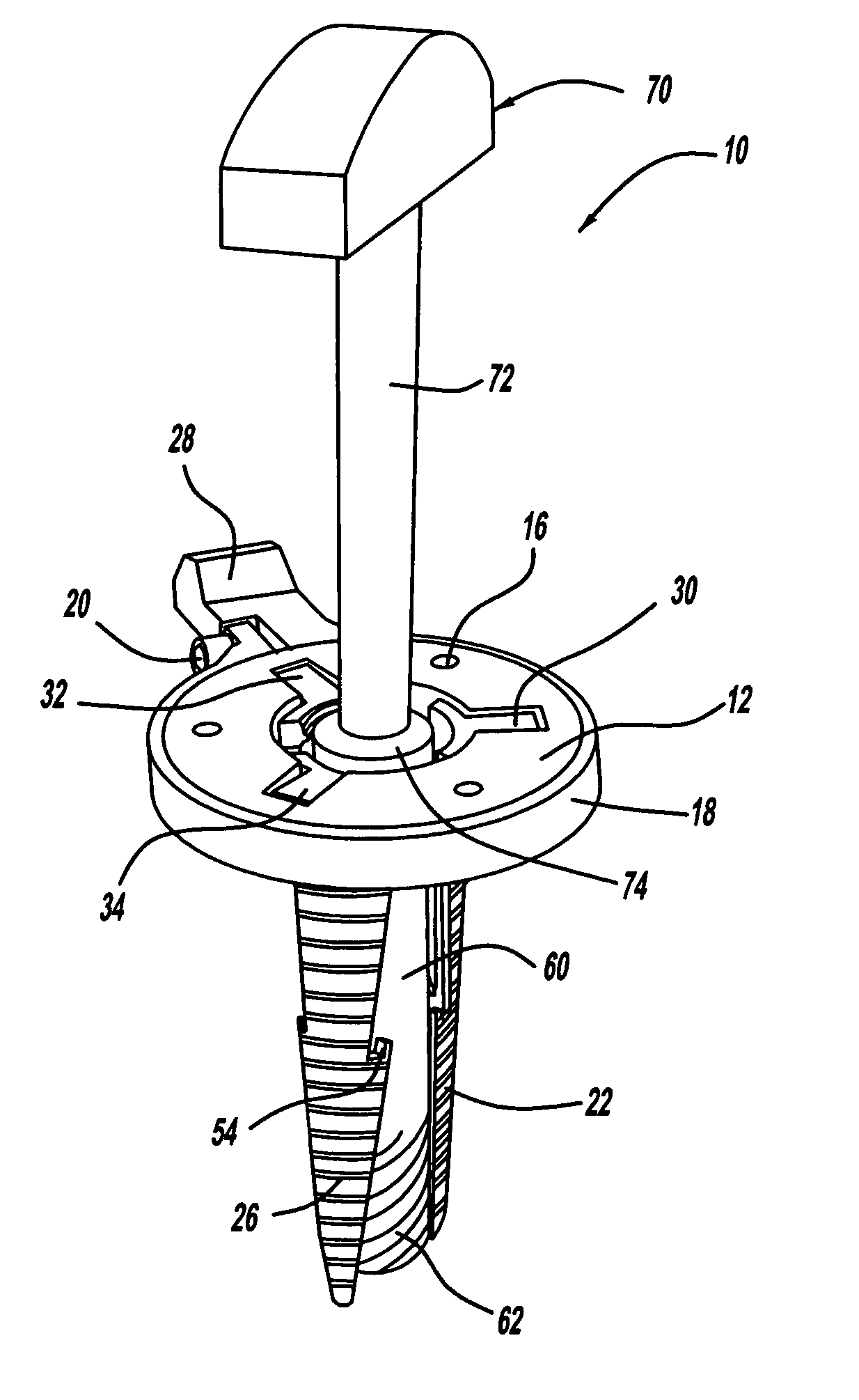
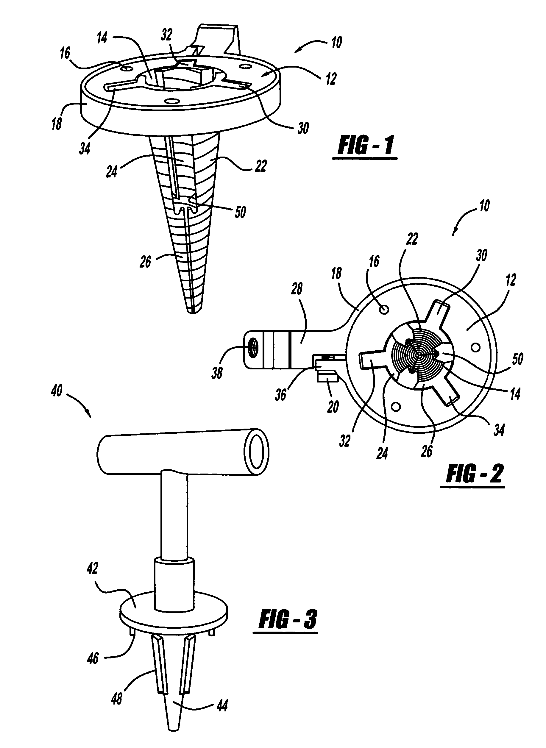
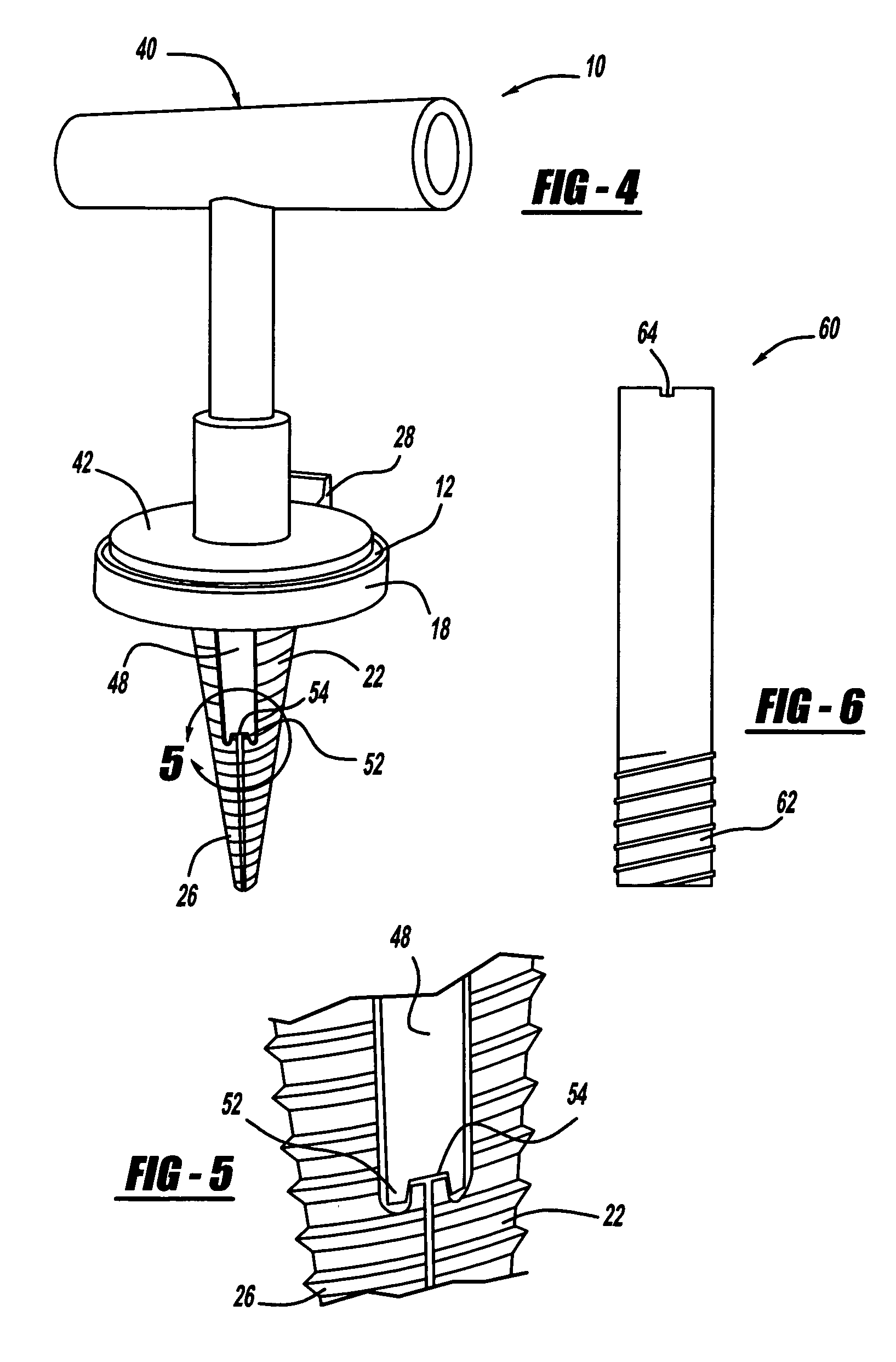
[0022]FIGS. 1-10 show various views of various components of a minimally invasive surgical access device assembly 10, according to an embodiment of the present invention. The various components of the assembly 10 can be fabricated and assembled by any suitable technique, such as molding, stamping, welding, etc., and can be made of any suitable material, such as aluminum, steel, radio-lucent carbon / graphite composites, etc.

[0023]As shown in FIGS. 1 and 2, the access dev...


PUM


Abstract

A minimally invasive surgical access device that allows access to a pathology being treated while significantly reducing the risk of damaging anatomical structures proximate the pathology. The access device includes a base portion having a central bore extending therethrough, and retractor blades pivotably mounted to the base portion. An insertion handle is coupled to the base portion to thread the retractor blades into the patient. The insertion handle is removed from the base portion, and a core hollow screw is threaded into the base portion to separate the retractor blades to gain access to the pathology through the hollow screw.

Description

BACKGROUND OF THE INVENTION[0001]1. Field of the Invention[0002]This invention relates generally to a minimally invasive access device for a surgical procedure and, more particularly, to a minimally invasive access device for spinal surgery, where the device includes retractor blades and an access tube, where the access tube is threaded into the retractor blades to cause them to separate, which provides a corridor for accessing a surgical pathology.[0003]2. Discussion of the Related Art[0004]Traditional surgical approaches for the human body involve the dissection of supporting structures, such as muscle, ligaments and / or bone, to access and expose the pathology being treated. These structures are usually vital to the long term health and function of the body. Typically, these structures are not involved in a disease process, but frequently need to be removed or dissected in order to gain visualization of the pathology.[0005]In the case of spinal disorders, the supporting muscle and...

Claims


Application Information

Patent Timeline
Patent Type & Authority Patents(United States)
IPC IPC(8): A61B1/32
CPCA61B17/02A61B1/32A61B2017/349
Inventor PEREZ-CRUET, MICK J.PEPPER, JOHN R.
Owner THOMPSON MIS
Learn More