
Detection of biospecific interactions using amplified differential time domain spectroscopy signal

a time domain and signal technology, applied in the field of bioanalytical technology, can solve the problems of disturbing the sample, requiring extra steps in sample preparation, and not being able to achieve the effect of simple bioassay procedur

Active Publication Date: 2008-05-06
RENESSELAER POLYTECHNIC INST
View PDF16 Cites 8 Cited by
  • Summary
  • Abstract
  • Description
  • Claims
  • Application Information

AI Technical Summary

Benefits of technology

This approach enhances sensitivity and reduces sample perturbation, allowing for non-invasive detection of biomolecular interactions, such as ligand-receptor binding and DNA hybridization, with improved signal quality and ease of use.

Problems solved by technology

Exploiting this inherent property for bioassay is not a simple procedure, however, because the emitted fluorescence signal is generally weak and often difficult to distinguish above a background signal.
Such addition disturbs the sample, however, and requires extra steps in sample preparation.
Other limitations of fluorescence indicators include fluorescence quenching by other solutes and the insensitivity of fluorescence to certain binding events.
In current solid-phase immunoassay procedures, wash steps are labor intensive and time consuming when performed manually and would require complicated and, therefore, expensive robotics in an automated format.
Unfortunately, compared to the existing biosensors in the marketplace, current THz-based systems suffer from poor signal-to-noise ratios and sensitivity.
Unfortunately, the complications associated with cryogenic detectors in FTIR and Raleigh lines in Raman are among the anomalies that hamper FTIR and Raman spectroscopy from being widely used.
Also, in general, FTIR has a relative poor signal-to-noise ratio for frequencies less than 1 THz.

Method used


Image

  • Detection of biospecific interactions using amplified differential time domain spectroscopy signal
  • Detection of biospecific interactions using amplified differential time domain spectroscopy signal
  • Detection of biospecific interactions using amplified differential time domain spectroscopy signal

Examples


example 1

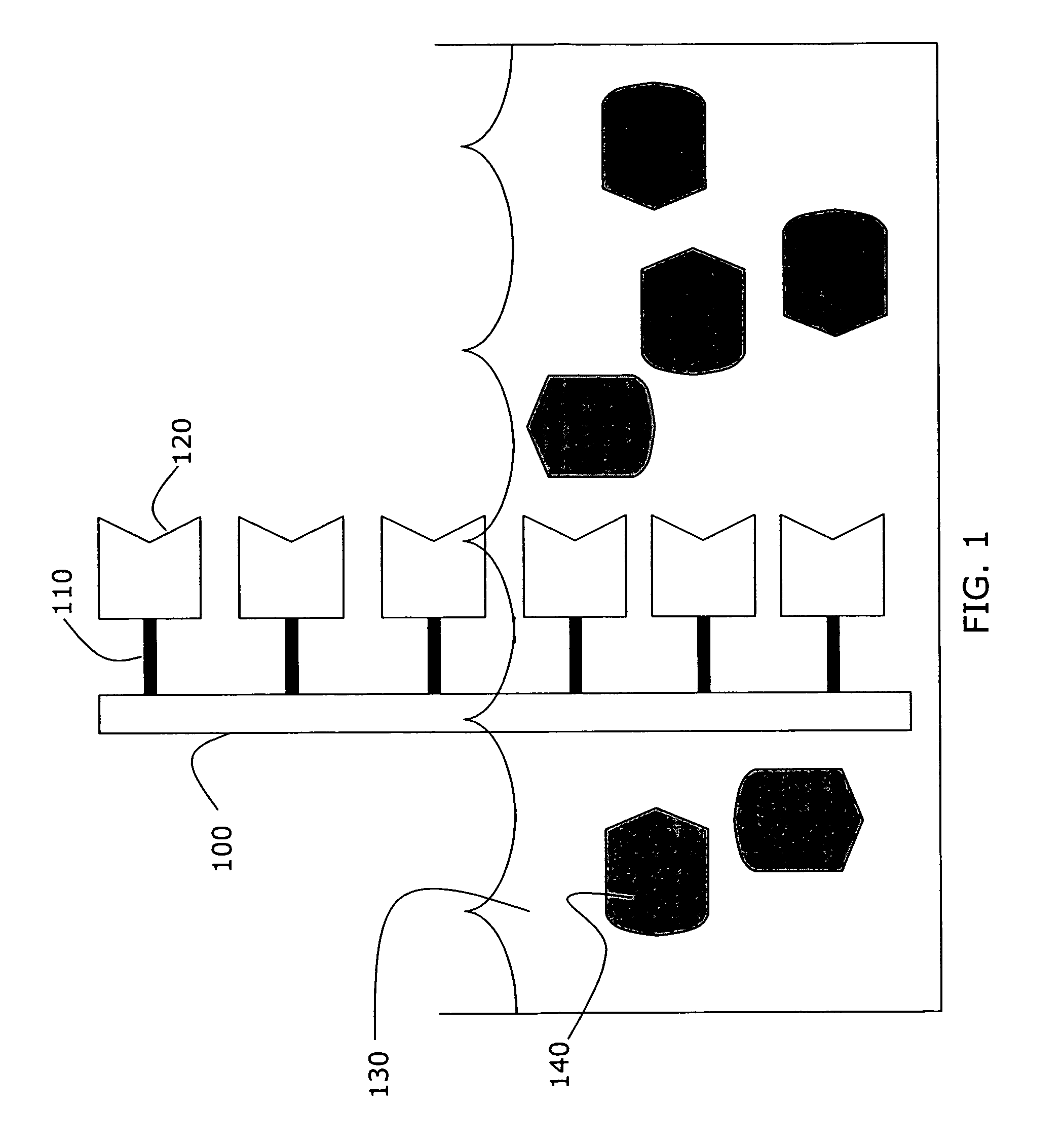
[0043]In an example of the present invention, the binding between the eggwhite glycoprotein, avidin, and vitamin H, biotin was detected by differential time domain THz spectroscopy. Avidin is a known protein characterized by its four identical binding sites for biotin. In this example, biotin is the tethered molecule, and is non-covalently attached to the support surface.

[0044]Prior to biotin deposition, the quartz slides were cleaned in 50% hot nitric acid for an hour, and then rinsed thoroughly in double distilled water. The quartz slides were further subjected to 1 mg / ml octadecanol solution for half an hour. The hydrophobic substrate was immobilized on the hydrophilic surface through molecular self-assembly. The spontaneous organization of the biotin lipid into the octadecanol self-assembled bilayers provides a certain level of stability to the octadecanol-biotin complexes.

[0045]After drying and washing the octadecanol-treated slides with double distilled water, the sample was d...

example 2

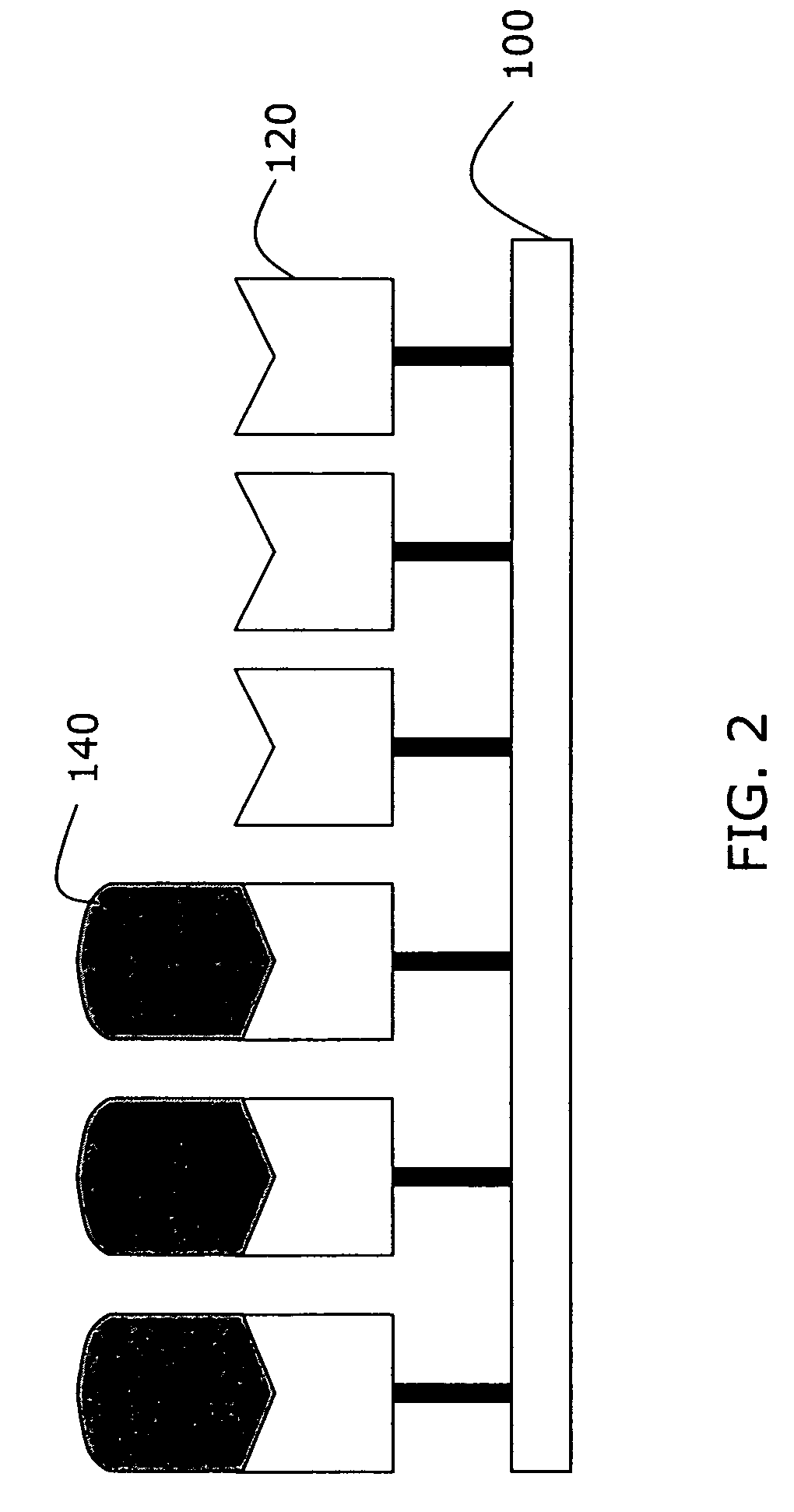
[0048]Similar results were achieved by securing avidin to the slide surface and exposing a portion of the avidin-treated slide to a solution of biotin. In this case, avidin was the tethered molecule, and was covalently attached to a glass slide. The procedure for covalent immobilization of avidin on glass slides included treating the surface of the glass slides with thiol-terminal silanes and a succinimde crosslinker. This treatment increased the density of bound avidin on the glass slides. See also S. Mickan, A. Menikh, J. Munch, D. Abbott & X.-C. Zhang, “Amplification and modeling of bioaffinity detection with terahertz spectroscopy,” Proceedings of SPIE—Biomedical Applications of Micro- and Nanoengineering, SPIE Vol. 4937, pp. 334-42 (Dan Nicolau ed., Melbourne, Australia, Dec. 16-18, 2002), the contents of which are incorporated in this document by reference.

[0049]In preparing the sample slide, the glass slides were cleaned, silanized, and crosslinked prior to deposition of the ...

example 3

[0055]A biotin-coated slide was prepared as in Example 1, and a portion of the slide was exposed to a solution containing avidin conjugated to agarose beads, as illustrated in FIG. 3. Agarose beads 340 bound to several avidin molecules 330 provided a greater change in the index of refraction upon binding to the tethered biotin molecules, thus amplifying the resulting difference signal detected through THz spectroscopy. The signal amplification caused by the agarose beads is apparent in FIG. 4, as the biotin-avidin-agarose signal 420 is approximately 35% greater than the unenhanced biotin-avidin complex.


PUM

PropertyMeasurementUnit
thickaaaaaaaaaa
thicknessaaaaaaaaaa
thicknessaaaaaaaaaa

Abstract

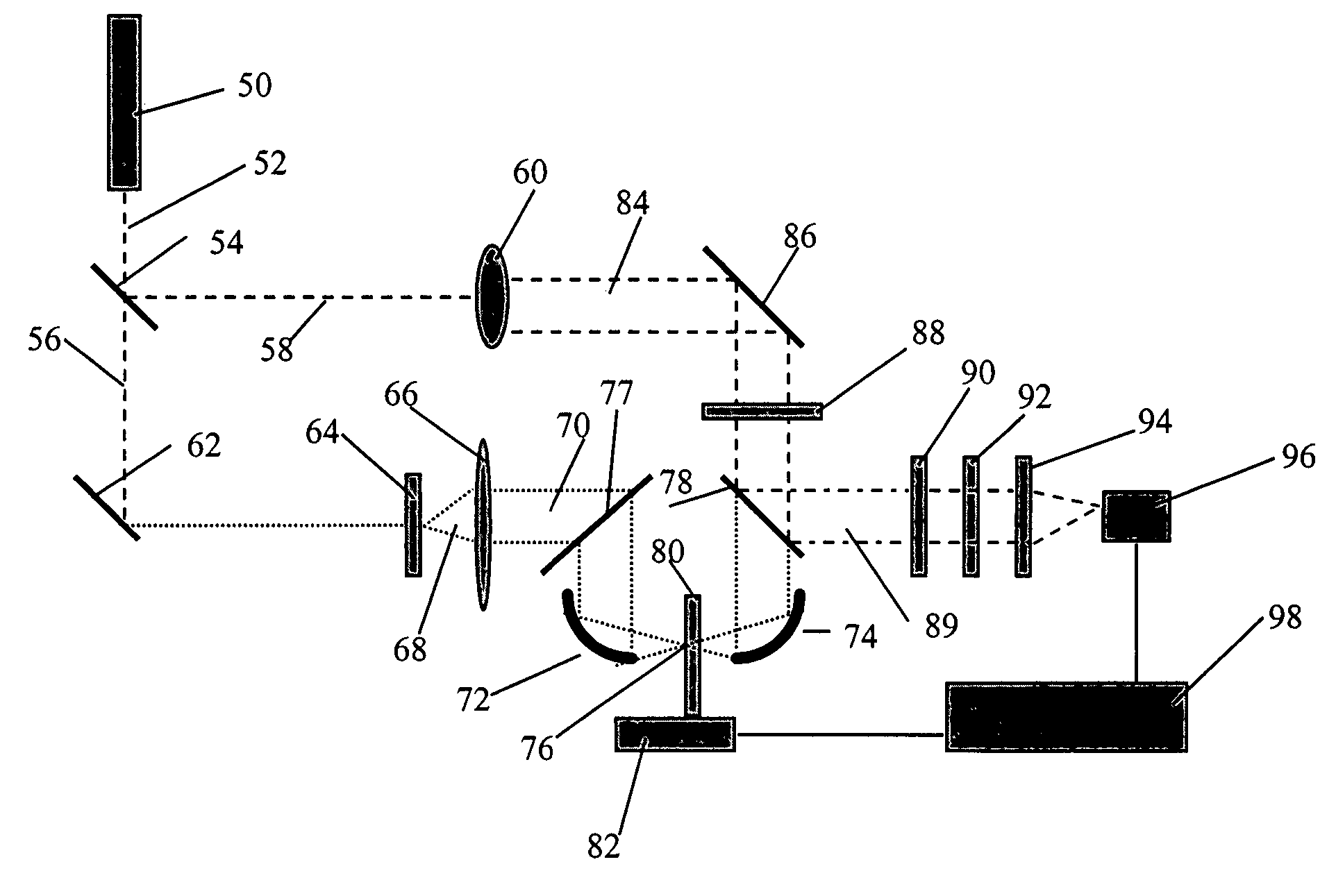
A method for detecting specific associations between a tethered molecule and an untethered target molecule. The method comprises (1) selecting a tethered molecule; (2) alternately impinging THz radiation onto the tethered molecule and onto a sample including the tethered and untethered target molecules; (3) detecting the radiation impinged on the tethered molecule to form a reference signal and the radiation impinged on the sample to form a sample signal; and (4) comparing the reference signal with the sample signal to generate a specimen signal indicative of an association between the selected tethered and target molecules. The method will detect whether a selected tethered molecule and the desired target exhibit any affinity or, in cases where the affinity is known, will detect the presence of the target molecule in a sample. Also provided is an apparatus for detecting specific associations between a tethered molecule and an untethered target molecule.

Description

[0001]This application is a U.S. National Phase Application of PCT International Application PCT / US 03 / 16122 filed May 22, 2003 which claims priority to U.S. Provisional Application 60 / 382,761 filed May 23, 2002.FIELD OF THE INVENTION[0002]This invention relates to bioanalytical technology and, more particularly, to a method and associated apparatus for detecting the affinity between a ligand and a biological specimen and using the detected affinity in a bioassay application.BACKGROUND OF THE INVENTION[0003]The accurate detection of minute amounts of chemical and biological substances has been a major goal in bioanalytical technology throughout the twentieth century. Over the years, a wide variety of biosensing strategies have been developed to satisfy numerous needs. The general aim is to develop a scheme that is able to simplify sample preparation steps, provide high selectivity and sensitivity, respond in a continuous and reversible manner, and accomplish measurements without sam...

Claims


Application Information

Patent Timeline
Patent Type & Authority Patents(United States)
IPC IPC(8): C12M1/34C12M3/00G01N21/35G01N33/543
CPCG01N33/54373G01N21/3586G01N21/3577
Inventor ZHANG, XI-CHENGMICKAN, SAMUEL PETERMENIKH, ABDELLAH
Owner RENESSELAER POLYTECHNIC INST
Learn More