
Endoscope with cleaning optics

a technology of endoscope and optics, which is applied in the field of medical instruments, can solve the problems of obstructing visualization, obstructing visualization, fogging of distal optical surface, etc., and achieves the effects of reducing manufacturing costs, simplifying endoscope design, and preventing barotrauma of the internal organs of patients

Inactive Publication Date: 2008-03-11
M S VISION
View PDF26 Cites 78 Cited by
  • Summary
  • Abstract
  • Description
  • Claims
  • Application Information

AI Technical Summary

Benefits of technology

[0012]The object of the present invention is to provide an endoscope with the complete visualization of patient's internal organs even in the most awkward cases.

Problems solved by technology

The dirtying of the distal optical member surface with patient's secretions such as blood, mucus, vomit, fat creates significant difficulties for the internal organs visualization.
These problems arise in the cases of bleeding an esophageal and gastrointestinal lesions, bleeding or copious secretions in airways, colonoscopy for poorly prepared patient, hysteroscopy during and after curettage and the like.
Another problem obstructing the visualization is fogging the distal optical surface due to the temperature difference between the optical surface and patient's cavity.
Therefore, cleaning the distal optical surface from patient's secretions and fog is one of the most important problems of current endoscope developments.
The disadvantage of these endoscopes is low effectiveness of optical surface cleaning from very adhesive substances such as blood, mucus, fat due to low air pressure and the direction of air / water flow, which does not collide with the optical surface and, therefore, cannot sweep off the dirt.
The supply of air / water flow of sufficient pressure is impossible, since this leads to barotrauma of patient internal organs by air / water jet.
Moreover, the internal organ barotrauma also can be caused by internal pressure during the operation in closed body cavity.
The devices do not have any means for the removal of excessive air / water from body cavity or for the dissipation of air / water jet.
Another disadvantage is excessive complexity and manufacturing cost of the endoscope system due to the dual (air and water) cleaning system and cumbersome supply and control system disposed beyond the endoscope device.
Another disadvantage is the arrangement of air / water nozzle in front of the optical surface (see U.S. Pat. Nos. 4,509,507, 4,667,655) restricting the vision field of the optical surface.
The disadvantage of this patent is very cumbersome and expensive design due to the dual cleaning system including the numerous equipment disposed outside the endoscope device.
However, upon sufficient air / water high pressure, the suction means and the barrier do not create a catcher completely able to catch air / water jets and to exclude deflection thereof towards patient internal organs, thus, barotrauma may occur.
Consequently, the capability of the effective cleaning of the optical surface is restricted.
Moreover, during water supply, the suction means cannot only remove water from a body cavity.
Inevitably, it also sucks in air creating an undesirable (and dangerous) vacuum in the body cavity.
Another disadvantage is the relatively great duration of the cleaning process consisting of washing the distal surface and successively drying it by air.
This somewhat simplifies the tubing, but complicates the supply control system.
On the other hand, the gap decreases the vacuum in the path of airflow going out of the nozzle and thereby lowers the possibility of its catch and removal.
Thus, the common disadvantages of the endoscope systems with cleaning optics are: their excessive complexity caused by dual (gas and liquid) cleaning systems and cumbersome and expensive pump and control means disposed outside the endoscope device; low cleaning effectiveness due to low pressure of cleaning fluid (gas and liquid); the absence of effective means for catching and removing the cleaning flow; the absence of reliable control means allowing to adjustment of the pressure in the body cavity required for surgical operations.
However, the device cannot provide cleaning of the optical surface from patient secretion due to low air pressure.
The device does not comprise any means preventing patient internal organs from barotrauma.
Therefore, its air pressure cannot be increased up to the value sufficient for effective cleaning the optical surface.
However, this oxygen supply cannot effectively clean the optical surface due to the low oxygen pressure and the absence of special oxygen flow directing towards the optical surface.
It is impossible to apply sufficient oxygen pressure here because the oxygen flow is directed distally and can cause the barotrauma of patient internal organs.
However, the application of high pressure oxygen for cleaning the optical surface is not disclosed in the prior art.
A main disadvantage of the device is that its application with the endoscope is a very cumbersome, expensive, and inconvenient combination including suction and pressurized gas supply means, pressure control system, vacuum and pressure pipes, as well as an endoscope cleaning system with identical equipment.
The disadvantage of known curettage devices is the absence of a visualization means eliminating the capability of operative estimate of curettage results.
This worsens the quality of the procedure, leads to patient traumatization and successive complications, and extends the operation duration.
The application of the visualization means immediately in the curettage device is hampered due to very significant dirtying of the optical surface by curettage products.
Another disadvantage of the curettage devices is the arrangement of the vacuum pump beyond the curettage device.
This increases the number of communication, heightens the manufacturing cost of the curettage system, and is inconvenient in the operation.

Method used


Image

  • Endoscope with cleaning optics
  • Endoscope with cleaning optics
  • Endoscope with cleaning optics

Examples


Embodiment Construction

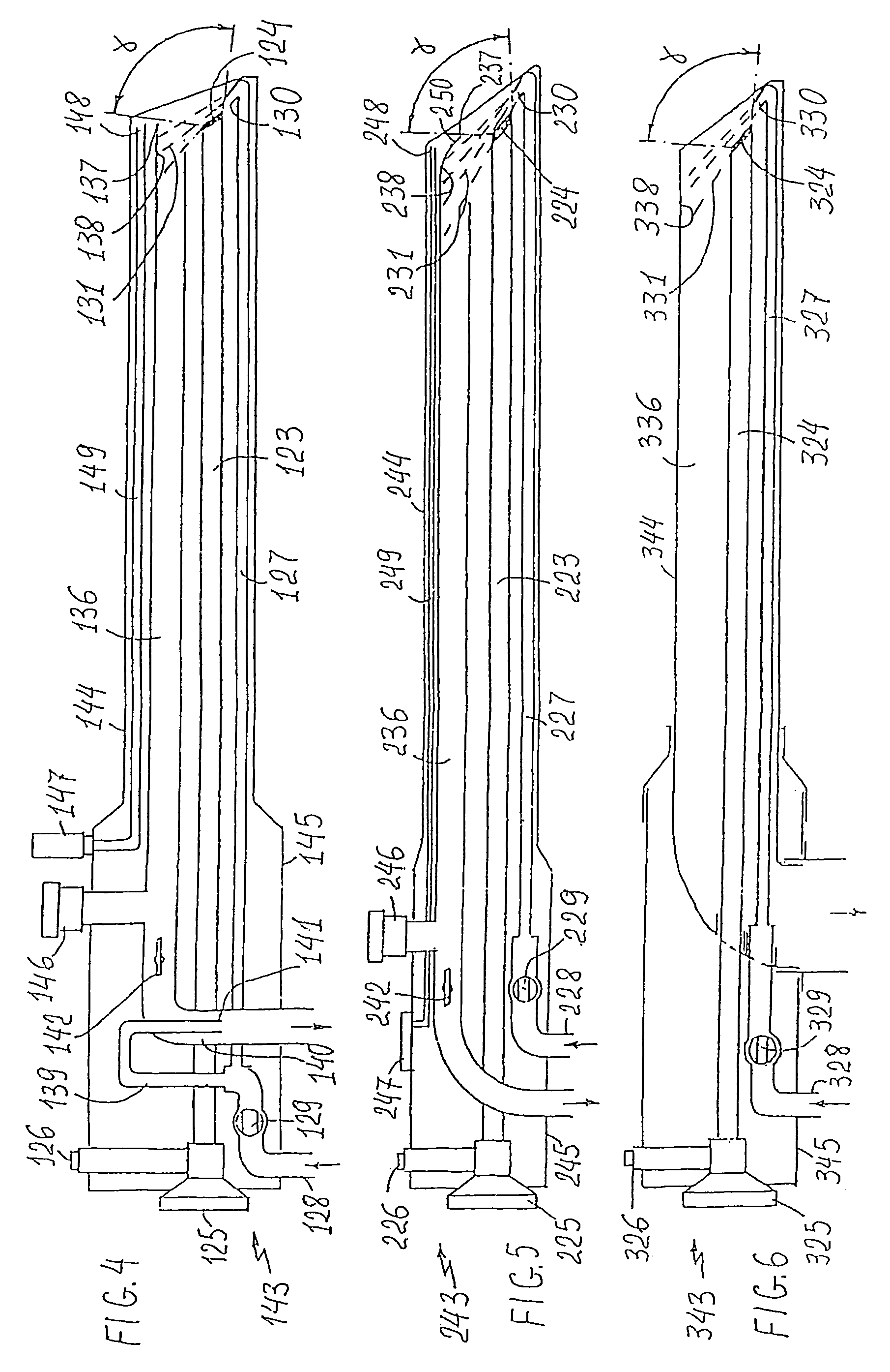
[0040]An explanation of the present invention is offered with reference made to the attached drawings in FIGS. 1 to 14.

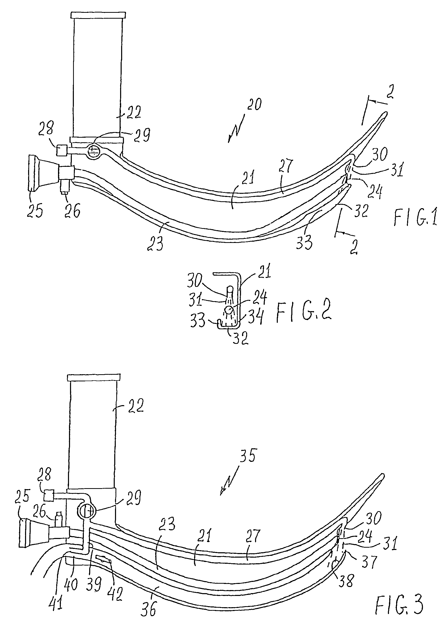
[0041]FIG. 1 shows a laryngoscope 20 with a blade 21, a handle 22, an optic channel 23 with a distal optical surface 24, an eyepiece 25 that can be connected to a display and a connector 26 for connection to a light source. There is also a pressurized gas supply channel 27 extending along the blade 21 and having an inlet port 28 connected with a pressurized gas source (not shown), disposed beyond laryngoscope 20, a valve 29 of type ON / OF, and an outlet nozzle 30 located at a distal end of channel 27. The nozzle 30 is directed towards the optical surface 24 so that a gas jet 31 going out of the nozzle 30 touches the optical surface 24 sweeping off the dirt from it. The pressure value of gas supplied to the nozzle 30 is sufficient to convert the gas jet 31 into a high pressure gas jet capable of completely cleaning the optical surface 24 of any patient secretions and ...


PUM


Abstract

The instrument has an original cleaning system, which includes a gas nozzle supplying a gas jet onto the optical surface under high pressure. This pressure is sufficient to enable the gas jet to be a single cleaning means for cleaning the optical surface in all clinical instances. A safety means prevents the patient's internal organs from barotrauma by the gas jet and includes a gas jet catcher and a suction pump of ejection type. The latter is arranged in the instrument's handle and can be used also for the removal of abundant secretions or body's small particles from the patient's cavity. Besides, the gas jet and the suction pump along with a control means form the system for pressure control within the patient's cavity.

Description

FIELD OF THE INVENTION[0001]This invention relates generally to medical instruments for the operation in patient's body cavity requiring the visualization of internal organs. More particularly, it relates to intubation laryngoscopes, bronchoscopes, endoscopes of various applications, vacuum curettage devices, and the like.BRIEF DESCRIPTION OF THE PRIOR ART[0002]All the endoscope systems applied for the visualization of patient's internal organs comprise at least one endoscope distal optical member disposed at the endoscope distal end. The dirtying of the distal optical member surface with patient's secretions such as blood, mucus, vomit, fat creates significant difficulties for the internal organs visualization. These problems arise in the cases of bleeding an esophageal and gastrointestinal lesions, bleeding or copious secretions in airways, colonoscopy for poorly prepared patient, hysteroscopy during and after curettage and the like. Another problem obstructing the visualization i...

Claims


Application Information

Patent Timeline
IPC IPC(8): A61B1/015A61B1/12A61B1/267A61B1/303A61B19/00A61M1/00A61M13/00
CPCA61B1/00091A61B1/126A61B1/127A61B1/267A61M1/0039A61M1/0076A61M13/003A61B1/303A61B19/34A61B2019/343A61B90/70A61B2090/701A61M1/76A61M1/804
Inventor SHALMAN, MICHAEL
Owner M S VISION
Learn More