Nogo receptor-mediated blockade of axonal growth
a nogo receptor and axonal growth technology, applied in the field of neuronal and molecular biology, can solve the problems of damage to the central nervous system, degradation of the myelin surrounding the axon, and the inability of damaged neurons to regenerate in the central, and achieve the effect of decreasing the nogo-dependent inhibition of axonal growth
- Summary
- Abstract
- Description
- Claims
- Application Information
AI Technical Summary
Benefits of technology
Problems solved by technology
Method used
Image
Examples
example 1
Identification of Nogo as a Member of the Reticulon Family of Proteins
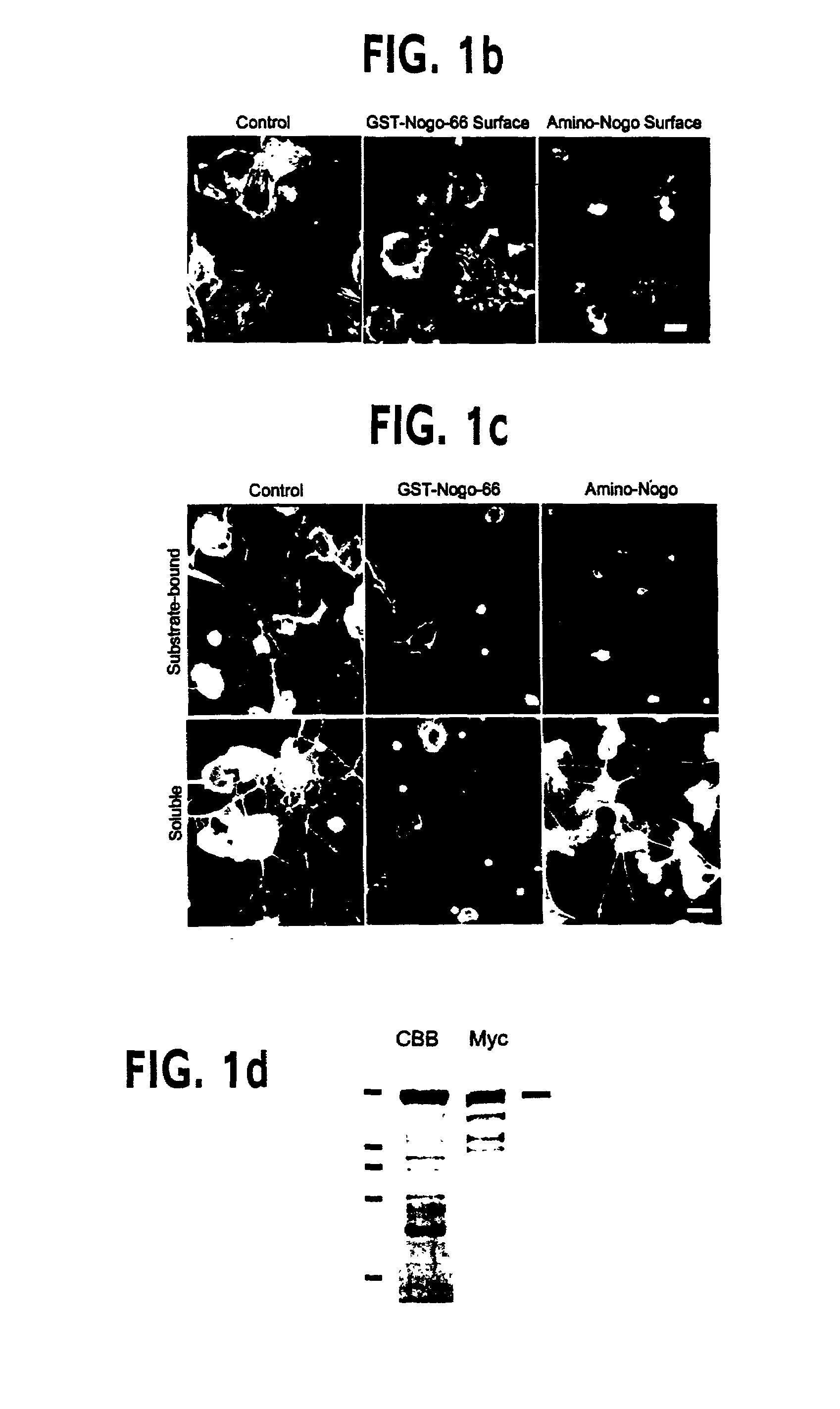
[0194]Adult mammalian axon regeneration is generally successful in the periphery but dismally poor in the CNS. However, many classes of CNS axons can extend for long distances in peripheral nerve grafts (Benfy & Aguayo (1982) Nature 296, 150–152). Comparison of CNS and peripheral nervous system (PNS) myelin has revealed that CNS white matter is selectively inhibitory for axonal outgrowth (Schwab & Thoenen (1985) J. Neurosci. 5, 2415–2423). Several components of CNS white matter, NI35, NI250 (Nogo) and MAG, with inhibitory activity for axon extension have been described (Wang et al., (1999) Transduction of inhibitory signals by the axonal growth cone, in Neurobiology of Spinal Cord Injury, Kalb & Strittmatter (editors) Humana Press; Caroni & Schwab, (1988) J. Cell Biol. 106, 1281–1288; Spillmann et al., (1998) J. Biol. Chem. 73, 19283–19293; McKerracher et al., (1994) Neuron 13, 805–811; Mukhopadhyay et al., (1994)...
example 2
Polyclonal Antibodies Against Nogo
[0201]The predicted intra-membrane topology of the two hydrophobic domains of Nogo indicates that the 66 amino acid residues between these segments is localized to the lumenal / extracellular face of the membrane. To explore this further, an antiserum directed against the 66 amino acid domain was generated.
[0202]For antibody production and immunohistology, anti-Myc immunoblots and immunohistology with the 9E10 antibody were obtained as described in Takahashi et al., (1998) Nature Neurosci., 1, 487–493 & Takahashi et al., (1999) Cell, 99, 59–69. The GST-Nogo fusion protein was employed as an immunogen to generate an anti-Nogo rabbit antiserum. Antibody was affinity-purified and utilized at 3 μg / ml for immunohistology and 1 μg / ml for immunoblots. To assess the specificity of the antiserum, staining was conducted in the presence of GST-Nogo protein at 0.1 mg / ml. For live cell staining, cells were incubated in primary antibody dilutions at 4° C. for one h...
example 3
Nogo Expression in the Central Nervous System
[0204]If Nogo is a major contributor to the axon outgrowth inhibitory characteristics of CNS myelin as compared to PNS myelin (Caroni & Schwab, (1988) J. Cell Biol. 106, 1281–1288; Spillmann et al., (1998) J. Biol. Chem. 73, 19283–19293; Bregman et al., (1995) Nature 378, 498–501), then Nogo should be expressed in adult CNS myelin but not PNS myelin. Northern blot analysis of Nogo expression was performed using probes derived from the 5′ Nogo-A / B-specific region and from the 3′ Nogo common region of the cDNA. A single band of about 4.1 kilobase was detected with the 5′ probe in adult rat optic nerve total RNA samples, but not sciatic nerve samples. The results indicate that the Nogo-A clone is a full length cDNA, and are consistent with a role for Nogo as a CNS-myelin-specific axon outgrowth inhibitor. Northern blot analysis with a 3′ probe reveals that optic nerve expresses high levels of the Nogo-A mRNA and much lower levels of Nogo-B a...
PUM
| Property | Measurement | Unit |
|---|---|---|
| Fraction | aaaaa | aaaaa |
| Fraction | aaaaa | aaaaa |
| Fraction | aaaaa | aaaaa |
Abstract
Description
Claims
Application Information
 - R&D
- Intellectual Property
- Life Sciences
- Materials
- Tech Scout
- Unparalleled Data Quality
- Higher Quality Content
- 60% Fewer Hallucinations
Browse by: Latest US Patents, China's latest patents, Technical Efficacy Thesaurus, Application Domain, Technology Topic, Popular Technical Reports.
© 2025 PatSnap. All rights reserved.Legal|Privacy policy|Modern Slavery Act Transparency Statement|Sitemap|About US| Contact US: help@patsnap.com


