Method and apparatus for collecting and processing physical space data for use while performing image-guided surgery

a technology of physical space and data collection, applied in the field of using imageguided surgery techniques, can solve the problems of frame discomfort for patients, dos-based systems without the continuing support of hardware vendors, etc., and achieve the effect of thorough investigation of the accuracy of all registration techniques

Inactive Publication Date: 2006-07-04
VANDERBILT UNIV
View PDF38 Cites 260 Cited by
  • Summary
  • Abstract
  • Description
  • Claims
  • Application Information

AI Technical Summary

Problems solved by technology

Unfortunately, frames are uncomfortable to patients, must be applied prior to imaging, and are cumbersome in the imaging environment and the operating room.
As the PC hardware evolved, however, it became apparent that DOS-based systems would not have the continuing support of hardware vendors.

Method used


Image

  • Method and apparatus for collecting and processing physical space data for use while performing image-guided surgery
  • Method and apparatus for collecting and processing physical space data for use while performing image-guided surgery
  • Method and apparatus for collecting and processing physical space data for use while performing image-guided surgery

Examples


Embodiment Construction

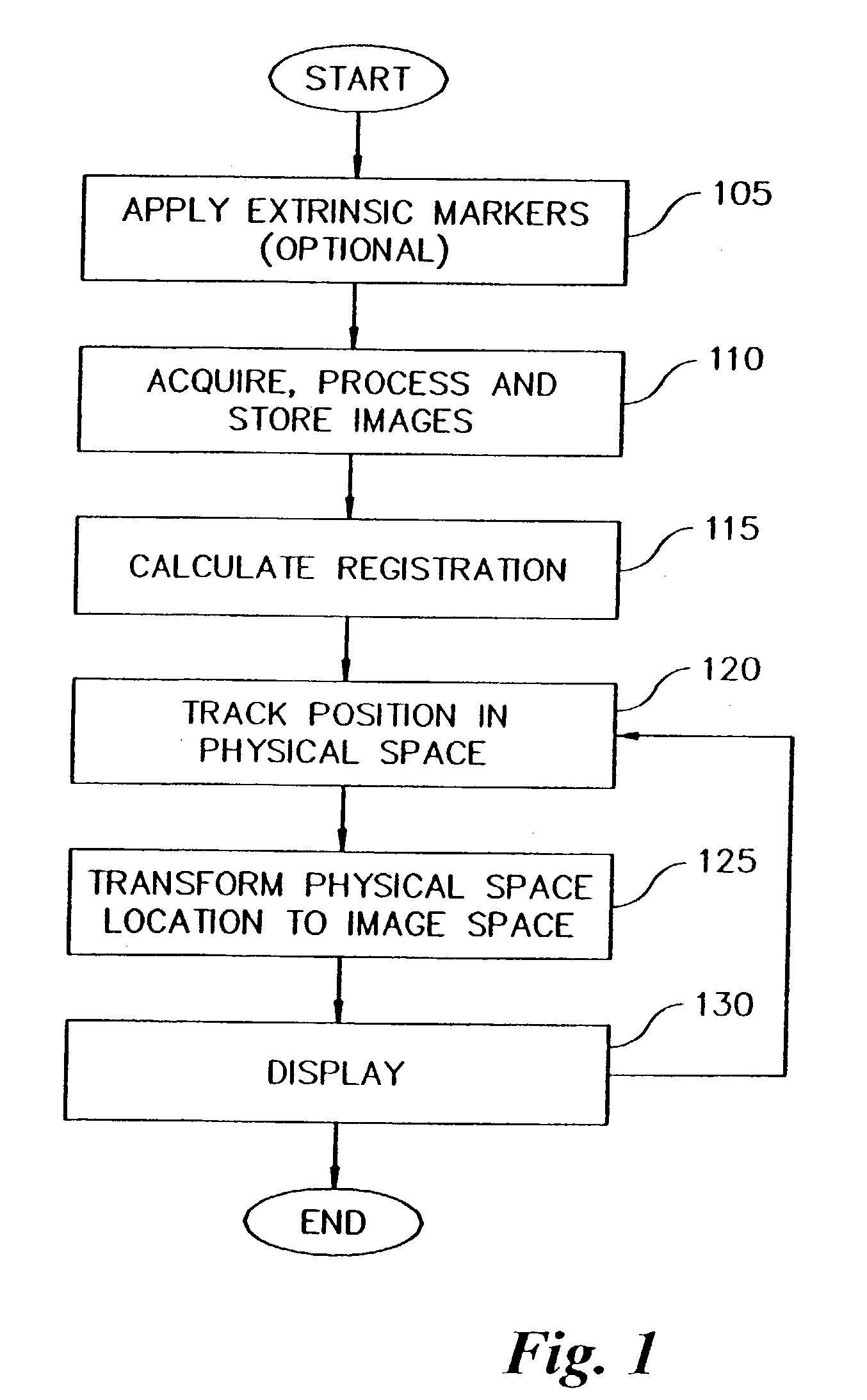
[0040]FIG. 1 shows some of the major events involved in preparing for and performing IIGS. In step 105, it is determined if any extrinsic markers will be attached to the patient. These makers, or fiducials, are designed to be imaged and then localized in both image space and physical space for use in a point-based registration algorithm. Appropriate image volumes for a patient are then acquired, stored and processed (step 110). Most image volumes are acquired as a set of slices, 256×256 or 512×512 pixels per slice at 2 bytes per pixel with 20–200 images per volume. These images are acquired on a computerized tomography (CT) scanner, a magnetic resonance imaging (MRI) scanner or a positron emission tomography (PET) scanner. Images are typically transferred from PACS servers in radiology to image-guided surgical computers, where they are processed for display before surgery if necessary. Transverse, sagittal, or coronal tomographic slices require minor processing before display. In or...


PUM


Abstract

A method and apparatus for collecting and processing physical space data used while performing image-guided surgery is disclosed. Physical space data is collected by probing physical surface points of surgically exposed tissue. The physical space data provides three-dimensional (3-D) coordinates for each of the physical surface points. Based on the physical space data collected, point-based registrations used to indicate surgical position in both image space and physical space are determined. In one embodiment, the surface of surgically exposed tissue of a living patient is illuminated with laser light. Light reflected from the illuminated surface of the exposed tissue is received and analyzed. In another embodiment, one or more magnetic fields of known shape and size are established in the proximity of the exposed tissue. Data associated with the strength of the magnetic fields is acquired and analyzed.

Description

CROSS-REFERENCE TO RELATED APPLICATIONS[0001]This application is a continuation-in-part of U.S. patent application Ser. No. 09 / 894,732, filed Jun. 27, 2001, now a U.S. Pat. No. 6,584,339 entitled “Method and Apparatus for Collecting and Processing Physical Space Data for Use While Performing Image-Guided Surgery,” which is incorporated herein by reference in its entirety.STATEMENT REGARDING FEDERALLY SPONSORED RESEARCH OR DEVELOPMENT[0002]This work was supported in part by grants from the National Institutes of Health (NIH) and the National Science Foundation (NSF) (NIH grants NIGMS GM52798, NRSA #1 F32 DK 09671-01 SB and R21 CA 91352-01; and NSF grants BES-9703714) and the U.S. Government may therefore have certain rights in this invention.BACKGROUND OF THE INVENTION[0003]1. Field of the Invention[0004]The present invention relates to using image-guided surgery techniques to collect data to insure accurate tracking of an ablation device.[0005]2. Background Information[0006]For over...

Claims


Application Information

Patent Timeline
Patent Type & Authority Patents(United States)
IPC IPC(8): A61B5/05A61BA61B8/00A61B8/12A61B8/14A61B18/02A61B18/14A61B19/00
CPCA61B19/52A61B19/5244A61B18/02A61B18/14A61B2019/5272A61B2019/507A61B2019/5255A61B2019/527A61B19/56A61B34/20A61B34/25A61B90/36A61B2034/107A61B2034/2055A61B2034/207A61B2034/2072
Inventor GALLOWAY, JR., ROBERT L.CHAPMAN, WILLIAM C.STEFANSIC, JAMES D.HERLINE, ALAN J.MIGA, MICHAEL I.CASH, DAVID M.SINHA, TUHIN K.
Owner VANDERBILT UNIV
Why Eureka
Try Eureka