
Automatic selection of the log-subtraction decomposition parameters for dual energy chest radiography

Inactive Publication Date: 2006-06-27
GE MEDICAL SYST GLOBAL TECH CO LLC
View PDF3 Cites 34 Cited by
  • Summary
  • Abstract
  • Description
  • Claims
  • Application Information

AI Technical Summary

Benefits of technology

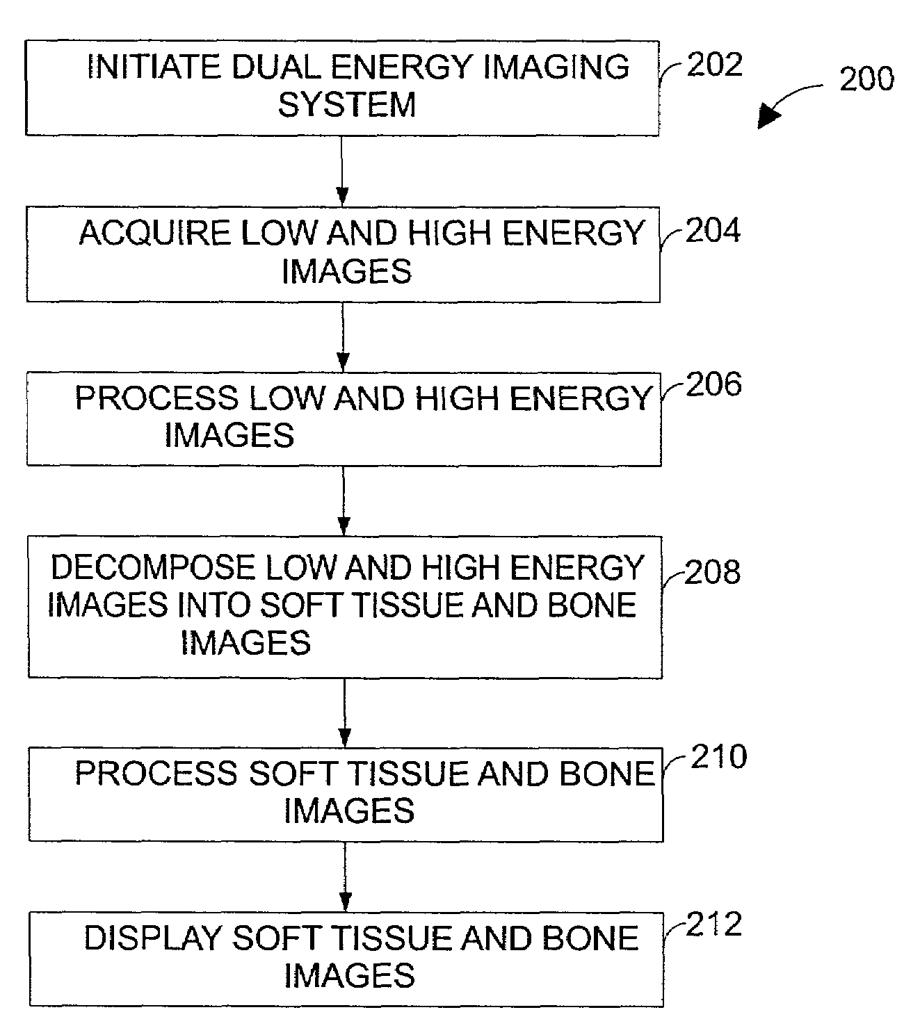
[0008]The present technique provides a variety of processing schemes for decomposing soft tissue and bone images more accurately from low and high-energy images acquired from an imaging system, such as a dual-energy digital radiography system using flat-panel technology. In particular, a parameter selection process is provided for automatically computing the decomposition parameters WS and WB to create soft tissue and bone images, respectively. The parameter selection process modifies a default decomposition parameter based on a variety of image and technique variables, such as intensity levels of the low and high-energy images, the patient size, and the collimator filtration setting. The parameter selection process avoids robustness problems associated with image-based algorithms, and the process may operate without any direct user interaction.

Problems solved by technology

Unfortunately, the soft tissue and bone decomposition parameters may be functions of several image and techniques variables, thereby complicating the selection of these parameters.
Moreover, the decomposed images typically have significant noise, contrast artifacts, and motion artifacts, which degrade the images and reduce the value of the images for medical diagnosis.
These artifacts are generally mitigated by post-decomposition processing techniques, yet the decomposed images still exhibit significant artifacts.
At relatively attenuated regions of the image, the foregoing dual-energy decomposition equations produce relatively noisy decomposed images.
For example, during a low-dose clinical data acquisition, the computationally efficient decomposition equations amplify noise and produce very noisy decomposed images at highly attenuated regions of the image.
However, the foregoing decomposition equations tend to amplify noise in the images, and the existing noise reduction techniques fail to mitigate the noise adequately.
Although the two images may be acquired over a relatively short time interval, such as 100–200 ms, these motion artifacts may significantly degrade the quality of the decomposed images.
The residual rib structure, which is present in about 30 percent of acquisitions, decreases the conspicuity of lung pathology and essentially defeats the purpose of generating soft tissue lung images by dual-energy imaging.
Traditional methods to correct for motion artifacts are relatively ineffective for dual-energy imaging, because the dual-energy images have significantly different local contrasts.

Method used


Image

  • Automatic selection of the log-subtraction decomposition parameters for dual energy chest radiography
  • Automatic selection of the log-subtraction decomposition parameters for dual energy chest radiography
  • Automatic selection of the log-subtraction decomposition parameters for dual energy chest radiography

Examples


Embodiment Construction

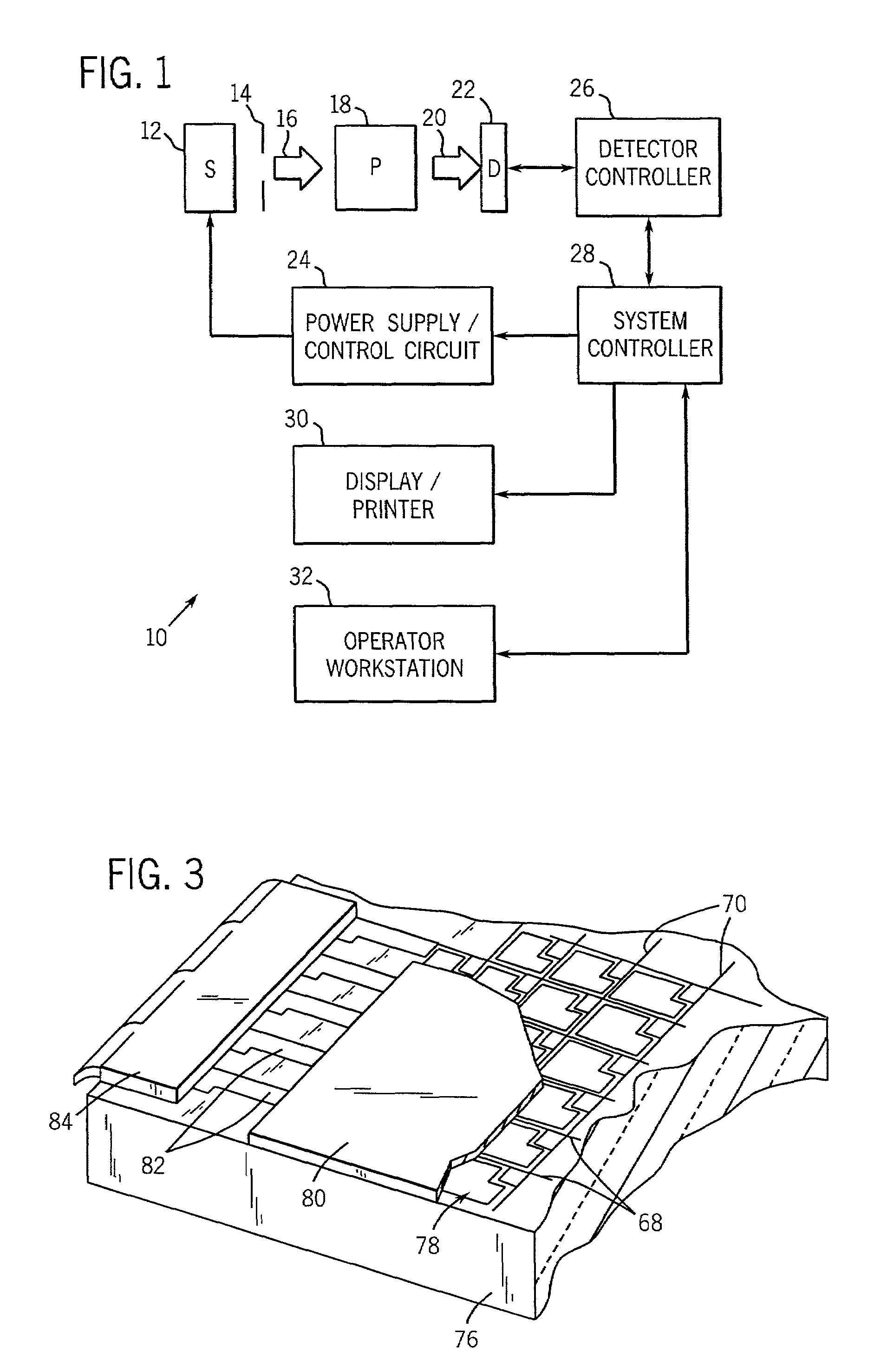
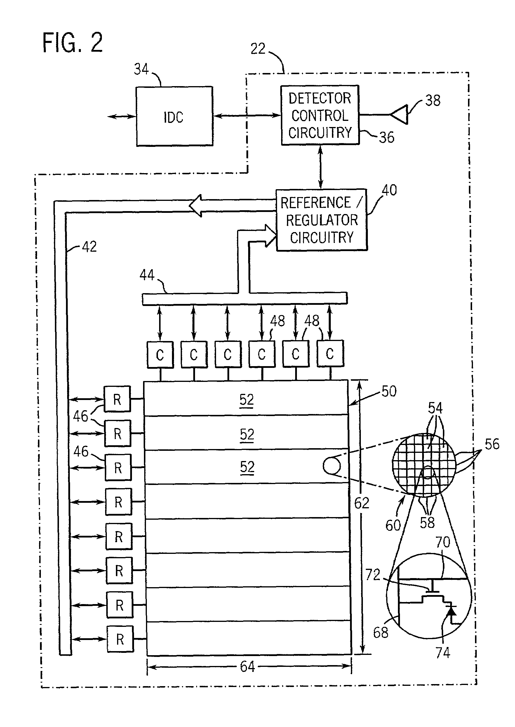
[0023]FIG. 1 illustrates diagrammatically an imaging system 10 for acquiring and processing discrete pixel image data. In the illustrated embodiment, system 10 is a digital X-ray system designed both to acquire original image data and to process the image data for display in accordance with the present technique. For example, the system 10 may acquire multiple images of a desired anatomy over a short time interval for comparison and processing, such as high and low-energy image exposures used for a dual-energy decomposition system. Accordingly, the system 10 may embody a dual-energy digital X-ray system, which is operable to decompose high and low-energy image exposures into soft tissue and bone images for further analysis of the desired anatomy. In the embodiment illustrated in FIG. 1, imaging system 10 includes a source of X-ray radiation 12 positioned adjacent to a collimator 14. Collimator 14 permits a stream of radiation 16 to pass into a region in which a subject, such as a hu...


PUM


Abstract

The present technique provides a variety of processing schemes for decomposing soft tissue and bone images more accurately from low and high-energy images acquired from an imaging system, such as a dual-energy digital radiography system using flat-panel technology. In particular, a parameter selection process is provided for automatically computing the decomposition parameters WS and WB to create soft tissue and bone images, respectively. The parameter selection process modifies a default decomposition parameter based on a variety of image and technique variables, such as intensity levels of the low and high-energy images, the patient size, and the collimator filtration setting. The parameter selection process avoids robustness problems associated with image-based algorithms, and the process may operate without any direct user interaction.

Description

BACKGROUND OF THE INVENTION[0001]The present invention relates generally to imaging systems, such as radiographic systems, and more particularly, to processing techniques for dual-energy radiography. Even more particularly, the present invention relates to a system and method for selecting decomposition parameters WS and WB for creating soft tissue and bone images from low and high-energy images acquired from an imaging system, such as a dual-energy digital radiography system using flat-panel technology.[0002]Medical diagnostic and imaging systems are ubiquitous in modern health care facilities. Currently, a number of modalities exist for medical diagnostic and imaging systems. These include computed tomography (CT) systems, x-ray systems (including both conventional and digital or digitized imaging systems), magnetic resonance (MR) systems, positron emission tomography (PET) systems, ultrasound systems, nuclear medicine systems, and so forth. Such systems provide invaluable tools f...

Claims


Application Information

Patent Timeline
IPC IPC(8): G06K9/00A61B6/00G06T5/00G06T5/50
CPCA61B6/405A61B6/482A61B6/505G06T5/50G06T2207/30061G06T2207/10116G06T2207/30008
Inventor JABRI, KADRI N.AVINASH, GOPAL B.SABOL, JOHN M.NICOLAS, FRANCOIS S.UPPALURI, RENUKAHAMADEH, MOHAMED A.
Owner GE MEDICAL SYST GLOBAL TECH CO LLC
Learn More