Single or multi-mode cardiac activity data collection, processing and display obtained in a non-invasive manner

a non-invasive technology, applied in the field of single or multi-mode cardiac activity data collection, processing and display obtained in a non-invasive manner, to achieve the effect of improving the signal-to-noise ratio (snr) and being easy to recogniz

Inactive Publication Date: 2006-05-09
TARJAN PETER P +2
View PDF3 Cites 182 Cited by
  • Summary
  • Abstract
  • Description
  • Claims
  • Application Information

AI Technical Summary

Benefits of technology

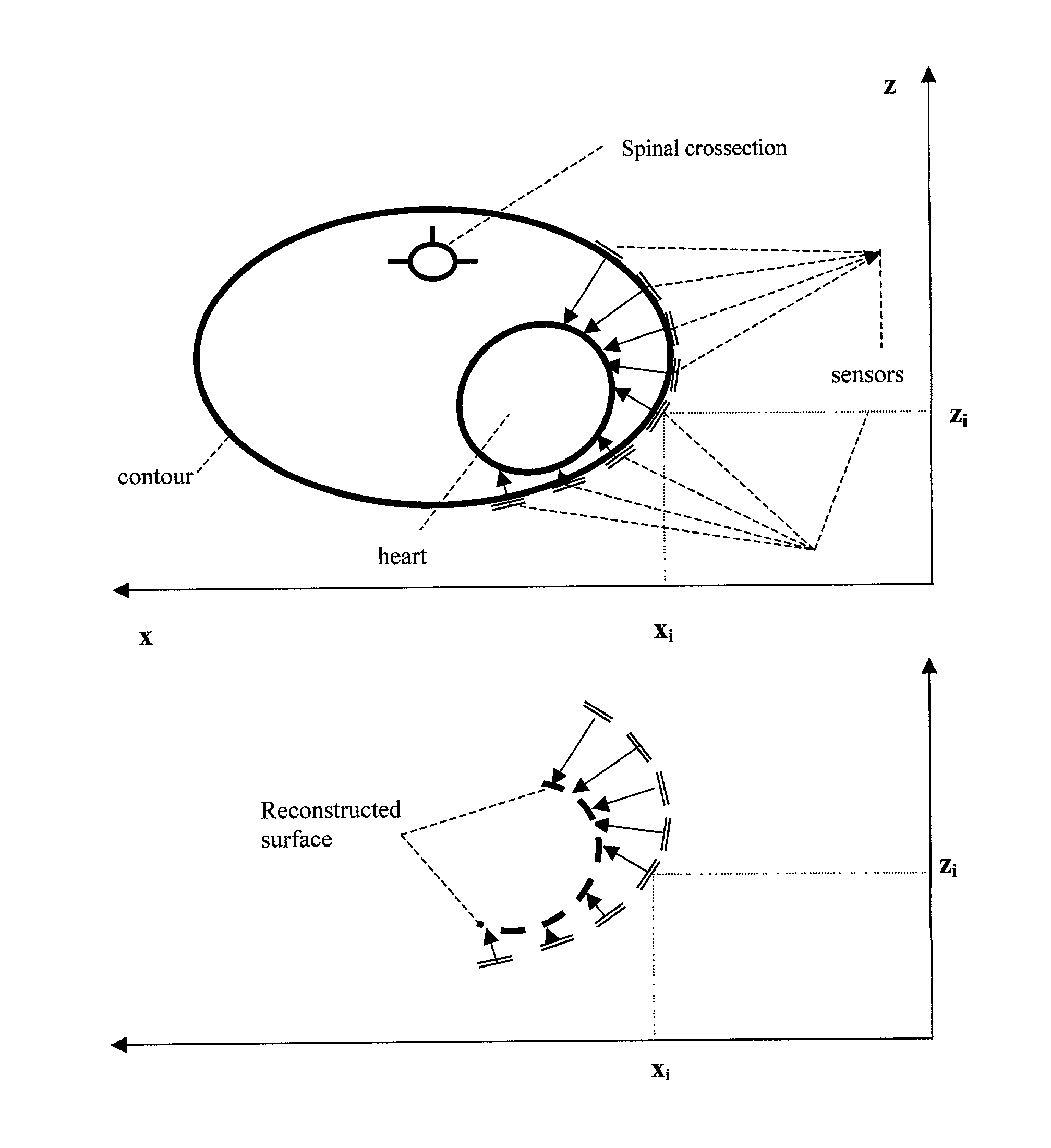
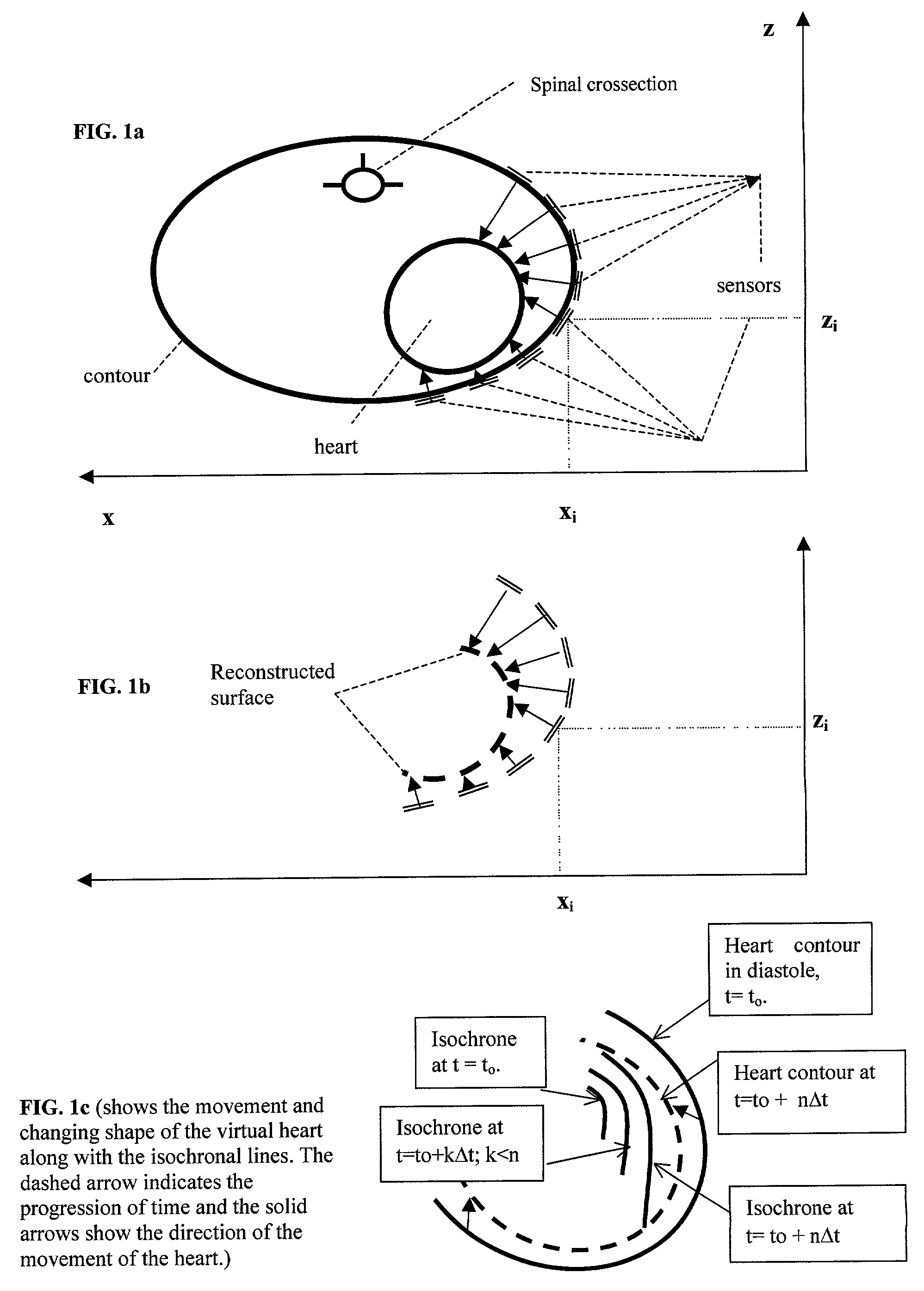
[0046]If one relies on the active sensors' far-field discriminatory behavior, then it is possible to postulate sources near the sensor, which will produce similar signals as the naturally produced data. This appears to be realizable if the instantaneous position of the source at the MOA is along the axis of the sensor. The distance from the sensor to the epicardium, the third part of the coordinate set for the dipole, may be obtained by using ultrasound. This implies that a partially open surface image of the epicardium of the left heart may be obtained non-invasively that depicts both the electrical activity and the instantaneous positions of the sources, as applicants expect to be able to provide the spatial information as well. Such a system provides a novel and economic way for the cardiologist to obtain monitoring and diagnostic data, as well as visual guidance in the delivery of interventional therapies, such as tissue ablation.
[0054]The isochrones are the curves on the chest surface which indicate the connecting line for simultaneously depolarized zones tissue . Their utility may be in detecting zones of delayed depolarization such as infracted or ischemic areas. The utility of such displays is in diagnosing the site and dimensions of an infarct. The display may also present valuable diagnostic data about the direction of propagation of the activity as those trajectories are orthogonal to the isochronal lines. Using a smaller number of sensors, such as seven sites, the eighth channel for a Lead II ECG allows combining LECGs recorded in sequence from the same sensors but placed in new locations, as long as it is assumed that the beat-to-beat rhythm is relatively stationary, within the range of respiration induced heart rate variability. This assumption is further justified when signal averaging is employed for each sensor to improve the signal-to-noise ratio (SNR). The trigger for the signal averaging process can be derived from a fiduciary point of the standard bipolar surface ECG, such as the peak of the “R-wave” of Lead II. The R-wave is the most pronounced feature of that signal and easy to recognize. Our prior work involved 30 to 60 second long recordings, whose SNR was improved by about a factor of 6 after averaging.
[0055]Another objective of the system of the present invention is to monitor the mechanical movement of the heart within the chest in its preferred embodiment or in other embodiments, electrically active biological or non-biological sources, especially with respect to the positions of the LECG sensors. The preferred embodiment is described, but the technology could easily be applied for other applications not described or expressed. The distance between the sensor and the closest part of the heart's surface can be monitored with ultrasonic echo detecting transducers. It is assumed that the speed of sound in the tissues from the body surface to the heart is known well enough to be able to compute the distance traveled from the time the echo from the heart returns to the transducer. This assumption is quite reasonable for the frontal and left lateral portions of the heart, based on the anatomic relationship between the heart and the left lung.
[0062]According to the teachings of the present invention, a display of the electrical activity of the heart as a function of space and time for medical use by a cardiologist is developed. Such display will enable the diagnostician to see whether the electromechanical behavior of the heart is normal, and if it is not, then where and when the aberrant behavior originates and how it propagates. The electrical activity of the heart may be viewed at the cellular level as the membranes of the muscle fibers depolarize and repolarize cyclically. The activity may also be viewed as depolarization and repolarization fronts moving through the heart, separating the tissues into polarized and depolarized compartments. As depolarization penetrates the heart muscle volume as a wavefront, the polarized compartment is pushed back, behind the advancing wavefront. However, after a finite period, typically ranging between 200 and 400 ms, the depolarized region returns to the polarized or “resting” state, awaiting the next depolarization wave. Cardiac arrhythmias, such as too rapid, too slow, or irregular rates of depolarization and contraction are associated with diseases of the heart and are studied in the electrophysiology (EP) laboratory by specialists with the aim to understand the aberrant mechanisms and treat those with drugs, devices or by surgical means. The EP studies are usually invasive, that is, electrodes are introduced into the heart or its vasculature to sense local activity. The positions of the sensing electrodes are ascertained by fluoroscopy. These procedures are very time-consuming and carry a variety of risks. The longer the procedure, the greater the risk for complications. Hence, preliminary information about the propagation of depolarization in space and time, even if insufficient for a definitive diagnosis and intervention, appears to be important in saving time, allowing a preliminary diagnosis, and reducing the time spent on the invasive study.Directly Obtained Laplacian Cardiac Electrograms (DOLCE)
[0069]The boundary of the body (unless it is immersed in a conductive fluid, such as seawater) prevents current flow across the surface, hence Jn the normal component of the current density at the surface is zero.

Problems solved by technology

This was based on theoretical considerations, but in the presence of noise the method needed improvement and more complex algorithms have been developed and tested The curves (or surfaces in three dimensions) connecting simultaneous MOAs are referred to as isochrones.

Method used


Image

  • Single or multi-mode cardiac activity data collection, processing and display obtained in a non-invasive manner
  • Single or multi-mode cardiac activity data collection, processing and display obtained in a non-invasive manner
  • Single or multi-mode cardiac activity data collection, processing and display obtained in a non-invasive manner

Examples


Embodiment Construction

[0077]Referring to the drawings in greater detail, FIG. 2a, represents the core idea in Laplacian recordings. The idea is that the “Laplacian” detects the second spatial partial derivative of the surface potential on the body. It is defined in Cartesian coordinates as:

[0078]∂2⁢V⁡(x,y)∂x2+∂2⁢V⁡(x,y)∂y2+∂2⁢V⁡(r)∂r2

[0079]where V(x,y) is the surface potential at x,y versus some reference potential, such as the right ankle.

[0080]The computational method adopted by Bin He at the University of Chicago approximates this Laplacian by measuring potentials (ECG values) at specific points (x,y) on a matrix and computing the differences at a given time.

[0081]

Δx=Δy

[0082]These small electrodes of about 2 to 5 mm diameter are placed on the chest surface. The distance, Δx, between adjacent electrodes is about 10 mm in He's setup.

[0083]The computed approximate Laplacian obtained is as follows:

[0084]V⁡(xi+1,yi)-V⁡(xi,yi)Δ⁢⁢x-V⁡(xi,yi)-V⁡(xi-1,yi)Δ⁢⁢x∝⁢∂V⁡(x,y)∂x⁢|x=xi+Δ⁢⁢x / 2⁢-∂V⁡(x,y)∂x⁢|x=xi-Δ⁢⁢x / 2∝⁢...


PUM


Abstract

The method of presenting concurrent information about the electrical and mechanical activity of the heart using non-invasively obtained electrical and mechanical cardiac activity data from the chest or thorax of a patient comprises the steps of: placing at least three active Laplacian ECG sensors at locations on the chest or thorax of the patient; where each sensor has at least one outer ring element and an inner solid circle element, placing at least one ultrasonic sensor on the thorax where there is no underlying bone structure, only tissue, and utilizing available ultrasound technology to produce two or three-dimensional displays of the moving surface of the heart and making direct measurements of the exact sites of the sensors on the chest surface to determine the position and distance from the center of each sensor to the heart along a line orthogonal to the plane of the sensor and create a virtual heart surface; updating the measurements at a rate to show the movement of the heart's surface; monitoring at each ultrasonic sensor site and each Laplacian ECG sensor site the position and movement of the heart and the passage of depolarization wave-fronts in the vicinity; treating those depolarization wave-fronts as moving dipoles at those sites to create images of their movement on the image of the beating heart's surface; and, displaying the heart's electrical activity on the dynamically changing image of the heart's surface with the goal to display an approximation of the activation sequence on the beating virtual surface of the heart

Description

BACKGROUND OF THE INVENTION[0001]1. Field of the Invention[0002]The present invention relates to the concurrent detection of the electrical and mechanical activity of the heart by non-invasive means, the processing of the data and its presentation to physicians and other health care providers with the objectives to diagnose a condition, monitor a condition, guide a therapeutic intervention, or provide prognosis regarding some pathology.[0003]2. Description of the Prior Art[0004]The heart is a complex pump that engineers may view as an electromechanical device. Its pumping performance varies from moment to moment or beat to beat as it reflects the dynamic and emotional state of the organism, a human or an animal, that it serves. The natural control of the heart is partly traced to the electrical activity of its tissues, which are partly influenced by the central nervous system, partly by chemical influences delivered through the blood, partly by the state of its own coronary circulat...

Claims


Application Information

Patent Timeline
IPC IPC(8): A61B5/0402A61B5/00A61B5/0408A61B8/08
CPCA61B5/0408A61B8/08A61B8/483A61B5/745A61B8/488A61B5/25A61B5/28
Inventor TARJAN, PETER P.LU, CHIH-CHENGBESIO, WALTER
Owner TARJAN PETER P
Why Eureka
Try Eureka