
Scanning-based detection of ionizing radiation for tomosynthesis

a technology of ionizing radiation and tomosynthesis, which is applied in the field of scanning-based apparatuses, can solve the problems of time-consuming tomosynthesis, and the plurality of images that have to be acquired at different angles, and achieves high spatial resolution, high signal-to-noise ratio, and high dynamic range

Active Publication Date: 2006-03-28
XCOUNTER AB
View PDF26 Cites 7 Cited by
  • Summary
  • Abstract
  • Description
  • Claims
  • Application Information

AI Technical Summary

Benefits of technology

This approach significantly reduces detection time while producing high-quality two-dimensional tomosynthesis images with high spatial resolution, signal-to-noise ratio, and low noise, even in the presence of overlaying tissue, making it reliable, accurate, and cost-effective.

Problems solved by technology

To use such a detector in tomosynthesis, wherein a plurality of images has to be acquired at different angles would be very time consuming.

Method used


Image

  • Scanning-based detection of ionizing radiation for tomosynthesis
  • Scanning-based detection of ionizing radiation for tomosynthesis
  • Scanning-based detection of ionizing radiation for tomosynthesis

Examples


Embodiment Construction

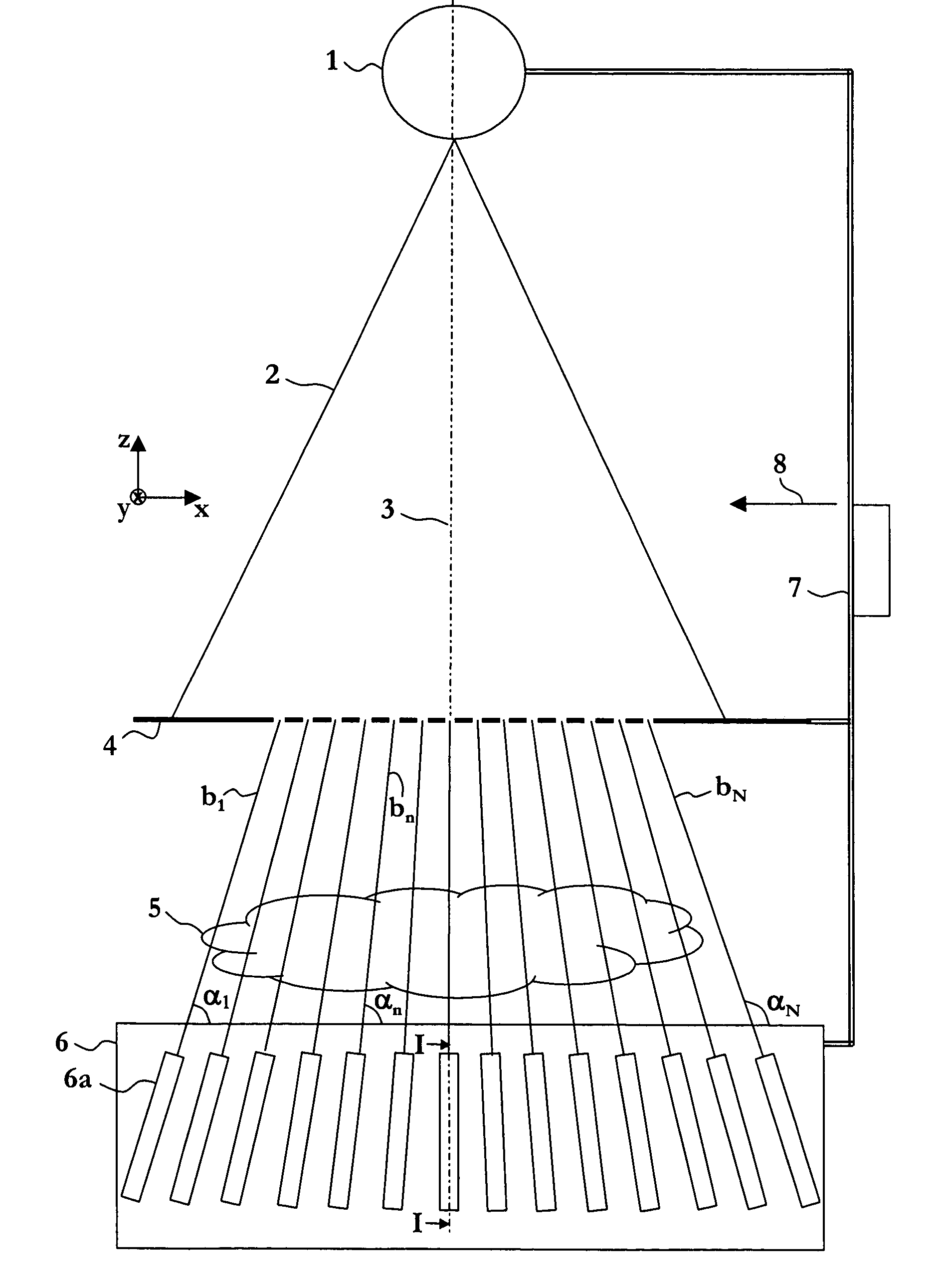
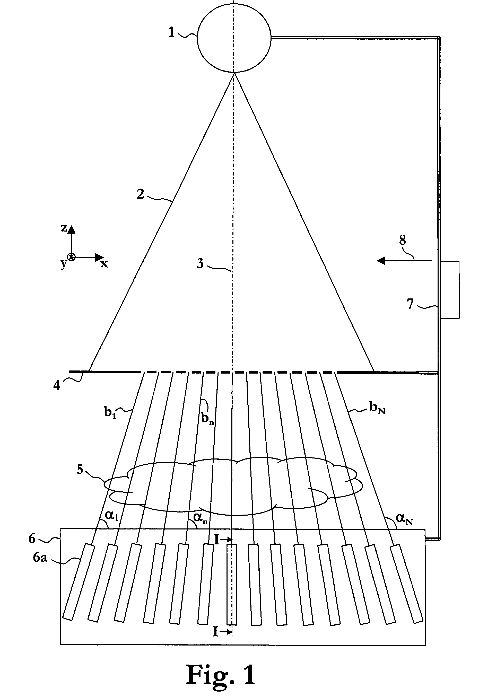
[0023]The apparatus of FIG. 1 comprises a divergent X-ray source 1, which produces X-rays 2 centered around an axis of symmetry 3 (parallel with the z axis), a collimator 4, a radiation detector 6, and a device 7 for rigidly connecting the X-ray source 1, the collimator 4, and the radiation detector 6 to each other and moving the X-ray source 1, the collimator 4, and the radiation detector 6 essentially linearly in direction 8 (typically parallel with the x axis) essentially orthogonal to the axis of symmetry 3 to scan an object 5, which is to be examined.

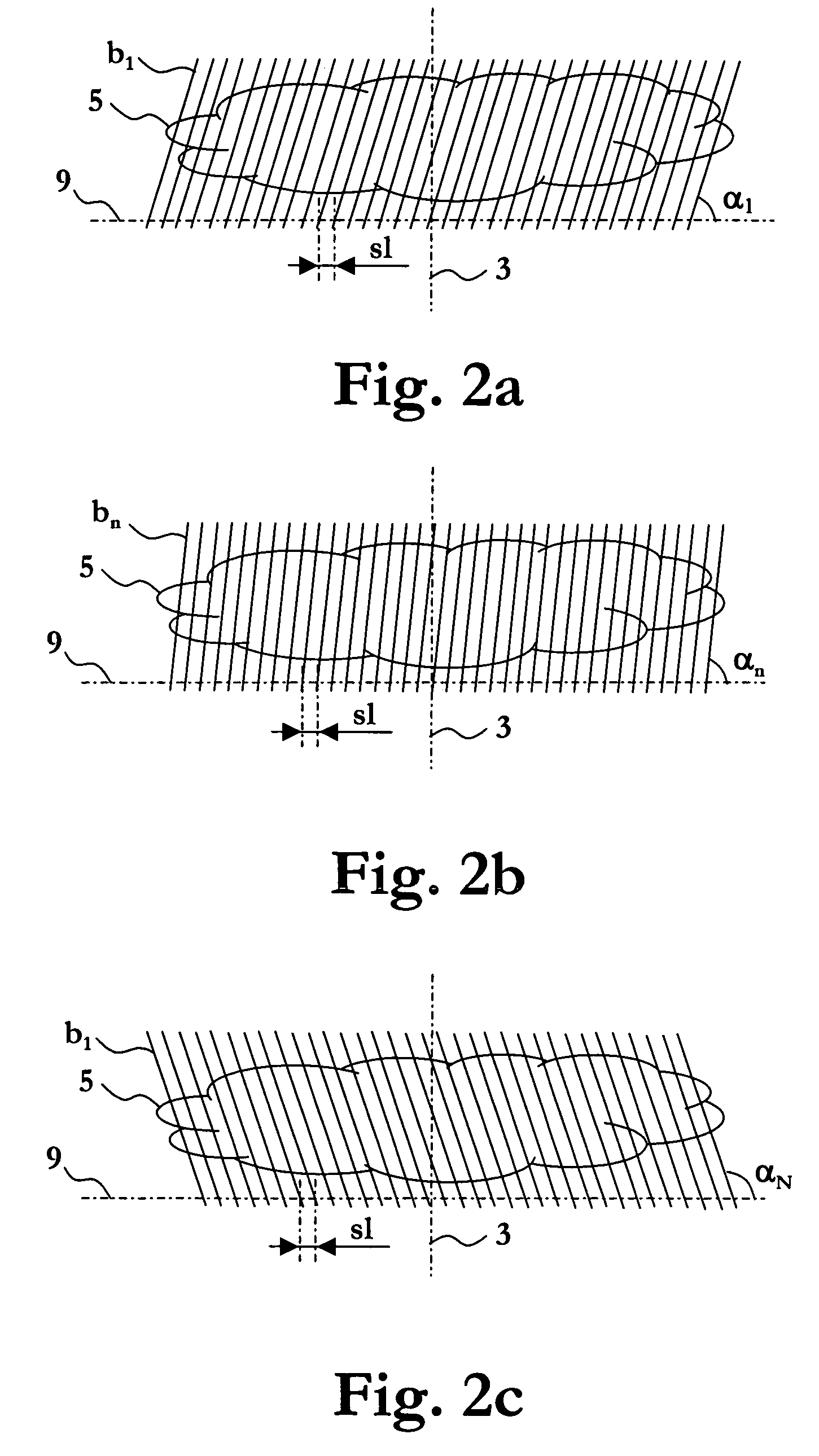
[0024]The radiation detector 6 comprises a stack of line detectors 6a, each being directed towards the divergent radiation source 1 to allow a respective ray bundle b1, . . . , bn, . . . , bN of the radiation 2 that propagates in a respective one of a plurality of different angles αl, . . . , αn, . . . , αN with respect to the front surface of the radiation detector 6 to enter the respective line detector 6a. The line detectors 6a ...


PUM

PropertyMeasurementUnit
anglesaaaaaaaaaa
anglesaaaaaaaaaa
anglesaaaaaaaaaa

Abstract

An apparatus for obtaining tomosynthesis data of an object comprises a source emitting radiation centered around an axis of symmetry; a radiation detector comprising a stack of line detectors, each being directed towards the source at a respective angle; and a device for moving the source and the radiation detector relative the object linearly in a direction orthogonal to the symmetry axis, while each of the line detectors is adapted to record line images of radiation as transmitted through the object in the respective angle. A device is provided for rotating the radiation detector around a rotation axis orthogonal to the symmetry axis, and the device for moving is further arranged to repeat the essential linear movement of the source and the radiation detector relative the object, while each of the line detectors is adapted to record a further plurality of line images of radiation as transmitted through the object.

Description

FIELD OF THE INVENTION[0001]The invention relates generally to scanning-based apparatuses and methods for obtaining tomosynthesis data for examination of an object.BACKGROUND OF THE INVENTION AND RELATED ART[0002]An X-ray medical diagnostic method such as mammography is a low-dose procedure that creates one or more images of a part of a patient such as a breast thereof, which is to be examined, e.g. for detection of early stages of cancer.[0003]The mammography diagnostic procedure generally includes obtaining two images of each of the patient's breasts, one from above and one from the side. A physician or radiologist then reviews the images of the breast, i.e., mammograms, to identify any breast cancer.[0004]While this procedure is one of the best methods of detecting early forms of breast cancer, it is still possible for the detection of breast cancer to be missed by a physician or radiologist reviewing the mammograms. For example, breast cancer may be missed by being obscured by r...

Claims


Application Information

Patent Timeline
Patent Type & Authority Patents(United States)
IPC IPC(8): G01N23/083G01N23/00A61B6/00A61B6/02A61B6/06
CPCA61B6/02A61B6/06A61B6/502A61B6/025
Inventor FRANCKE, TOMSOKOLOV, SKIFF
Owner XCOUNTER AB
Learn More