
Method and apparatus for performing quantitative analysis and imaging surfaces and subsurfaces of turbid media using spatially structured illumination

a spatial structure and illumination technology, applied in the field of optical measurement of turbid media, can solve the problems of affecting the accuracy of the data from the prediction of the model, affecting the accuracy of the data, and difficult to maintain

Active Publication Date: 2005-10-25
RGT UNIV OF CALIFORNIA
View PDF2 Cites 55 Cited by
  • Summary
  • Abstract
  • Description
  • Claims
  • Application Information

AI Technical Summary

Benefits of technology

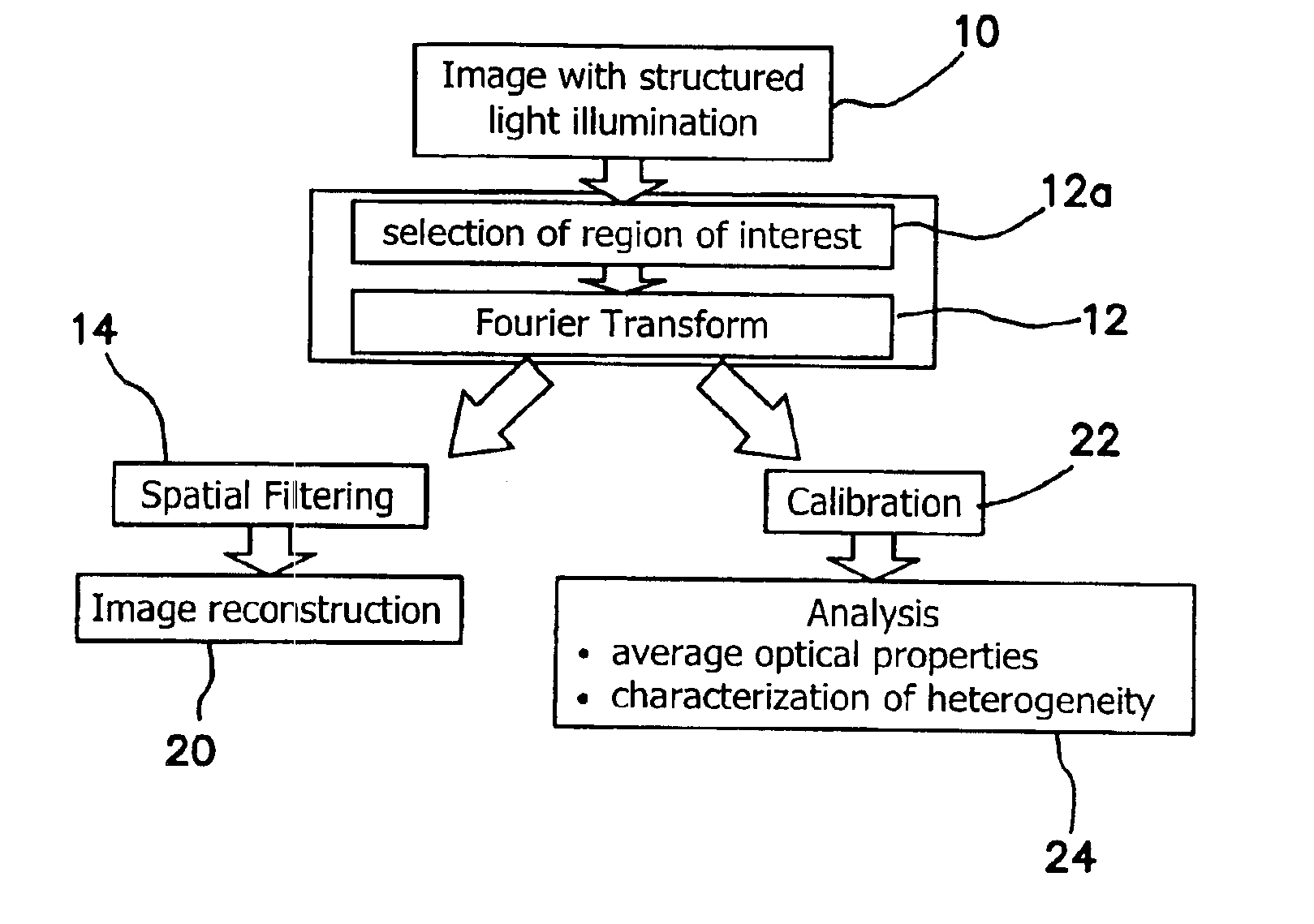
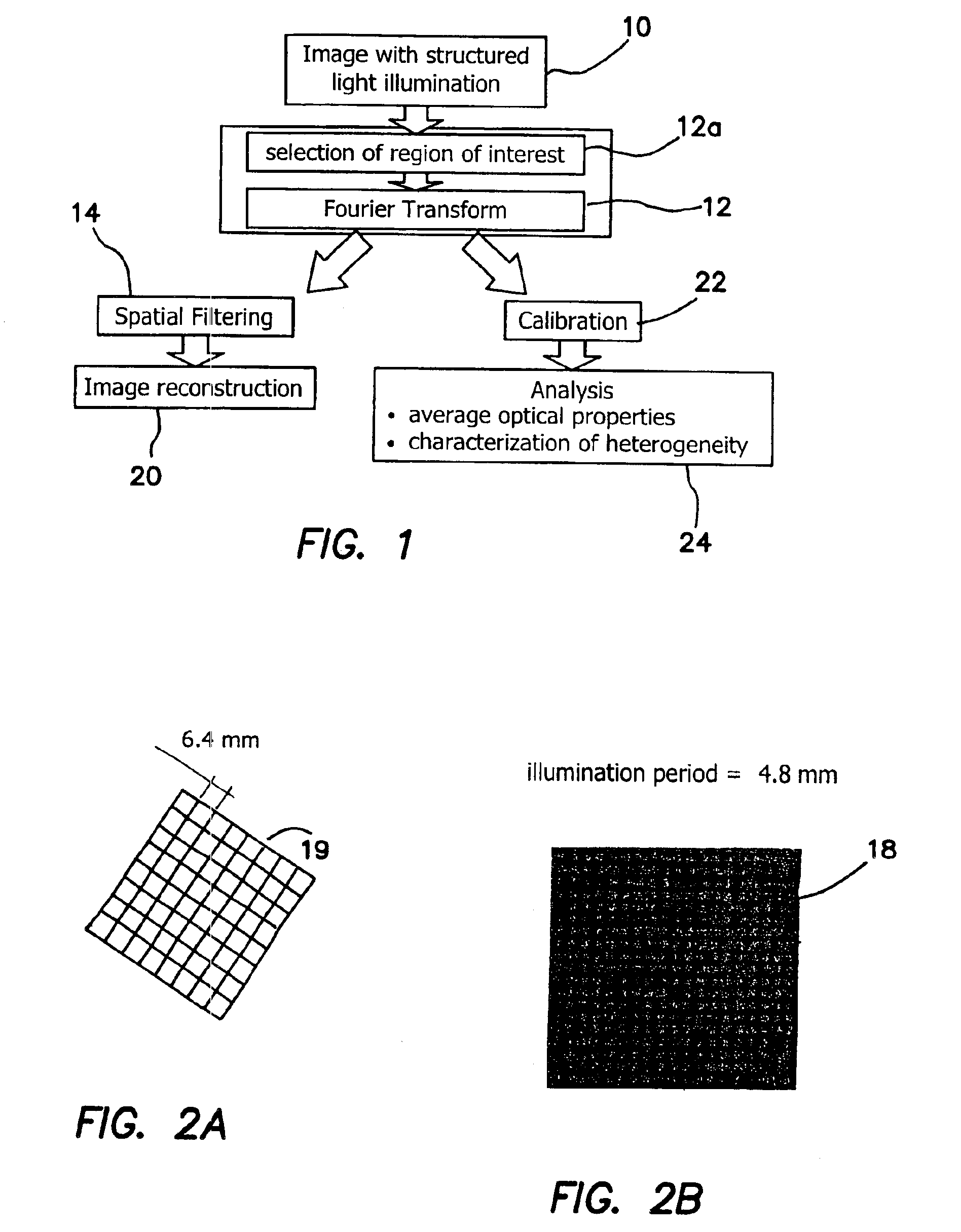
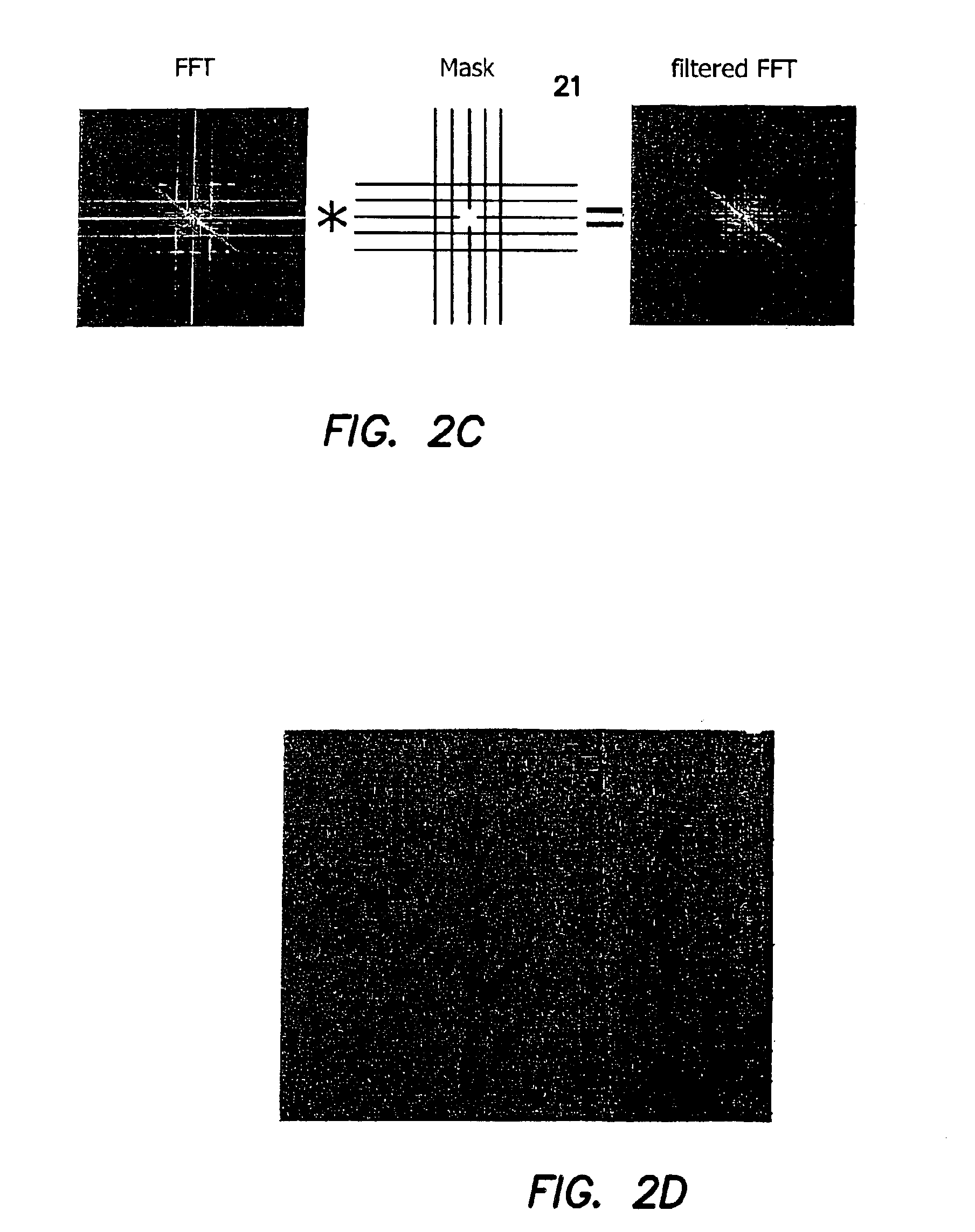
[0014]Typically, a square grid of dots or lines, of various spatial frequencies, is used as the patterned illumination. The transformation of the illumination pattern, which is a consequence of the sample composition, contains the optical property information, which can be deduced according to previously known analytical algorithms. For example, sample composition, can also be deduced, based on capturing data in the structure illumination geometry, using methods based on multivariate calibration approach. It is also possible to recover optical properties from frequency domain phase measurement (FDPM) data using a chemometric approach which is amendable to modulated imaging. Two types of analysis can be applied and they are not mutually exclusive. According to the invention both types of analysis can be performed separately or together. Subsurface imaging can be performed by filtering the spatial pattern of the illumination in the Fourier domain. Analysis in Fourier space is one possibility, but it is expressly contemplated as being within the scope of the invention that wavelet based filtering, filtering using principle components, other mathematical spaces can be equivalently substituted. The average depth probed by the patterned illumination differs from uniform illumination. The depth sensitivity, which is a function of the details of the sample composition, can be optimized by changing the spatial frequency of illumination. Moreover, the combination of several of images performed with various spatial frequencies can allow for the reconstruction of the three dimensional volume of the sample.

Problems solved by technology

Unsurprisingly, these three techniques have different merits and limitations.
The larger the spread of distances probed, the more likely that heterogeneities, such as those found in biological tissue, will distort the data from the predictions of the model.
Because such techniques require sources that can be pulsed or modulated rapidly, covering a large wavelength range requires a tunable laser or an extensive collection of laser diodes, both of which can be expensive, difficult to maintain, and slow to cover the entire spectrum.
This is an important drawback, because the quantification of chromophore concentrations can be significantly affected by use of a limited number of wavelengths.
Nevertheless, most of these techniques as described above reply on “point spectroscopy” and measure only a single, small area of tissue at a time.
Scanning or multiplexing can be used to overcome such a disadvantage, but is typically slow and cumbersome to implement.

Method used


Image

  • Method and apparatus for performing quantitative analysis and imaging surfaces and subsurfaces of turbid media using spatially structured illumination
  • Method and apparatus for performing quantitative analysis and imaging surfaces and subsurfaces of turbid media using spatially structured illumination
  • Method and apparatus for performing quantitative analysis and imaging surfaces and subsurfaces of turbid media using spatially structured illumination

Examples


Embodiment Construction

[0034]Illumination with patterned or structured light, such as described below in connection with FIG. 2b, allows for subsurface imaging of turbid media such as tissue and allows for the determination of the optical properties over a large area. Both the average and the spatial variation of the optical properties can be determined. The invention provides a fast, non-contact, scan-free method to image and quantify the optical properties of a tissue. In general, the method does not have to be a non-contacting methodology, although to be able to perform the method in a non-contact mode is a significantly enhanced benefit and utility.

[0035]The method is based on spatially structured or patterned illumination, and can be thought of as a generalization of spatially-resolved optical measurements with a single source. Moreover, structured or patterned illumination has the advantage of providing for integration of subsurface imaging, quantitative determination of scattering and absorption pr...


PUM

PropertyMeasurementUnit
depthsaaaaaaaaaa
optical propertiesaaaaaaaaaa
depthaaaaaaaaaa

Abstract

Illumination with a pattern of light allows for subsurface imaging of a turbid medium or tissue, and for the determination of the optical properties over a large area. Both the average and the spatial variation of the optical properties can be noninvasively determined. Contact with the sample or scanning is not required but may be desired. Subsurface imaging is performed by filtering the spectrum of the illumination in the Fourier domain but other filtering approaches, such as wavelet transform, principle component filter, etc may be viable as well. The depth sensitivity is optimized by changing the spatial frequency of illumination. A quantitative analysis of the average optical properties and the spatial variation of the optical properties is obtained. The optical properties, i.e. reduced scattering and absorption coefficients are determined from the modulated transfer function, MTF.

Description

RELATED APPLICATIONS[0001]The present application is related to U.S. Provisional Patent Application Ser. No. 60 / 365,578, filed Mar. 19, 2002, which is incorporated herein by reference and to which priority is claimed pursuant to 35 USC 119.GOVERNMENT RIGHTS[0002]This invention was made with Government support under Grant No. RR001192, awarded by the National Institutes of Health. The Government has rights in this invention.BACKGROUND OF THE INVENTION[0003]1. Field of the Invention[0004]The present invention relates to optical measurement of turbid media and in particular to optical measurement tissue absorption and scattering parameters and tissue imaging. The invention may also be used as a means to monitor a chemical process, for example, a pharmaceutical slurry.[0005]2. Description of the Prior Art[0006]Reflectance spectroscopy is a technique for characterizing turbid media that has become widely used in medical diagnostics. In many cases access to quantitative information, for e...

Claims


Application Information

Patent Timeline
Patent Type & Authority Patents(United States)
IPC IPC(8): G01N21/47G01N21/49
CPCG01N21/49
Inventor BEVILACQUA, FREDERICCUCCIA, DAVIDDURKIN, ANTHONY J.TROMBERG, BRUCE J.
Owner RGT UNIV OF CALIFORNIA
Learn More