Eureka AIR delivers breakthrough ideas for toughest innovation challenges, trusted by R&D personnel around the world.

Methods of enhancing radiation effects with metal nanoparticles

a metal nanoparticle and radiation effect technology, applied in the field of enhanced radiotherapy, can solve the problems of limited irradiation, serious damage to normal tissue, and radiation not generally very specific to tumors, and achieve the effect of enhancing the cancer-killing effect of irradiation, high use, and better respons

Inactive Publication Date: 2005-10-18
NANOPROBES
View PDF7 Cites 131 Cited by
  • Summary
  • Abstract
  • Description
  • Claims
  • Application Information

AI Technical Summary

Benefits of technology

[0014]In particular, the present invention provides methods for enhancing the effects of radiation directed to a tissue or cells in or from an animal by administering an amount of metal nanoparticles to the animal or to the tissue or cells ex vivo, then irradiating the animal with radiation directed to the tissue or cells, or irradiating the tissue or cells ex vivo. The methods of the present invention are useful for ablating unwanted tissues in an animal without unacceptable damage to surrounding normal tissues or substantial toxicity to the animal.
[0024]In one embodiment of the present invention, the enhanced radiation therapy is applied to an animal such as a human individual, to ablate or destruct an unwanted tissue, e.g., a tumor. Metal nanoparticles can be administered to an animal prior to irradiation by standard methods, e.g., intravenous or intra-arterial injection, direct injection into a target tissue (e.g., tumor), and implantation of a reservoir device capable of a slow release of metal nanoparticles. In general, nanoparticles are administered in an amount to achieve a concentration in the animal of at least about 0.05 to 10% metal by weight, preferably 0.1 to 5% metal by weight, and more preferably 0.3% to 2% metal by weight, in order to achieve radiation enhancement.

Problems solved by technology

Unfortunately, such radiations are not generally very specific for the tumor.
Tumoricidal doses of radiation often results in serious damage to normal tissue, thus limiting irradiation to lower doses that are not curative.
Although the mechanisms are not completely understood, some data suggest that these bases may be incorporated into DNA or RNA leading to functional disruption, or that they may inhibit enzymes, such as thymidylate synthetase, thus disrupting DNA synthesis.
5-iododeoxyuridine and 5-bromodeoxyuridine are being used in clinical trials, but have shown only limited efficacy.
These chemical methods of radiation enhancement are not widely used in standard clinical radiotherapy treatments because of the unpredictability and variability of response in different tumor types, the toxicity of the agents, and some disappointing clinical trials.
These compounds absorb visible light and result in formation of toxic free radicals.
A disadvantage of this phyotodynamic therapy (PDT) is that it requires visible (laser) light to penetrate the tumor and is thus limited to superficial tissue, or those tissues that are optically accessible, generally superficial malignancies.
Uniformity of dose delivery is also a problem due to the high absorbance of the light by tissue.
These restrictions limit the use of PDT to only a few tumor types and situations.
This method requires a costly source of slow neutrons such as a low-power nuclear reactor or a high-current of energetic protons impinging on a cooled lithium target.
Moreover, the boron-containing compounds available so far to treat human tumors do not have extraordinary affinities for tumors.
These factors have hindered widespread development and use of BNCT.
Unfortunately, this level of substitution is not practical in vivo, and it would be difficult to selectively make such changes only in tumor cells.
Unfortunately, it would be impractical to place bulk metal surfaces throughout all tumor vessels and tissues.
Therefore, a few stents in a tumor would not be enough to treat the whole tumor.
However, there is no showing of delivery of sufficient metal to the tumor by means of these particles for effective treatment.
In addition, the form of radiation taught by Regulla is restricted to less than 400 keV, which could not treat tumors at depth.
Histological examination showed gold particles predominantly in the interstitial fluid, and “no gold particles were found in zones of tightly packed tumor cells, suggesting that it would be difficult to achieve uniform delivery of particles .
. . throughout a tumour volume by direct injection.” Since the dose is only increased in about a 100 micron region around the gold, this method would leave many tumor cells without enhanced treatment.
Nor has there been any attempt of using a nanoscale approach to selectively ablate the unwanted cells or tissue.

Method used


Image

  • Methods of enhancing radiation effects with metal nanoparticles
  • Methods of enhancing radiation effects with metal nanoparticles
  • Methods of enhancing radiation effects with metal nanoparticles

Examples


example 1

High Loading of Immunotargeted Gold Nanoparticles into Tumor Cells by Receptor Mediated Endocytosis

[0106]An anti-Epidermal Growth Factor receptor (EGFr) antibody, MAb225, was expressed in cell culture, then purified by protein A column chromatography. This antibody has been shown to have good tumor to non-tumor localization of drug and radioisotope conjugates. The antibody was checked for purity by gel electrophoresis and its activity was verified by targeting to A431 cells using 15 nm colloidal gold antibody conjugates and silver enhancement. A431 cells, which highly express EGFr, and a control cell line with low expression of EGFr, MCF7, were cultured. Gold-antibody conjugates were added to the growth medium and 2 days later, the medium was changed and more gold-antibody was added. The uptake of colloidal gold by A431 cells was obvious after one day, since a cell pellet now had a black appearance, and examination under the light microscope revealed visible black dots in the cytopl...

example 2

Dose Enhancement Factor of 4.5 Achieved in Vitro

[0108]A431 cells were targeted with the gold nanoparticle-anti-EGFr antibody conjugate as described in Example 1. The cells were then washed several times and resuspended in fresh medium, then aliquots of cell suspension were exposed to 250 kVp x-rays with doses between 0 and 16 Gray. A431 cells not exposed to gold nanoparticles were similarly irradiated and used as controls. After irradiation, cells were plated and grown for one week. A metabolic cell viability test was then done to determine the surviving fraction. Cells containing gold showed lower survival at all doses. Analysis of the data showed that the dose required to kill one-half of the cells was 4.5 times less for cells containing the gold nanoparticles. The dose enhancement factor was therefore 4.5.

example 3

Toxicity of Gold Nanoparticle Found to be Low

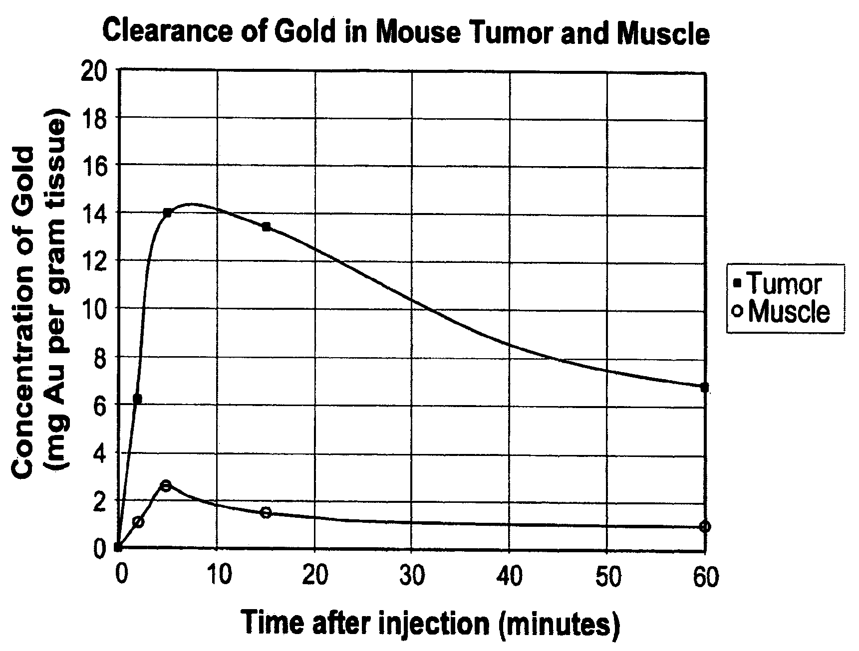
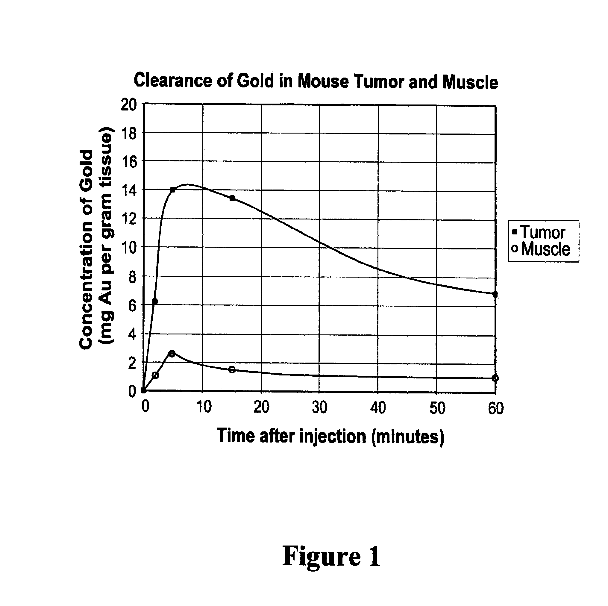
[0109]Balb / C mice with hindleg tumors were intravenously injected in the tail vein with gold nanoparticles with an average gold diameter of about 2 nm to produce a concentration in the tumor of approximately 0.3% or 0. 15% gold by weight. After two weeks necropsy was performed which showed no unusual findings. Blood was drawn for hematology and chemistry, and yielded the results shown in Table 1. These preliminary results indicate there was no acute toxicity at these doses.


PUM

PropertyMeasurementUnit
diameteraaaaaaaaaa
sizeaaaaaaaaaa
atomic numberaaaaaaaaaa

Abstract

The present invention provides methods of using metal nanoparticles 0.5 to 400 nm in diameter to enhance the dose and effectiveness of x-rays or of other kinds of radiation in therapeutic regimes of ablating a target tissue such as tumor. The metal nanoparticles can be administered intravenously, intra-arterially, or locally to achieve specific loading in and around the target tissue. The metal nanoparticles can also be linked to chemical and / or biochemical moieties which bind specifically to the target tissue. The enhanced radiation methods can also be applied to ablate unwanted tissues or cells ex vivo.

Description

FIELD OF THE INVENTION[0001]This invention relates in general to methods of enhanced radiotherapy. In particular, the present invention relates to methods of enhancing the therapeutic effects of radiation in promoting the shrinkage and / or elimination of tissues targeted for destruction, e.g., tumors, by using nanoparticles of gold and / or other metals, without unacceptable damage to surrounding normal tissues.BACKGROUND OF THE INVENTION[0002]Radiation of various forms, for example x-rays, laser light, and microwaves, as well as particle beams of, for example, neutrons, electrons, and protons, have been used to treat tumors. Unfortunately, such radiations are not generally very specific for the tumor. Tumoricidal doses of radiation often results in serious damage to normal tissue, thus limiting irradiation to lower doses that are not curative.[0003]Radiosensitizer compounds are drugs that act in combination with radiation to produce improved response, usually by making DNA more suscep...

Claims


Application Information

Patent Timeline
Patent Type & Authority Patents(United States)
IPC IPC(8): A61N5/00A61N5/06A61B6/00A61K41/00A61K47/48A61K49/22A61N1/40A61N5/10
CPCA61K41/0038A61K41/0052A61K47/48561A61K47/48861A61K47/48884A61K49/222A61K49/226A61N1/406A61N5/00B82Y5/00A61N5/062A61N5/10A61N2005/1098Y10S977/923Y10S977/927Y10S977/928Y10S977/93Y10S977/929Y10S977/728Y10S977/91Y10S977/777Y10S977/911A61K47/6849A61K47/6923A61K47/6929
Inventor HAINFELD, JAMES F.SLATKIN, DANIEL N.
Owner NANOPROBES
Why Eureka
Eureka Blog
Learn More