
Interactive computer-aided diagnosis method and system for assisting diagnosis of lung nodules in digital volumetric medical images

a computer-aided diagnosis and digital volumetric technology, applied in image enhancement, tomography, instruments, etc., can solve the problems of large amount of data for the physician to examine, inability to accurately diagnose cancer or other diseases, and difficulty in identifying cancer patients, etc., to achieve rapid computation of numeric values and increase the acceptance of physicians

Inactive Publication Date: 2005-09-13
SIEMENS MEDICAL SOLUTIONS USA INC
View PDF16 Cites 38 Cited by
  • Summary
  • Abstract
  • Description
  • Claims
  • Application Information

AI Technical Summary

Benefits of technology

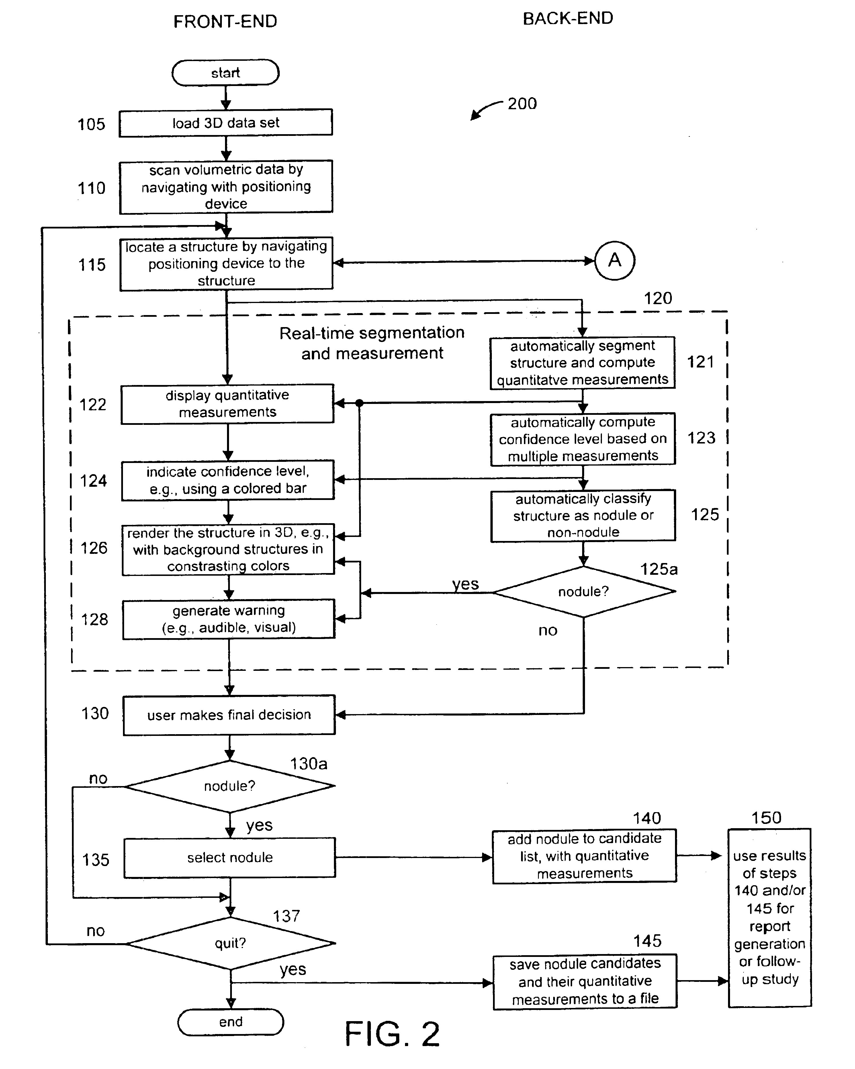
[0011]The method and system mesh seamlessly with physicians in their current work practices. By incorporating the physicians' years of training into the present invention, the invention takes advantage of their knowledge, augmenting it with the strengths of the computer in rapid computation of numeric values. Such an invention is an “open box”, increasing its acceptance among physicians. The invention provides results in “real-time” so that it does not add a time delay to the physician's examination process.

Problems solved by technology

In current clinical practice, cancer or other diseases may be missed during a physician's un-aided examination of medical image data, in part because of the large volume of data.
This is particularly a problem for screening applications, since there is generally little time to devote to the examination of each patient's data, and the entire range of the data must be examined to make sure it is free from disease.
However the change from two dimensions to three, especially to large volume 3-D data (such as image volumes produced by multi-detector high resolution CT scanners), results in a much larger amount of data for the physician to examine.
Furthermore, the low-dose imaging for cancer screening poses additional challenges to the traditional manual clinical reading.
Unfortunately, many current CADx systems are not readily accepted by physicians, because their aid is seen as more of a distraction than a help.
Many such systems present the results of the computer's diagnosis to the physicians by marks, such as a red circle or arrow on the softcopy, which some physicians believe can create a bias in their interpretation of the data.
Furthermore, too many systems are perceived as a “black box”, where physicians feel they do not have any understanding of how such systems work and how they generate their diagnoses.

Method used


Image

  • Interactive computer-aided diagnosis method and system for assisting diagnosis of lung nodules in digital volumetric medical images
  • Interactive computer-aided diagnosis method and system for assisting diagnosis of lung nodules in digital volumetric medical images
  • Interactive computer-aided diagnosis method and system for assisting diagnosis of lung nodules in digital volumetric medical images

Examples


Embodiment Construction

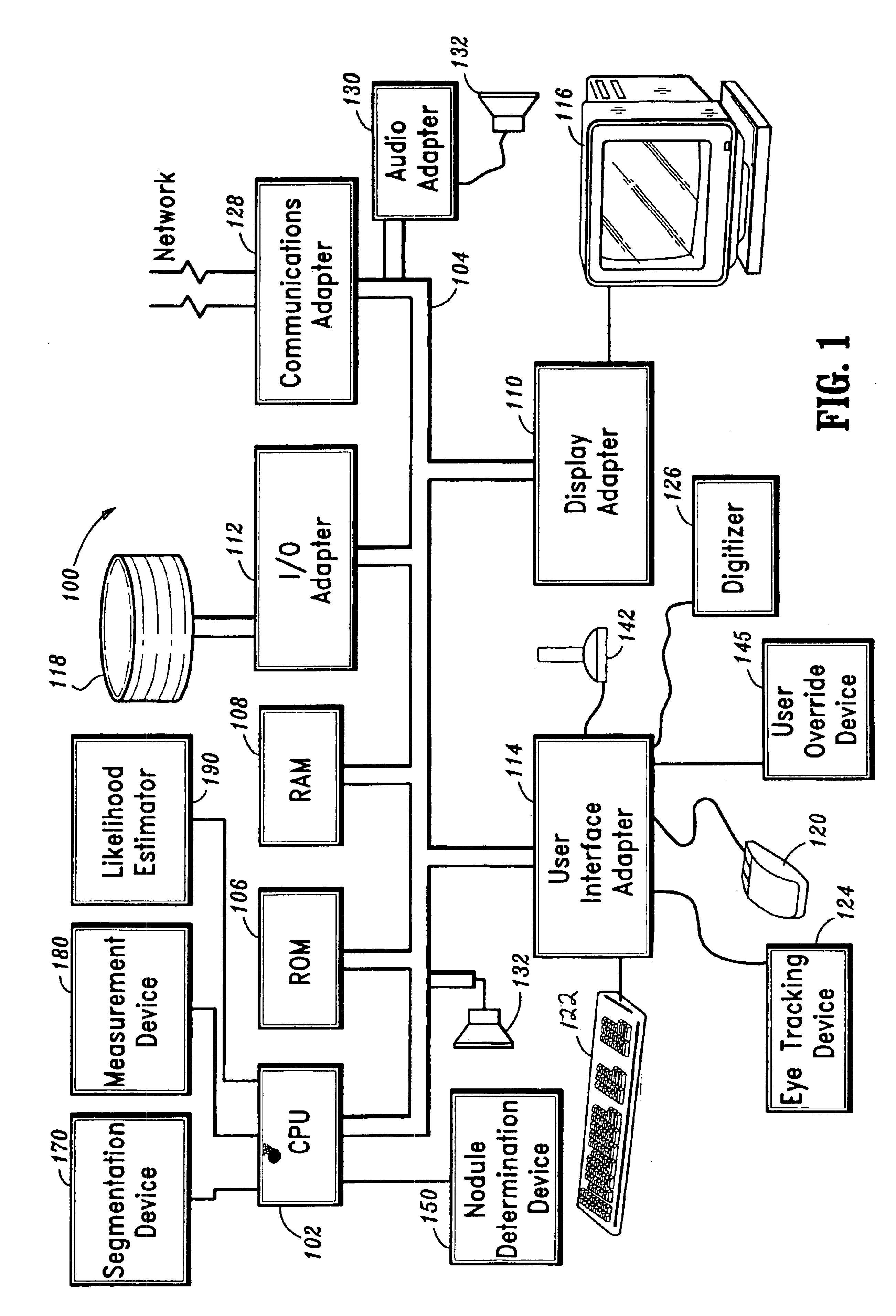
[0026]The present invention is directed to an interactive computer-aided diagnosis (ICAD) method and system for assisting diagnosis of lung nodules in digital volumetric medical images.

[0027]It is to be understood that the present invention may be implemented in various forms of hardware, software, firmware, special purpose processors, or a combination thereof. Preferably, the present invention is implemented as a combination of hardware and software. Moreover, the software is preferably implemented as an application program tangibly embodied on a program storage device. The application program may be uploaded to, and executed by, a machine comprising any suitable architecture. Preferably, the machine is implemented on a computer platform having hardware such as one or more central processing units (CPU), a random access memory (RAM), and input / output (I / O) interface(s). The computer platform also includes an operating system and microinstruction code. The various processes and func...


PUM


Abstract

A computer-assisted diagnosis method for assisting diagnosis of anatomical structures in a digital volumetric medical image of at least one lung includes identifying an anatomical structure of interest in the volumetric digital medical image. The anatomical structure of interest is automatically segmented, in real-time, in a predefined volume of interest (VOI). Quantitative measurements of the anatomical structure of interest are automatically computed, real-time. A result of the segmenting step and a result of the computing step are displayed, in real-time. A likelihood that the anatomical structure of interest corresponds to a disease or an area warranting further investigation is estimating, in real-time, based on predefined criteria and the quantitative measurements. A warning is generated, in real-time, when the likelihood is above a predefined threshold.

Description

CROSS-REFERENCE TO RELATED APPLICATIONS[0001]This application claims the benefit of provisional application 60 / 230,772 filed on Sep. 7, 2000.[0002]This application is related to U.S. patent application, Ser. No. 09 / 840,266, entitled “Method and System for Automatically Detecting Lung Nodules from High Resolution Computed Tomography (HRCT) Images”, filed on Apr. 23, 2001, which is commonly herewith, and the disclosure of which is incorporated herein by reference. This application is also related to U.S. patent application Ser. No. 09 / 606,564, entitled “Computer-aided Diagnosis of Three Dimensional digital image data”, filed on Jun. 29, 2000, and U.S. Pat. No. 6,697,506, entitled “Mark-Free Computer-Assisted Diagnosis Method and System for Assisting Diagnosis of Abnormalities in Digital Medical Images Using Diagnosis Based Image Enhancement”, issued on Feb. 24, 2002, which are commonly assigned herewith, and the disclosures of which are incorporated herein by reference.BACKGROUND[0003...

Claims


Application Information

Patent Timeline
IPC IPC(8): G06K9/00G06T5/00G06T7/00G06F19/00A61B6/03
CPCG06F19/321G06F19/324G06F19/3437G06F19/3487G06K9/00127G06T7/0012G06T7/0081G06F19/322G06F19/3406G06T2207/10081G06T2207/20012G06T2207/20132G06T2207/20148G06T2207/20156G06T2207/30064G06T7/11G06T7/136G16H40/63G16H10/60G16H50/50G16H15/00G16H30/40G16H70/60G06V20/69
Inventor NOVAK, CAROL L.FAN, LIQIAN, JIANZHONG
Owner SIEMENS MEDICAL SOLUTIONS USA INC
Learn More