
Cellular arrays and methods of detecting and using genetic disorder markers

a technology of genetic disorder and cellular array, applied in the field of tissue sample screening and genomic regions, can solve the problems of limited amount of diagnostic or molecular information, slow and tedious process, and impede the development of new molecular markers of clinical importance, and achieve rapid and accurate detection and high resolution

Inactive Publication Date: 2005-06-14
UNITED STATES OF AMERICA
View PDF10 Cites 22 Cited by
  • Summary
  • Abstract
  • Description
  • Claims
  • Application Information

AI Technical Summary

Benefits of technology

[0014]The invention also features a method of analyzing a biological sample for a genetic disorder by exposing a genosensor comparative genomic hybridization (gCGH) array of genomic regions to a nucleic acid sample from a cell with a known specific genetic disorder, and identifying as a biomarker a genomic region to which the nucleic acid hybridizes; obtaining a candidate probe that hybridizes to the biomarker; exposing the candidate probe to a tissue specimen array to determine a statistical measure of hybridization of the candidate probe; selecting a candidate probe having a statistically significant measure of hybridization; and using a selected candidate probe to analyze a biological sample for the genetic disorder. This analysis of the biological sample can provide diagnostic or prognostic information.

Problems solved by technology

Although this microscopic examination and classification of tumors has improved medical treatment, the microscopic appearance of a tissue specimen stained by standard methods (such as hematoxylin and eosin) can often reveal only a limited amount of diagnostic or molecular information.
The development of new molecular markers of clinical importance has been impeded by the slow and tedious process of evaluating biomarkers in large numbers of clinical specimens.

Method used


Image

  • Cellular arrays and methods of detecting and using genetic disorder markers
  • Cellular arrays and methods of detecting and using genetic disorder markers
  • Cellular arrays and methods of detecting and using genetic disorder markers

Examples


example 1

ecimens

[0090]A total of 645 breast cancer specimens were used for construction of a breast cancer tumor tissue microarray. The samples included 372 fresh-frozen ethanol-fixed tumors, as well as 273 formalin-fixed breast cancers, normal tissues and fixation controls. The subset of frozen breast cancer samples was selected at random from the tumor bank of the institute of Pathology, University of Basel, which includes more than 1500 frozen breast cancers obtained by surgical resections during 1986-1997. Only the tumors from this tumor bank were used for molecular analyses. This subset was reviewed by a pathologist, who determined that the specimens included 259 ductal, 52 lobular, 9 medullary, 6 mucinous, 3 cribriform, 3 tubular, 2 papillary, 1 histiocytic, 1 clear cell, and 1 lipid rich carcinoma. There were also 15 ductal carcinomas in situ, 2 carcinosarcomas, 4 primary carcinomas that had received chemotherapy before surgery, 8 recurrent tumors and 6 metastases.

[0091]Histological g...

example 2

tochemistry

[0092]After formation of the tissue array and sectioning of the donor block, standard indirect immunoperoxidase procedures were used for immunohistochemistry (ABC-Elite, Vector Laboratories). Monoclonal antibodies from DAKO (Glostrup, Denmark) were used for detection of p53 (DO-7, mouse, 1:200), erbB-2 (c-erbB-2, rabbit, 1:4000), and estrogen receptor (ER ID5, mouse, 1:400). A microwave pretreatment was performed for p53 (30 minutes at 90° C.) and erbB-2 antigen (60 minutes at 90° C.) retrieval. Diaminobenzidine was used as a chromogen. Tumors with known positivity were used as positive controls. The primary antibody was omitted for negative controls. Tumors were considered positive for ER or p53 if an unequivocal nuclear positivity was seen in at least 10% of tumor cells. The erbB-2 staining was subjectively graded into 3 groups: negative (no staining), weakly positive (weak membranous positivity), strongly positive (strong membranous positivity).

example 3

nt in situ Hybridization (FISH)

[0093]Two-color FISH hybridizations were performed using Spectrum-Orange labeled cyclin D1, myc, or erbB2 probes together with corresponding FITC labeled centromeric reference probes (Vysis). One-color FISH hybridizations were done with spectrum orange-labeled 20q13 minimal common region (Vysis, and see Tanner et al., Cancer Res. 54:4257-4260 (1994)), mybL2 and 17q23 probes (Barlund et al., Genes Chrom. Cancer 20:372-376 (1997)). Before hybridization, tumor array sections were deparaffinized, air dried and dehydrated in 70, 85, and 100% ethanol followed by denaturation for 5 minutes at 74° C. in 70% formamide-2×SSC solution. The hybridization mixture contained 30 ng of each of the probes and 15 μg of human Cot1-DNA. After overnight hybridization at 37° C. in a humidified chamber, slides were washed and counterstained with 0.2 μM DAPI in an antifade solution. FISH signals were scored with a Zeiss fluorescence microscope equipped with double-band pass fi...


PUM

PropertyMeasurementUnit
diametersaaaaaaaaaa
diametersaaaaaaaaaa
diametersaaaaaaaaaa

Abstract

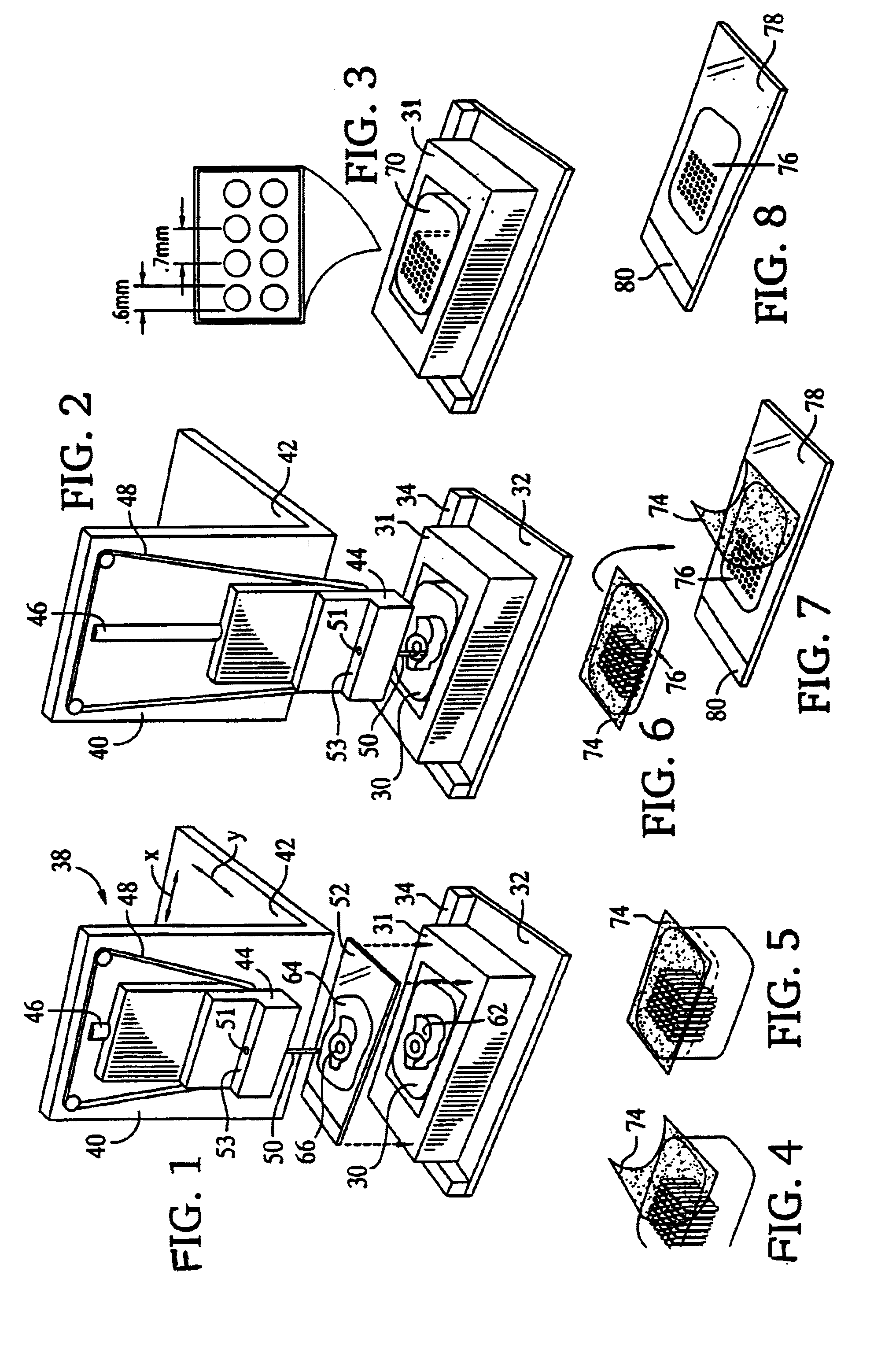
A method is disclosed for rapid molecular profiling of tissue or other cellular specimens by placing a donor specimen in an assigned location in a recipient array, providing copies of the array, and performing a different biological analysis of each copy. The results of the different biological analyses are compared to determine if there are correlations between the results of the different biological analyses at each assigned location. In some embodiments, the specimens may be tissue specimens from different tumors, which are subjected to multiple parallel molecular (including genetic and immunological) analyses. The results of the parallel analyses are then used to detect common molecular characteristics of the genetic disorder type, which can subsequently be used in the diagnosis or treatment of the disease. The biological characteristics of the tissue can be correlated with clinical or other information, to detect characteristics associated with the tissue, such as susceptibility or resistance to particular types of drug treatment. Other examples of suitable tissues which can be placed in the matrix include tissue from transgenic or model organisms, or cellular suspensions (such as cytological preparations or specimens of liquid malignancies or cell lines).

Description

CROSS-REFERENCE TO RELATED APPLICATIONS[0001]This application is a Continuation of U.S. patent application Ser. No. 09 / 429,448, filed on Oct. 28, 1999, now abandoned which claims priority from U.S. Provisional Application Ser. No. 60 / 106,038, filed on Oct. 28, 1998, and U.S. Provisional Application Ser. No. 60 / 150,493, filed on Aug. 24, 1999. The content of these applications are incorporated herein by reference in their entirety.FIELD OF THE INVENTION[0002]The invention relates to the screening of tissue samples and genomic regions to discover markers for genetic disorders such as cancer.BACKGROUND OF THE INVENTION[0003]Biological mechanisms of many diseases have been clarified by microscopic examination of tissue specimens. Histopathological examination has also permitted the development of effective medical treatments for a variety of illnesses. In standard anatomical pathology, a diagnosis is made on the basis of cell morphology and staining characteristics. Tumor specimens, for...

Claims


Application Information

Patent Timeline
Patent Type & Authority Patents(United States)
IPC IPC(8): B01L3/00C12Q1/68G01N1/30G01N1/31G01N33/50G01N33/543G02B21/34G01N1/04G01N1/08C12N15/09C12M1/00G01N1/28G01N1/36
CPCG02B21/34G01N33/54306G01N33/5005G01N1/312G01N1/2813B01L3/5085C12Q1/6809C12Q1/6837C12Q1/6841C12Q1/6886B01J2219/00659B01J2219/00702G01N2001/368G01N2001/288G01N2001/282G01N1/08C12Q2600/118C12Q2600/112B01J2219/00743C12Q2600/106C12Q2543/10C12Q2539/103C12Q2537/157C12Q2565/501C12Q2600/156
Inventor KALLIONIEMI, OLLI-PMULLER, UWE RICHARDSAUTER, GUIDOKONONEN, JUHABARLUND, MAARIT
Owner UNITED STATES OF AMERICA
Learn More