Angiogenic tissue implant systems and methods
a technology of angiogenesis and tissue, applied in the field of angiogenesis tissue implant systems and methods, can solve the problem that the animal host cannot naturally provide these new vascular structures, and achieve the effect of small area, sustained cell viability, and compact area
- Summary
- Abstract
- Description
- Claims
- Application Information
AI Technical Summary
Benefits of technology
Problems solved by technology
Method used
Image
Examples
example 1
demonstrates a methodology that can be followed to identify for other cell types the applicable metabolic transit value that assures cell survival during the ischemic period after implantation.
The absolute permeability and porosity values that constitute a given metabolic transport value will depend upon the type of cell and the methodologies of determining permeability and porosity. Different conditions will give different absolute values. Still, regardless of the test conditions, the relative differences in permeability and porosity values derived under constant, stated conditions will serve as an indicator of the relative capabilities of the boundaries to support implanted cell viability.
Tables 1 and 2 also show that good tissue survival occurs even with membrane materials that are subject to the formation of an avascular fibrotic response (the so-called "foreign body capsule"). The fact that these membrane materials create this response has, in the past, led to the widely held v...
example 2
In an experiment, the practitioner grew RAT-2 fibroblasts (ATCC CRL 1764) in 20% Fetal Bovine Serum, 2 mM 1-glutamine, and DMEM (Sigma) (high glucose) until 100% confluent. The RAT-2 cells were split 1:2 in the above media, 16 to 24 hours before surgery.
On the day of surgery, the cells were washed with 15 ml of HBSS (no ions) and trypsinized off the culture flask. The practitioner neutralized the trypsin by adding 5 ml of the above media. The practitioner pelleted the cells by centrifugation (1000 rpm, 10 minutes, at 22 degrees C).
The pelleted cells were counted and resuspended in media in three concentrations: 5.3.times.10.sup.3 cells / 10 .mu.l; 5.8.times.10.sup.5 cells / 10 .mu.l; and 5.8.times.10.sup.6 cells / 10 .mu.l.
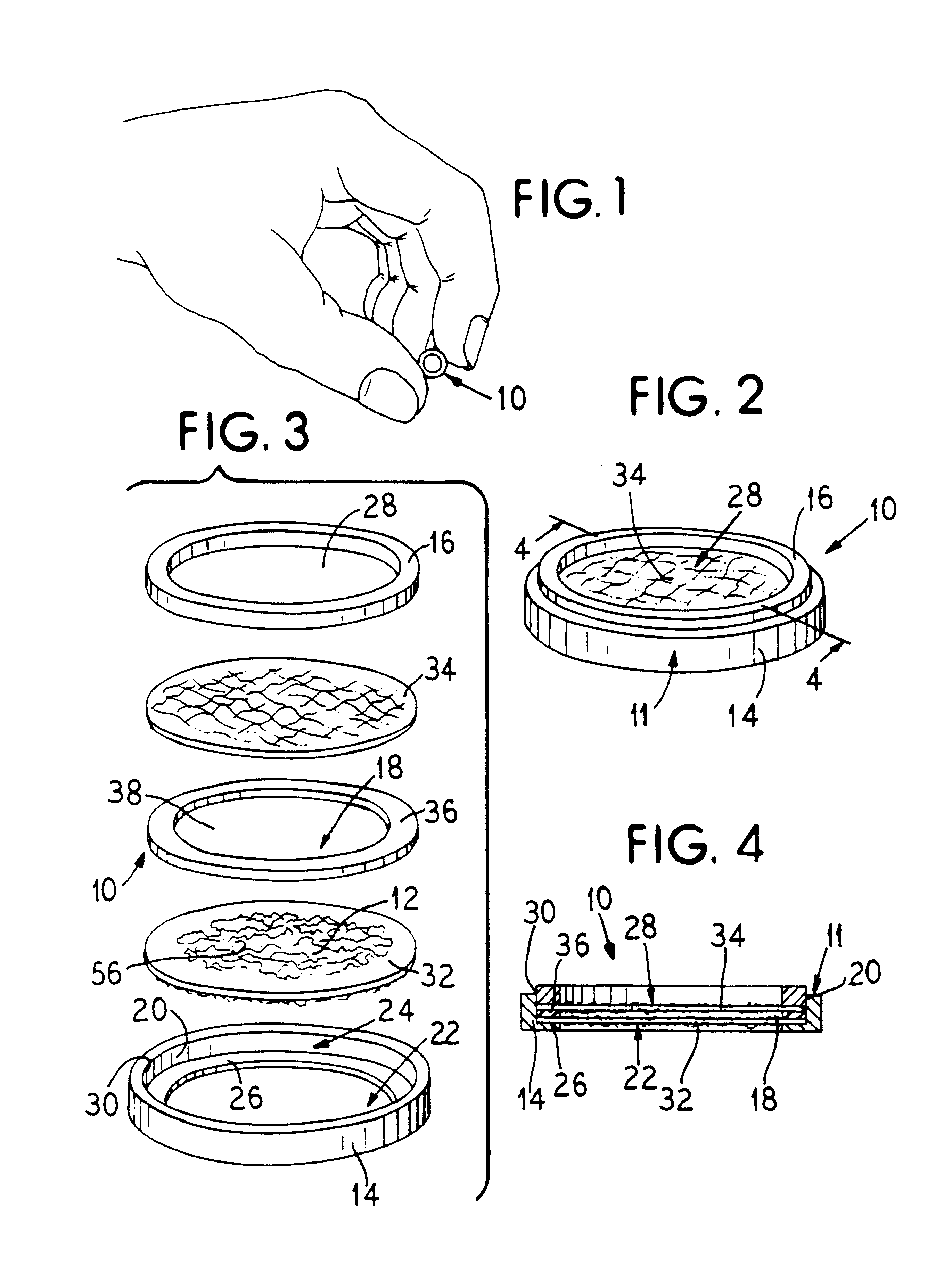
Implant assemblies like that shown in FIGS. 1 to 4 having boundaries of differing permeability values were made. The permeability values ranged from 0.2.times.10.sup.-4 cm / sec to 9.times.10.sup.4 cm / sec (see Tables 1 and 2 to follow). The total boundary area for each as...
example 3
Assemblies like that shown in FIGS. 1 to 4 and constructed according to the foregoing process have been successfully used to accomplish complete correction of diabetes in partially pancreatectomized and streptozotocin-treated rat hosts. The animals were corrected up to 293 days. Upon removal of the implants, the animals reverted to a diabetic state. Histology of the implants revealed the presence of vascular structures close to the boundary.
These assemblies presented a boundary area of about 0.77 cm.sup.2. Each assembly sustained an initial cell load of about 600 pancreatic islets (or about 600,000 pancreatic cells).
When implanted, the assemblies sustained cell densities of about 200,000 islets / cm.sup.3. These assemblies, made and used in accordance with the invention, supported 8 times more pancreatic islets in a given volume than the CytoTherapeutics assemblies (having cell densities of only 25,000 islets / cm.sup.3).
Deriving a Therapeutic Loading Factor
As earlier described, one asp...
PUM
| Property | Measurement | Unit |
|---|---|---|
| Length | aaaaa | aaaaa |
| Length | aaaaa | aaaaa |
| Length | aaaaa | aaaaa |
Abstract
Description
Claims
Application Information
 - R&D
- Intellectual Property
- Life Sciences
- Materials
- Tech Scout
- Unparalleled Data Quality
- Higher Quality Content
- 60% Fewer Hallucinations
Browse by: Latest US Patents, China's latest patents, Technical Efficacy Thesaurus, Application Domain, Technology Topic, Popular Technical Reports.
© 2025 PatSnap. All rights reserved.Legal|Privacy policy|Modern Slavery Act Transparency Statement|Sitemap|About US| Contact US: help@patsnap.com


