Eureka AIR delivers breakthrough ideas for toughest innovation challenges, trusted by R&D personnel around the world.

Capsular polysaccharide adhesin antigen, preparation, purification and use

a polysaccharide adhesin and antigen technology, applied in the field of exopolysaccharide adhesin, can solve the problems that the microorganism(s) that permit these normal skin commensals to become noso-comial pathogens have not been well characterized

Inactive Publication Date: 2004-06-01
THE BRIGHAM & WOMEN S HOSPITAL INC
View PDF33 Cites 30 Cited by
  • Summary
  • Abstract
  • Description
  • Claims
  • Application Information

AI Technical Summary

Problems solved by technology

Although the virulence of coagulase-negative staphylococci clearly is enhanced in the presence of a foreign body, the microbial factor(s) that permit these normal skin commensals to become noso-comial pathogens have not been well characterized.

Method used


Image

  • Capsular polysaccharide adhesin antigen, preparation, purification and use
  • Capsular polysaccharide adhesin antigen, preparation, purification and use
  • Capsular polysaccharide adhesin antigen, preparation, purification and use

Examples


example ii

Isolation of Strain PR-62A Teichoic Acid

Teichoic acid was recovered from the Zeta-prep 250 cartridge in the fraction eluting with 0.6M NaCl. This material was digested with nuclease enzymes as described above, heated at 100.degree. C., pH 4.0, for 1 h, then chromatographed on a 2.6.times.90 cm column of Sepharose CL-4B in 0.2M ammonium carbonate. Serologically active fractions eluting with a Kav of 0.33-0.57 (peak=0.48) were pooled, dialyzed against deionized water, and lyophilized.

example iii

Chemical Components of Crude Extract, Teichoic Acid Fraction of Slime, and Purified Adhesin

Utilizing the methodology described above, a fraction isolated from the culture supernatant of S. epidermidis strain RP-62A that appeared to have the properties of an adhesin was analyzed. The chemical components of the crude extract, the isolated teichoic acid, and the purified adhesin are shown in Table 1.

Crude extract contained numerous components, of which carbohydrate and phosphate were predominant. The teichoic acid fraction of slime was composed principally of phosphate, glycerol, glucose, and glucosamine. The purified adhesin was principally composed of carbohydrate with only low to non-detectable levels of protein, nucleic acids, and phosphate. No lipids were detected in the purified adhesin. The principal monosaccharides identified were galactose, glucosamine and galactosamine; glucose was absent. In addition, a complex chromatogram of monosaccharides indicated the presence of galact...

example iv

Serological Properties of Crude Extract, Teichoic Acid, and Purified Adhesin

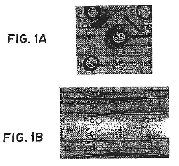
Serologically, crude extract gave three precipitin lines in double diffusion when tested against a rabbit antisera raised against whole cells of strain RP-62A (FIG. 1A), while teichoic acid and the purified adhesin gave single precipitin lines. By immunoelectrophoresis (FIG. 1B), the crude extract had multiple precipitin lines against antisera to whole cells. In contrast, purified adhesin gave a single precipitin line which did not move in the electric field. Purified teichoic acid gave a strong precipitin line migrating towards the anodal end of the gel, as well as a weaker, more negatively charged line when high concentrations of antigen were used. A mixture of teichoic acid and purified adhesin resulted in two precipitin lines corresponding to the individual, purified components.


PUM


Abstract

A substantially pure capsular exopolysaccharide adhesin of coagulaso-negative staphylococcal strains, and a general method to prepare such adhesins, are described. Vaccines composed of such adhesins, and uses of such adhesins to produce polyclonal and mono-clonal antibodies against such adhesins, are also disclosed. The adhesins are useful in coating polymeric medical materials to prevent colonization by coagulase-negative staphylococcal strains, and as a probe in selecting desirable polymeric medical materials. Such adhesin antibodies are useful in vivo to prevent infection by noso-comial coagulase-negative staphylococcal strains, in assays for the detection of such bacteria, in assays for the estimation of such adhesins in complex mixtures, and as an affinity chromatography matrix.

Description

1. Field of the InventionThe present invention relates to a substantially pure exopolysaccharide adhesin isolated from a particular strain of Staphylococcus epidermidis, to a general method capable of isolating this compound in substantially pure form, and to uses of said purified adhesin product as a vaccine for the production of antibodies effective against the binding of homologous bacterial cells to polymeric materials, and as a probe for the development of polymeric materials useful as catheters and medical prostheses.2. Description of the Background ArtBoth Staphylococcus aureus (coagulase-positive) and Staphylococcus epidermidis (coagulase-negative) have a characteristic propensity for invading skin and adjacent tissues at the site of prosthetic medical devices, including intravascular catheters, cerebrospinal fluid shunts, hemodialysis shunts, vascular grafts, and extended-wear contact lenses. Within 48 to 72 hours, relatively large numbers of staphylococci are demonstrable ...

Claims


Application Information

Patent Timeline
Patent Type & Authority Patents(United States)
IPC IPC(8): A61K39/085C07K16/12C12Q1/06A61K38/00C12N15/02A61K39/00A61K39/09A61P31/04C07K14/00C07K16/00C12N5/10C12P21/00C12P21/08C12R1/44C12R1/45C12R1/91
CPCA61K39/085C07K16/1271C12Q1/06C12R1/46A61K38/00A61K39/00G01N2333/31Y10S435/82A61P31/04C12R2001/46C12N1/205
Inventor PIER, GERALD B.
Owner THE BRIGHAM & WOMEN S HOSPITAL INC
Why Eureka
Eureka Blog
Learn More