Method for the fluorescent detection of a DNA sequence in real time

a dna sequence and real-time detection technology, applied in the field of real-time detection of dna sequences using fluorescence, can solve the problems of delay for autoradiographic exposure time, short useful life of probes, and inability to detect dna sequences in real-tim

Inactive Publication Date: 2001-03-06
US DEPT OF HEALTH & HUMAN SERVICES
View PDF1 Cites 18 Cited by
  • Summary
  • Abstract
  • Description
  • Claims
  • Application Information

AI Technical Summary

Problems solved by technology

There are several disadvantages associated with the use of a radioactively labeled probe.
For example, radioactive probes are dangerous to handle and when labeled with .sup.32 P, which is the standard protocol, the probe has a short useful life (a few weeks at most).
In addition, the use of a radioactive probe prevents detection of the results in real time, that is, a delay for an autoradiographic exposure time is required.
The shift in migration results in a change in the length of time required for the probe to be driven past the reader.

Method used


Image

  • Method for the fluorescent detection of a DNA sequence in real time
  • Method for the fluorescent detection of a DNA sequence in real time
  • Method for the fluorescent detection of a DNA sequence in real time

Examples


Embodiment Construction
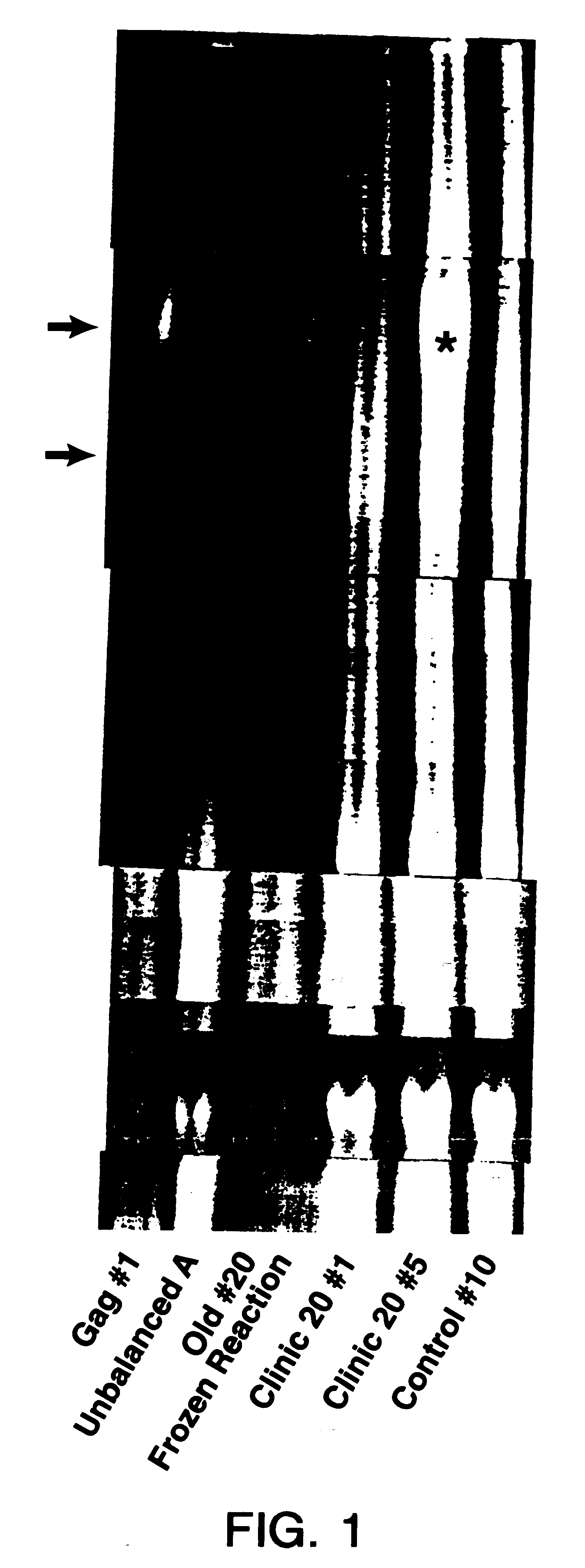
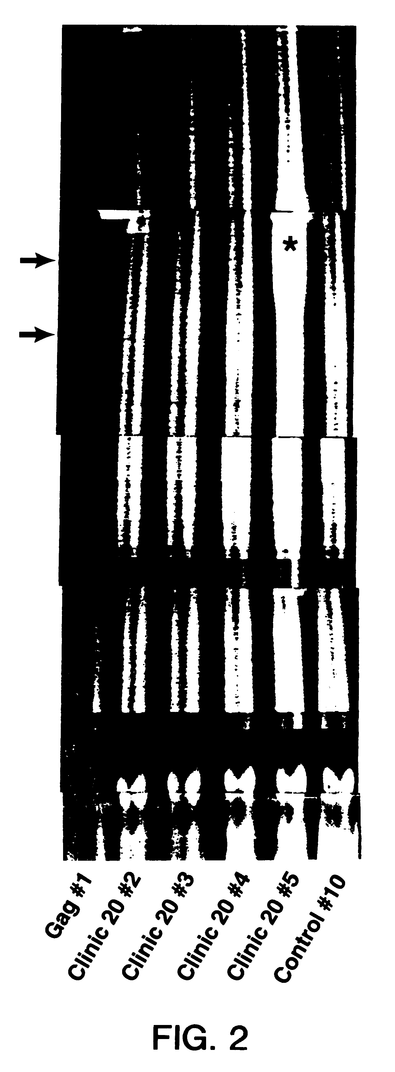
Cell Lysates

Samples were prepared as previously described [Kumar et al., AIDS Research and Human Retroviruses 5, 345-353 (1989)]. Clinical samples and HIV-1 and human T cell leukemia virus (HTLV-1) infected cells were handled in a Biological Safety level 3 laboratory following NIH safety guidelines. The T lymphocyte cell line H9 was used as a negative control in some experiments. Most clinical samples were peripheral blood mononuclear cells, separated from heparinized blood samples. The cells were viably cryopreserved in a controlled-rate freezer and stored at -70.degree. C. DNA substrates for PCR analysis were prepared by washing cells in phosphate-buffered saline (PBS), pH 7.5, three times, followed by suspending the cells in cold distilled water at the desired concentration. For the PCR reaction the cells were suspended at 1-5.times.10.sup.6 cells per ml. Cells were lysed, and virus was inactivated by heating at 95.degree. C. for 20 minutes. Lysates were stored at -20.degree. C.

D...


PUM

PropertyMeasurementUnit
Sizeaaaaaaaaaa
Fluorescenceaaaaaaaaaa

Abstract

The present invention relates to a method of detecting a DNA sequence by means of a DNA:DNA hybrid in real time using fluorescence. The present invention eliminates the need to use radioactive probes to detect the DNA and eliminates the delay needed for autoradiographic exposure of the X-ray to the radioactive label.

Description

1. Field of the InventionThe present invention relates to the detection of a DNA sequence in real time using fluorescence.2. Background InformationThe polymerase chain reaction (PCR) developed by Saiki et al. [Science 230, 1350-1354 (1985)] provided a method for rapidly amplifying small specific segments of DNA. The PCR technique has greatly simplified the analysis of DNA sequences at the genomic level.Detection or identification of a segment of DNA involves a two step procedure. The first step is the PCR amplification reaction using carefully selected primers. The second step identifies whether the desired DNA segment was amplified.Prior to the present invention, the second step has required the use of a radioactively labeled DNA probe to precisely identify a PCR amplified DNA segment. There are several disadvantages associated with the use of a radioactively labeled probe. For example, radioactive probes are dangerous to handle and when labeled with .sup.32 P, which is the standar...

Claims


Application Information

Patent Timeline
IPC IPC(8): C12Q1/68C12Q1/70G01N21/78G01N27/447G01N33/58
CPCC12Q1/6844C12Q2565/125C12Q2563/107C12Q2531/107C12Q2561/113
Inventor HUGHES, STEPHEN H.KUMAR, RAMESHBRUMBAUGH, JOHN
Owner US DEPT OF HEALTH & HUMAN SERVICES
Why Eureka
Try Eureka