Methods and systems for performing thoracoscopic coronary bypass and other procedures

a thoracoscopic and coronary bypass technology, applied in the field of thoracoscopic methods for performing cardiac procedures, can solve problems such as the release of emboli from the aortic lumen

Inactive Publication Date: 2000-02-22
EDWARDS LIFESCIENCES LLC
View PDF31 Cites 180 Cited by
  • Summary
  • Abstract
  • Description
  • Claims
  • Application Information

AI Technical Summary

Benefits of technology

Such partitioning facilitates isolation of the heart, and in particular permits the heart to be stopped while the patient is supported by cardiopulmonary bypass. Once the patient's heart is stopped, a variety of surgical procedures can be performed using percutaneously introduced instruments in a minimally invasive fashion. Thus, the present invention contemplates, at least in its preferred embodiments, the possibility of effective ascending aortic occlusion, cardioplegia, venting, right heart deflation and topical cooling in association with extracorporeal cardiopulmonary by-pass all without necessitating a median sternotomy or other thoracic incision.
The method according to the present invention may be carried out on humans or other mammalian animals. The method is of particular applicability in humans as it allows an alternative approach to open heart surgery and the development of closed cardioscopic surgery. The method according to the invention enables a percutaneous by-pass system to be associated with cardioplegia, venting and cooling of the heart which subverts the need for median sternotomy. This may, in turn, reduce the complications of the surgery.
The ascending aorta is preferably occluded by a balloon catheter introduced percutaneously through the femoral artery. This catheter must carry adjacent its tip an inflatable cuff or balloon of sufficient size that upon being inflated it is able to completely occlude the ascending aorta. The length of the balloon should preferably not be so long as to impede the flow of blood or other solution to the coronary arteries or to the brachiocephalic, left carotid or left subclavian arteries. A balloon length of about 40 mm and diameter of about 35 mm is suitable in humans. The balloon may be of a round, cylindrical, or other appropriate shape to fully and evenly accommodate the lumen of the ascending aorta. This maximizes the surface area contact with the aorta, and allows for even distribution of occlusive pressure.
The methods of the present invention provide a minimally-invasive approach for forming coronary artery bypass grafts with an efficacy equal to or greater than conventional open surgical bypass techniques. The methods of the present invention can be adapted to create anastomoses of a variety and type similar to those created by open surgical techniques, while greatly reducing patient trauma since there is no need to perform a sternotomy. Moreover, the preferred use of an endovascular catheter to partition the aorta and isolate the heart offers a substantial advantage over open surgical techniques where external clamps are placed on the aorta. External clamps can damage the aorta and may frequently cause the release of emboli from the aortic lumen. Additionally, since the sternum does not need to heal after the procedure, both internal mammary arteries can frequently be used in a single procedure to provide multiple bypass routes. Heretofore, one internal mammary artery was often left in place to provide blood flow to promote healing of the sternum in many open-surgical procedures.

Problems solved by technology

External clamps can damage the aorta and may frequently cause the release of emboli from the aortic lumen.

Method used


Image

  • Methods and systems for performing thoracoscopic coronary bypass and other procedures
  • Methods and systems for performing thoracoscopic coronary bypass and other procedures
  • Methods and systems for performing thoracoscopic coronary bypass and other procedures

Examples


Embodiment Construction

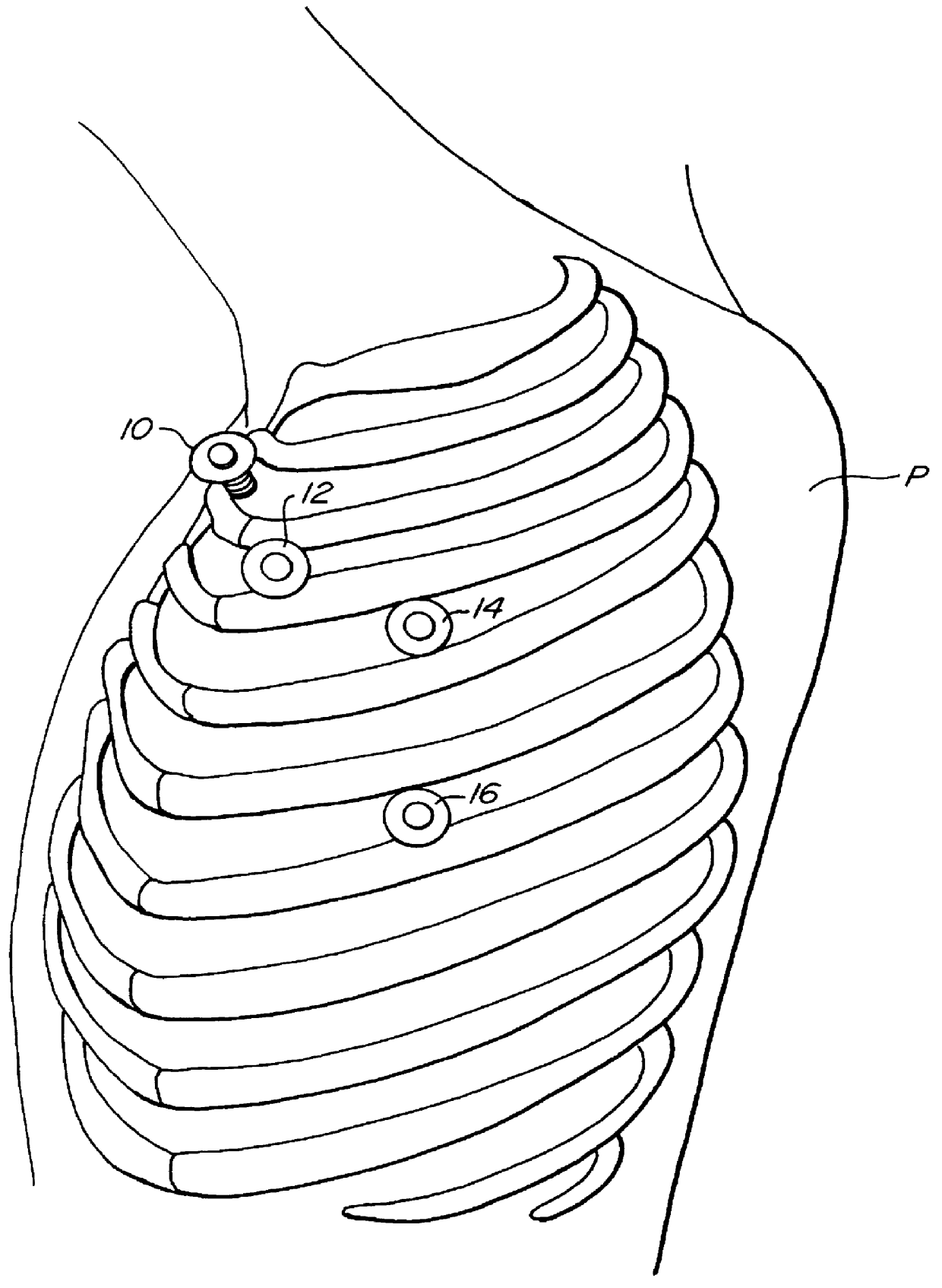
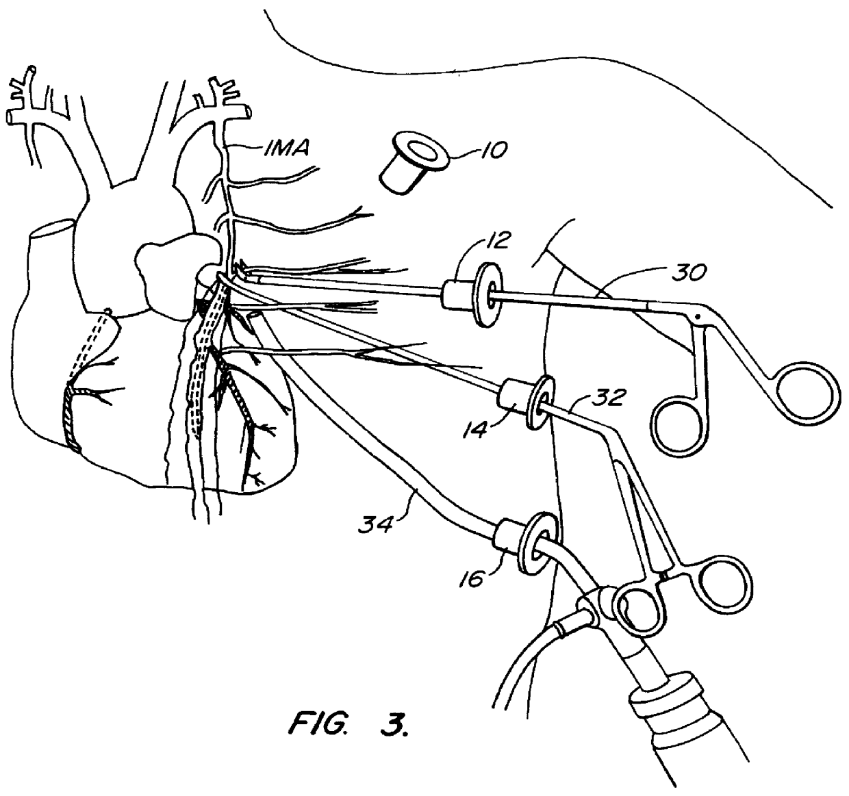
The methods of the present invention are suitable for performing a variety of surgical cardiac procedures where the heart will be stopped and the patient supported by cardiopulmonary bypass. The procedures will be minimally invasive and be performed using surgical instruments introduced through a plurality of trocar sheaths placed through the patient's chest. A viewing scope, such as a thoracoscope, will be placed through at least one of the trocar sheaths, and selected surgical instruments will be placed through others of the trocar sheaths and their manipulation viewed by the treating physician using the viewing scope. The term "viewing scope" as used herein is intended to encompass conventional endoscopes, laparoscopes, thoracoscopes. and other video-based visualization devices, as well as other types of devices that facilitate direct or indirect visualization of a body cavity through a small percutaneous penetration.

Microscope-based and other types of direct visualization system...


PUM


Abstract

A method for closed-chest cardiac surgical intervention relies on viewing the cardiac region through a thoracoscope or other viewing scope and endovascularly partitioning the patient's arterial system at a location within the ascending aorta. The cardiopulmonary bypass and cardioplegia can be induced, and a variety of surgical procedures performed on the stopped heart using percutaneously introduced tools. The method of the present invention will be particularly suitable for forming coronary artery bypass grafts, where an arterial blood source is created using least invasive surgical techniques, and the arterial source is connected to a target location within a coronary artery while the patient is under cardiopulmonary bypass and cardioplegia.

Description

1. Field of the InventionThe present invention relates generally to thoracoscopic methods for performing cardiac procedures. More particularly, the present invention relates to thoracoscopic methods for performing procedures externally on or internally within the heart while the patient's chest is unopened, the patient's heart is stopped, and the patient is supported by cardiopulmonary bypass.Coronary artery disease remains the leading cause of morbidity and mortality in Western societies. Coronary artery disease is manifested in a number of ways. For example, disease of the coronary arteries can lead to insufficient blood flow resulting in the discomfort and risks of angina and ischeria. In severe cases. acute blockage of coronary blood flow can result in myocardial infarction, leading to immediate death or damage to the myocardial tissue.A number of approaches have been developed for treating coronary artery disease. In less severe cases, it is often sufficient to treat the sympto...

Claims


Application Information

Patent Timeline
IPC IPC(8): A61B18/14A61F2/24A61M25/00A61B17/00A61B17/02A61M1/00A61B17/30A61B17/34A61M25/10A61B17/04A61B1/00A61B17/06A61B17/064A61B17/068A61B17/12A61B17/122A61B17/128A61B17/28A61B19/00A61F2/958A61F7/00A61F7/10
CPCA61B17/00234A61B17/0218A61B17/0469A61B17/06061A61B17/122A61B17/1227A61B17/1285A61B17/29A61B18/1442A61B18/1492A61B19/26A61B19/52A61F2/2427A61F7/10A61M39/0247A61B17/0057A61B17/0467A61B17/064A61B17/068A61B17/12013A61B17/2909A61B17/30A61B17/3403A61B2017/00243A61B2017/00247A61B2017/00637A61B2017/00663A61B2017/0225A61B2017/0237A61B2017/0243A61B2017/047A61B2017/0472A61B2017/0474A61B2017/0475A61B2017/2912A61B2017/2927A61B2017/2932A61B2017/2943A61B2017/306A61B2017/308A61B2017/3405A61B2017/3427A61B2017/3492A61B2018/00214A61B2018/00232A61B2018/00261A61B2018/00291A61B2018/00363A61B2018/00392A61B2018/00577A61B2019/464A61B2019/5206A61F2007/0001A61F2007/0054A61F2007/101A61M25/0023A61M25/0032A61M25/1011A61M2025/0003A61M2025/0031A61M2025/028A61M2025/1052A61M2039/0279A61M2202/047A61M2205/3344A61M2205/3355A61M2205/366A61M2210/127A61M2230/005A61B90/36A61B90/50A61B2090/064A61B2090/306
Inventor STERMAN, WESLEY D.SIEGEL, LAWRENCE C.CURTIS, PATRICIA E.STEVENS, JOHN H.MACHOLD, TIMOTHY R.
Owner EDWARDS LIFESCIENCES LLC
Why Eureka
Try Eureka