Novel photocleavable mass-tags for multiplexed mass spectrometric imaging of tissues using biomolecular probes
a mass spectrometric imaging and biomolecular probe technology, applied in the field of immunohistochemistry and in situ hybridization, can solve the problems of incomplete cycling, limited fluorescence microscopy, laborious, etc., and achieve the effect of avoiding cross-reaction of the probes with each other and avoiding artifacts in tissue imaging
- Summary
- Abstract
- Description
- Claims
- Application Information
AI Technical Summary
Benefits of technology
Problems solved by technology
Method used
Image

Examples
example 1
15-Plex PC-MT-Ab Based MSI Using Bead-Arrays as a Model System
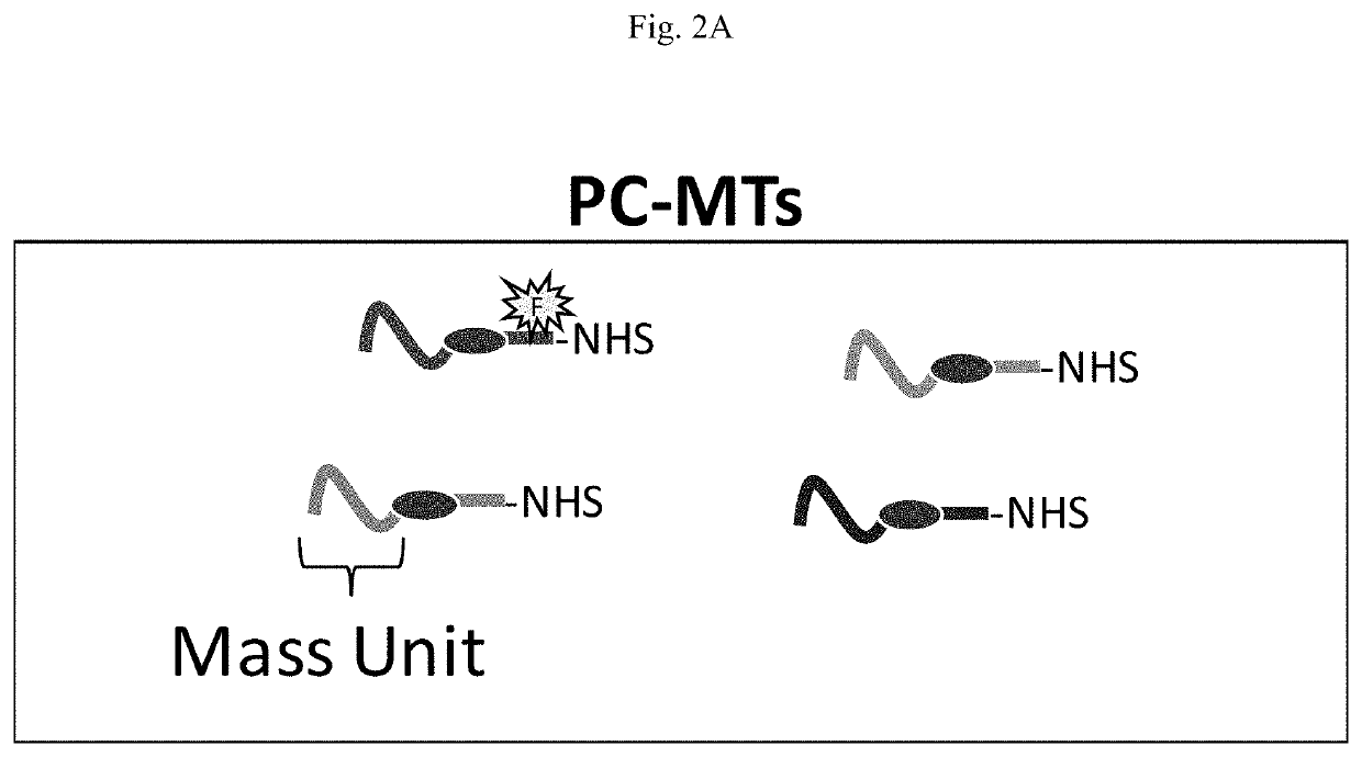
[0107]PC-MTs.
[0108]The peptide based PC-MTs were produced using standard Fmoc amino acid solid-phase peptide synthesis (SPPS) [Behrendt, White et al. (2016) J Pept Sci 22: 4-27]. An Fmoc-protected photocleavable amino acid linker (see Fmoc-PC-Linker in FIG. 3) was introduced into the peptide chain in the same manner as the other amino acids and the N-terminal α-amine of the peptide was acetylated with acetic anhydride using standard procedures. The NHS-ester probe-reactive moiety was created on the ε-amine of a lysine (K) included in the spacer unit (see FIG. 3 Step 1, NHS-ester). Conversion of the ε-amine of the lysine (K) to an NHS-ester was achieved using disuccinimidyl suberate (DSS). The use of bifunctional succinimidyl esters such as DSS or DSC (disuccinimidyl carbonate) has been previously reported for conversion of primary amines to NHS-esters [Morpurgo, Bayer et al. (1999) J Biochem Biophys Methods 38: 17-28]. PC...
example 2
5-Plex MIHC Using PC-MT-Abs on Mouse Brain Sagittal FFPE Tissue Sections
[0122]Immunostaining with PC-MT-Abs
[0123]PC-MTs and PC-MT-Abs were prepared as in Example 1 and used as follows in MIHC: For deparaffinization and hydration, FFPE tissue sections were treated as follows (each treatment step in separate staining jars): 3× with xylene for 5 min each; 1× with xylene:ethanol (1:1) for 3 min; and then hydrate 2× with 100% ethanol for 2 min each, 2× with 95% ethanol for 3 min each, 1× with 70% ethanol for 3 min, 1× with 50% ethanol for 3 min and 1× with TBS for 10 min.
[0124]Antigen retrieval was achieved in 200 mL of 1× Citrate Buffer (pH 6.0; see Materials) in a beaker preheated in a water bath at 95° C. for 1 hr and cooled in the same beaker for 30 min at room temperature. Slides were then blocked in a staining jar for 1 hr with 50 mL Tissue Blocking Buffer (2% [v / v] normal serum [rabbit and mouse] and 5% (w / v) BSA in TBS-T; note TBS-T is TBS supplemented with 0.05% [v / v] Tween-20)....
example 3
MISH Using PC-MT-NA miRNA Hybridization Probes on FFPE Tissue Sections
[0130]Preparation of PC-MT Nucleic Acids (PC-MT-NAs)
[0131]Amine-modified Locked Nucleic Acid (LNA) probes (see Materials) were labeled with PC-MT as follows: To 100 μL of LNA probe solution (10 μM in 200 mM sodium chloride and 200 mM sodium bicarbonate) sufficient PC-MT was added from a 10 mM stock in anhydrous DMF for a 200-fold molar excess relative to the LNA probe (2 μL of 10 mM stock added every 30 min for a total of 5 additions). The reaction was carried out for total 2.5 hr protected from light and with gentle mixing. To remove unreacted PC-MT, the resultant PC-MT-NA was processed on NAP-5 Sephadex G-25 Columns according to the manufacturer's instructions using TE-150 mM NaCl (10 mM Tris, pH 8.0, 1 mM EDTA and 150 mM NaCl) as the pre-equilibration buffer.
[0132]In Situ Hybridization with PC-MT-NAs
[0133]For deparaffinization and hydration, FFPE tissue sections were processed in the same manner as Example 2. T...
PUM
| Property | Measurement | Unit |
|---|---|---|
| Acidity | aaaaa | aaaaa |
| Mass | aaaaa | aaaaa |
| Structure | aaaaa | aaaaa |
Abstract
Description
Claims
Application Information
 - R&D
- Intellectual Property
- Life Sciences
- Materials
- Tech Scout
- Unparalleled Data Quality
- Higher Quality Content
- 60% Fewer Hallucinations
Browse by: Latest US Patents, China's latest patents, Technical Efficacy Thesaurus, Application Domain, Technology Topic, Popular Technical Reports.
© 2025 PatSnap. All rights reserved.Legal|Privacy policy|Modern Slavery Act Transparency Statement|Sitemap|About US| Contact US: help@patsnap.com