
Method and system for augmented imaging in open treatment using multispectral information

Pending Publication Date: 2022-01-13
DEUTES KREBSFORSCHUNGSZENT STIFTUNG DES OFFENTLICHEN RECHTS
View PDF0 Cites 4 Cited by
  • Summary
  • Abstract
  • Description
  • Claims
  • Application Information

AI Technical Summary

Benefits of technology

The invention relates to a method for training a machine learning based regressor or classifier to predict tissue parameters such as absorption, scattering, and reflectance from images. The method involves using different simulated light sources to train the machine learning based regressor or classifier. The training data is obtained by simulating the light using various techniques that make assumptions about the spectral composition of the light. The assumptions are made based on the spectral composition of the training light source. The machine learning based regressor or classifier can then be trained based on this adapted training data, allowing the machine to accurately predict tissue parameters even when the illumination light source is different from the training light source. Using this method, the machine learning based regressor or classifier can provide a confidence value for the predicted tissue parameters, indicating its accuracy. This training process is faster than traditional methods and can be carried out in practical timeframes.

Problems solved by technology

The stopping or reduction in perfusion is known as “ischemia” and can lead to irreversible damage.
Depending on the organ, this damage will occur within minutes to hours.
The damage is caused by the imbalance between supply and demand, since cells still consume oxygen and other nutrients, but no new oxygenated blood is arriving.
Ischemia can also be an unwanted side effect after surgery.
While it is in principle known that oxygenation can be detected using multispectral imaging, known methods suffer from severe drawbacks.
One deficiency in existing methods is that they are not able to provide “live” augmented images, i.e. images created in real time at a rate of e.g. 25 Hz or above, such as to give immediate feedback to the surgeon and to avoid motion artefacts or blurring.
A further deficiency is the lack of precision in the functional information conveyed by the images.
However, while these methods are fast, they are restricted to estimating oxygenation, but no perfusion, they require small multispectral bandwidths and they also rely on a number of unrealistic assumptions regarding tissue composition that lead to imprecise results.
The drawback of these methods is that they are too slow for real-time estimation of tissue parameters when dealing with high-resolution multispectral images; they are thus not suited for application during surgery or interventional procedures.
So far, it has been difficult to apply this technique under varying lighting conditions, which currently limits the versatility and practical applicability of the method.

Method used


Image

  • Method and system for augmented imaging in open treatment using multispectral information
  • Method and system for augmented imaging in open treatment using multispectral information
  • Method and system for augmented imaging in open treatment using multispectral information

Examples


examples

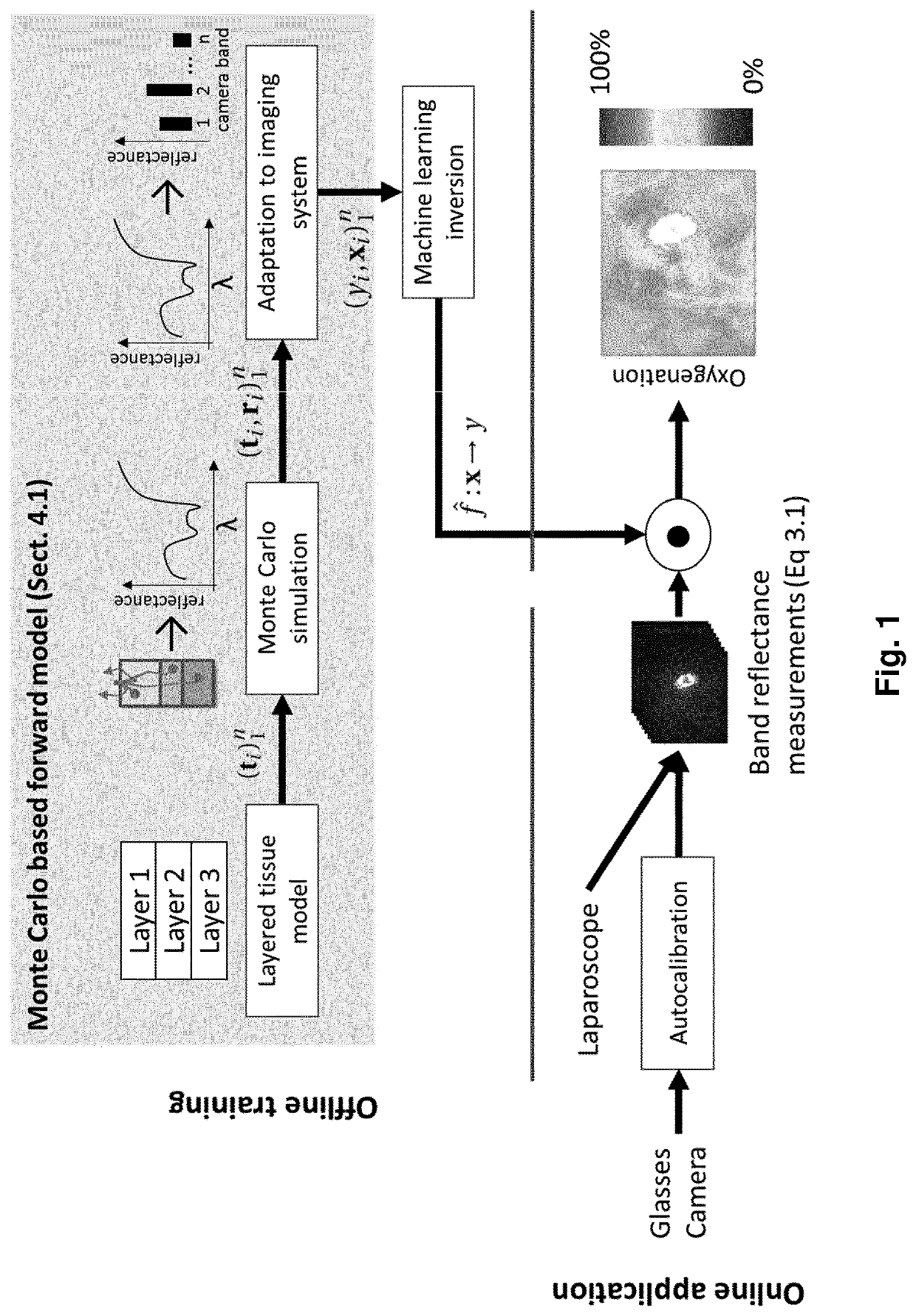
[0367]The approach to OoD detection explained above has been validated by the inventors in various examples both in silico as well as in vivo use cases.

[0368]An in silico quantitative validation based on simulations is described first. In the simulation framework used, multispectral imaging pixels are again generated from a vector t of tissue properties, which are assumed to be relevant for the image formation process. Plausible tissue samples t are drawn from a layered tissue model as explained in the dedicated section of the same name above. The simulation framework was used to generate a data set Xraw, consisting of 550,000 high resolution spectra and corresponding ground truth tissue properties. It was split in a training Xrawtr and a test set Xrawte, comprising 500,000 and 50,000 spectra respectively.

[0369]For the in silico quantitative validation, the (high resolution) spectra of the simulated data sets were converted to plausible camera measurements using the filter response ...


PUM


Abstract

Disclosed herein is a method of generating augmented images of tissue of a patient undergoing open treatment, in particular open surgery, wherein each augmented image associates at least one tissue parameter with a region or pixel of the image of the tissue, said method comprising the following steps: estimating a spectral composition of light illuminating a region of interest of the tissue, obtaining one or more multispectral images of the region of interest, applying a machine learning based regressor or classifier to the one or more multispectral images, or an image derived from said multispectral image, to thereby derive one or more tissue parameters associated with image regions or pixels of the corresponding multispectral image, wherein said regressor or classifier has been trained to predict the one or more tissue parameters from a multispectral image under a given spectral composition of illumination, wherein the regressor or classifier employed is made to match the estimated spectral composition of light illuminating said region of interest of the tissue.

Description

FIELD OF THE INVENTION[0001]The present invention is in the field of medical technology. In particular, the present invention relates to a method and system for augmented imaging of tissue using multispectral information.BACKGROUND OF THE INVENTION[0002]In open or minimally invasive surgery, there is a need for augmented imaging of the tissue under treatment. Augmented imaging as understood herein indicates providing an image of the tissue which indicates additional information other than the film image of the tissue itself. This additional information is generally referred to as “tissue parameter” in the present disclosure.[0003]One important example of such additional information could be functional information, in which information related to the functional state of the tissue is provided. Particularly important functional states to be indicated to the surgeon is the perfusion or the oxygenation of the tissue. Functional images in a broader sense may also comprise images providin...

Claims


Application Information

Patent Timeline
IPC IPC(8): A61B90/00A61B5/1455A61B5/145A61B5/00A61B5/02A61B5/0205G06T7/00G06K9/62G06K9/46G16H30/40G16H50/20G16H50/70G16H20/40G06V10/141G06V10/60G06V10/764
CPCA61B90/361G06K2009/4666A61B5/14546A61B5/443A61B5/4875A61B5/4547A61B5/02042A61B5/0205A61B5/489A61B5/4893A61B5/7275A61B5/749A61B5/0002G06T7/0012G06K9/627G06K9/4661G16H30/40G16H50/20G16H50/70G16H20/40A61B2090/365G06T2207/20081G06K2209/051A61B5/14551A61B5/0037A61B5/0075A61B5/1455A61B5/1495A61B5/4842A61B5/4881A61B5/4887A61B5/7267A61B2505/05A61B2560/0223A61B2560/0247A61B2576/00A61B5/0071G06T2207/10068G06T2207/20008G06T2207/20084G06V10/141G06V10/467G06V10/60G06V2201/031G06V10/764G06F18/2413
Inventor MAIER-HEIN, LENAWIRKERT, SEBASTIAN JOSEFVEMURI, ANANT SURAJMENJIVAR, LEONARDO ANTONIO AYALASEIDLITZ, SILVIAKIRCHNER, THOMASADLER, TIM
Owner DEUTES KREBSFORSCHUNGSZENT STIFTUNG DES OFFENTLICHEN RECHTS
Learn More