
Spectral dual-layer ct-guided interventions

a dual-layer ct and guidance technology, applied in the field of spectral imaging, can solve the problems of no or little rapid vanishing of contrast, and declining concentration of injected contrast agents, and achieve the effect of improving the visual contrast of liver lesion and increasing the success ra

Pending Publication Date: 2021-08-19
KONINKLJIJKE PHILIPS NV
View PDF0 Cites 0 Cited by
  • Summary
  • Abstract
  • Description
  • Claims
  • Application Information

AI Technical Summary

Benefits of technology

The present invention provides a spectral imaging method for enhancing the visual contrast of liver lesions during interventional procedures, which improves the safety and success rates of such interventions. The method dynamically adapts a displayed image modality based on the uptake of the contrast agent, compensating for changing conditions in the uptake due to the complex dynamics of contrast agents in organs with complex contrast agent dynamics. The method also allows for the selection of a region of interest to reduce processing resources and facilitate the sharing and portability of standard imaging protocols for a large variety of lesions encountered in image-guided interventions. The method requires only reconstruction of one second image modality from the second spectral scan, as compared to the more time-consuming process of obtaining at least three different image modalities according to the method of the first aspect.

Problems solved by technology

Conventional CT imaging techniques are sometimes facing the problem of no or little visual contrast of a liver lesion over the surrounding tissue, a problem which is peculiar to organs like the liver or the kidney.
Contrast-enhanced CT mitigates this problem partially, but, in turn, faces the problems of declining concentrations of injected contrast agents due to renal excretion, complex contrast agent dynamics during different uptake conditions, as well as washout of the contrast agent in a delayed phase after injection.
In consequence, a visual contrast may be vanishing quickly or may become inappropriate under a changing uptake condition at a moment of time, which may cause the failure of interventions which target the contrast-enhanced lesion and use CT for guidance, in particular liver or kidney interventions.

Method used

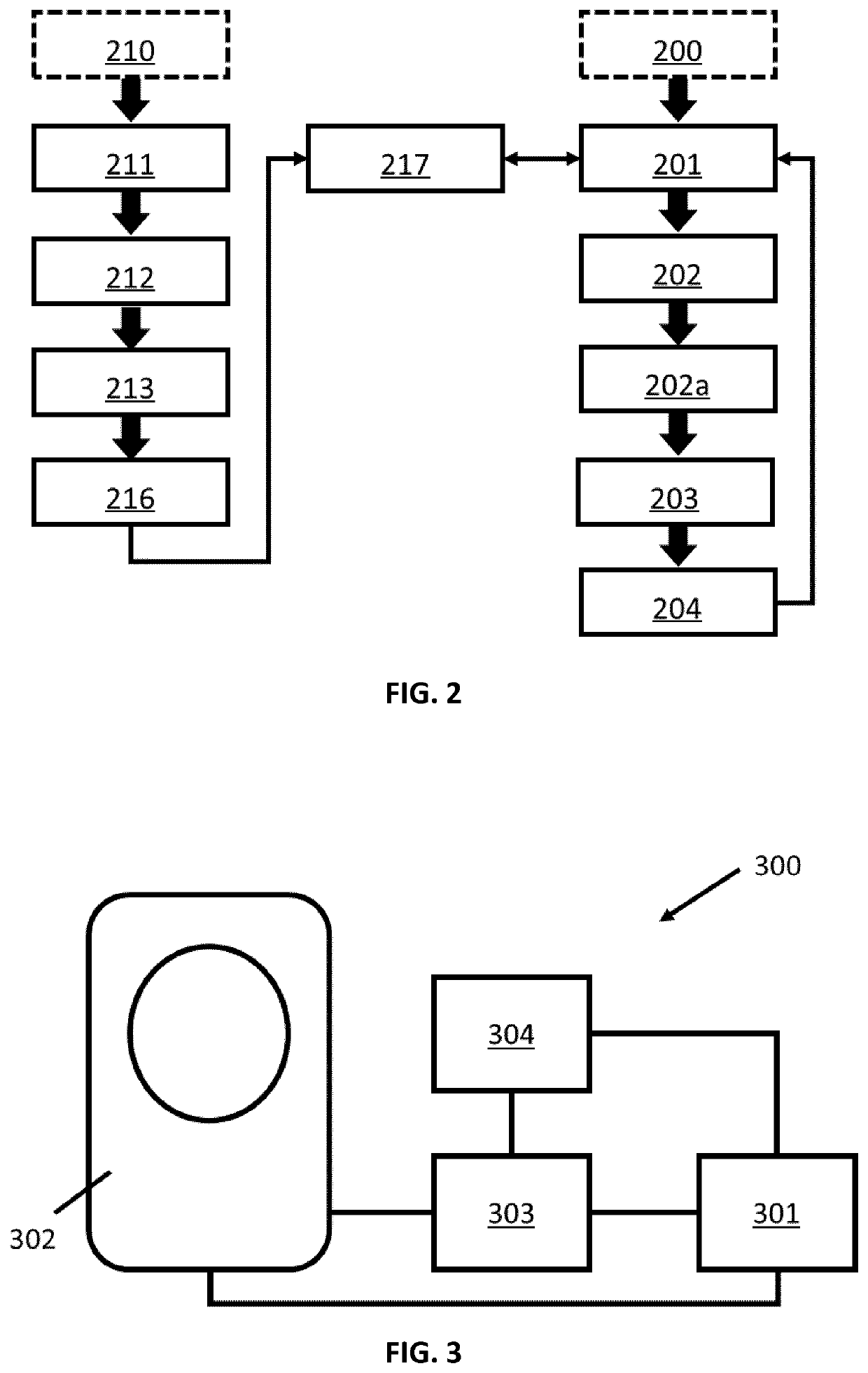

Image

  • Spectral dual-layer ct-guided interventions
  • Spectral dual-layer ct-guided interventions

Examples


Embodiment Construction

[0025]The present invention will be described with respect to particular embodiments and with reference to certain drawings but the invention is not limited thereto but only by the claims.

[0026]It is to be noticed that the term “comprising”, used in the claims, should not be interpreted as being restricted to the means listed thereafter; it does not exclude other elements or steps. It is thus to be interpreted as specifying the presence of the stated features, integers, steps or components as referred to, but does not preclude the presence or addition of one or more other features, integers, steps or components, or groups thereof. Thus, the scope of the expression “a device comprising means A and B” should not be limited to devices consisting only of components A and B. It means that with respect to the present invention, the only relevant components of the device are A and B.

[0027]Reference throughout this specification to “one embodiment” or “an embodiment” means that a particular...


PUM


Abstract

A method of spectrally imaging an organ during an image-guided intervention is disclosed. Based on at least one first scan, a first image modality is obtained. A selection of a region of interest is obtained within the first image modality. Based on at least one second spectral scan of the organ, at least three second image modalities are obtained. A contrast value is calculated over the previously selected region of interest for all obtained second image modalities. The second image modality with the largest contrast value is selected for display. Alternatively, image modalities of the organ are displayed over time and the modalities for display are selected according to an imaging protocol which associates a lesion type and timestamp of a spectral scan with a modality having optimal contrast.

Description

FIELD OF THE INVENTION[0001]The present invention relates to a spectral imaging method generally and to spectral imaging by dual-layer CT for guidance more particularly.BACKGROUND OF THE INVENTION[0002]Conventional CT imaging techniques are sometimes facing the problem of no or little visual contrast of a liver lesion over the surrounding tissue, a problem which is peculiar to organs like the liver or the kidney. Contrast-enhanced CT mitigates this problem partially, but, in turn, faces the problems of declining concentrations of injected contrast agents due to renal excretion, complex contrast agent dynamics during different uptake conditions, as well as washout of the contrast agent in a delayed phase after injection. In consequence, a visual contrast may be vanishing quickly or may become inappropriate under a changing uptake condition at a moment of time, which may cause the failure of interventions which target the contrast-enhanced lesion and use CT for guidance, in particular...

Claims


Application Information

Patent Timeline
Patent Type & Authority Applications(United States)
IPC IPC(8): A61B34/20A61B6/03A61B6/00
CPCA61B34/20A61B6/032A61B6/54A61B6/50A61B6/461A61B6/481A61B6/482A61B6/507
Inventor PROKSA, ROLANDFINGERLE, ALEXANDER ANDRÉ
Owner KONINKLJIJKE PHILIPS NV
Learn More