
Assembloid - 3D mimetic tissue structure based on patient- derived multiple cell types and method of manufacturing the same

a tissue structure and 3d technology, applied in the field of 3d mimetic tissue structure, can solve the problems of inability to explain factors including a microenvironment associated with a disease in vivo, and the inability of psc-derived organoids to be applied to tumor models,

Pending Publication Date: 2021-03-18
POSTECH ACAD IND FOUND
View PDF1 Cites 7 Cited by
  • Summary
  • Abstract
  • Description
  • Claims
  • Application Information

AI Technical Summary

Benefits of technology

The present invention provides a method of manufacturing a 3D mimetic tissue structure, which includes an organoid and microenvironment-constituting cells. The organoid may be derived from stem cells or tumor cells, and the microenvironment-constituting cells may include fibroblasts, endothelial cells, immune cells, and muscle layers. The manufacturing process involves culturing the organoid in a medium containing fibroblasts and endothelial cells, and then culturing it in a medium containing muscle cells. The resulting 3D mimetic tissue structure is a bladder tissue or bladder tumor tissue that can be used for various applications such as tissue engineering or drug testing.

Problems solved by technology

Although these organoids represent the potential for a variety of biological studies on normal tissues and cancer, questions remain because numerous factors including a microenvironment associated with a disease in vivo cannot be explained.
Also, there is a problem in that PSC-derived organoids cannot be applied to a tumor model.
To overcome these problems, recent studies have reported the structures of organotypic organoids including various cell components, but these organotypic organoids still do not precisely mimic in vivo tissue because they use a traditional co-culture system or a simple mixture of cellular components without an organized structure, and moreover, there is a limitation in which most of the cellular components that constitute a tissue microenvironment or stroma are lacking.

Method used


Image

  • Assembloid - 3D mimetic tissue structure based on patient- derived  multiple  cell  types  and  method  of  manufacturing the same
  • Assembloid - 3D mimetic tissue structure based on patient- derived  multiple  cell  types  and  method  of  manufacturing the same
  • Assembloid - 3D mimetic tissue structure based on patient- derived  multiple  cell  types  and  method  of  manufacturing the same

Examples


example 1

on and Methods for Experiments

[0070]1-1. Mice

[0071]For lineage tracing experiments, CK5CreERT2 (JAX: 018394) mice were crossed with R26Rainbow / Rainbow mice to obtain CK5CreERT2; R26Rainbow / Rainbow mice. Unless particularly stated, in all other experiments, C57BL / 6 mice were used. In each experiment, mice were randomly selected from a cage for drug treatment. Procedures were performed under isoflurane anesthesia using a standard vaporizer. All procedures were performed according to the protocols approved by the Institutional Animal Care and Use Committee at POSTECH (IACUC number: POSTECH-2019-0055).

[0072]1-2. Human Bladder Tumor Samples

[0073]Human bladder tumor samples were obtained from the tissue bank of Seoul National University Hospital (SNUH). 0.5 to 1 cm3 specimens of bladder tumor tissue were acquired from patients undergoing TURB or a cystectomy according to a protocol approved by the SNUH Institutional Review Board (IRB). The tumor samples were transported to POSTECH after c...

example 2

ion of Mimetic Property of the Urothelium of Native Bladder by Long-Term Cultured Bladder Organoid

[0107]2-1. Confirmation of Mature Bladder Tissue Differentiation of Urothelial Stem Cell

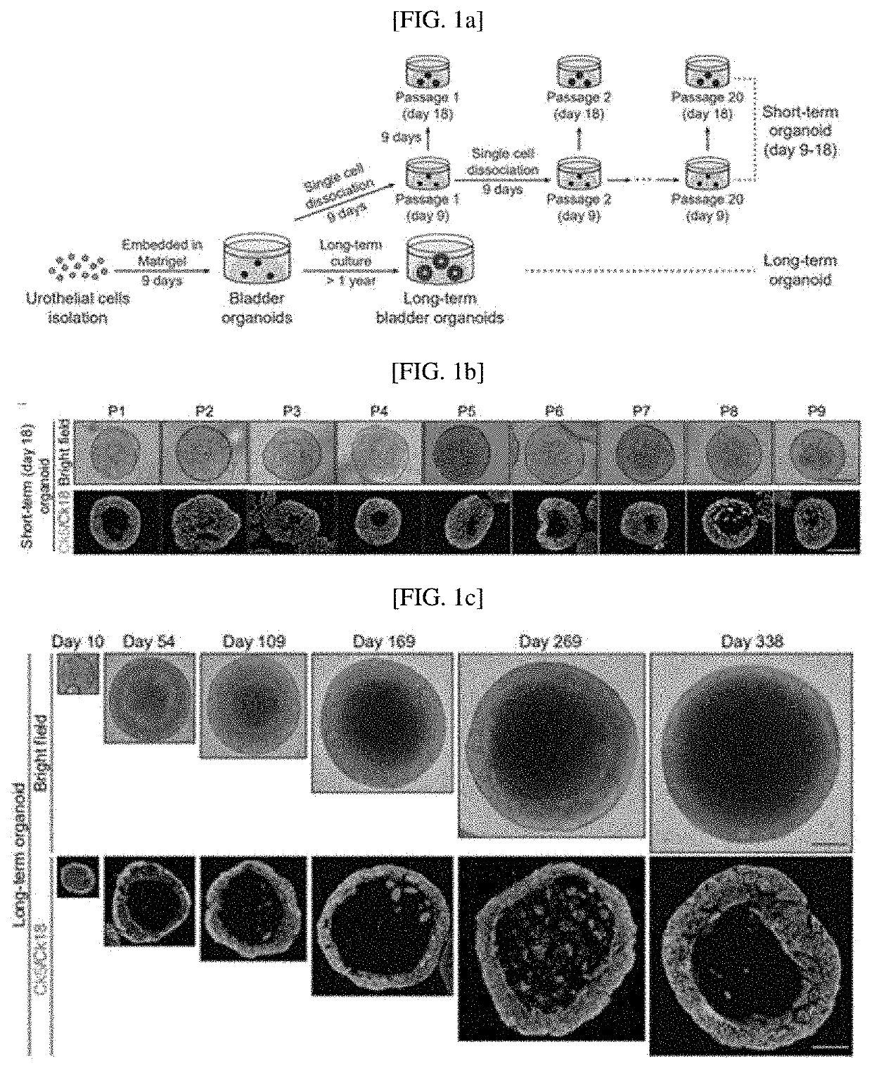
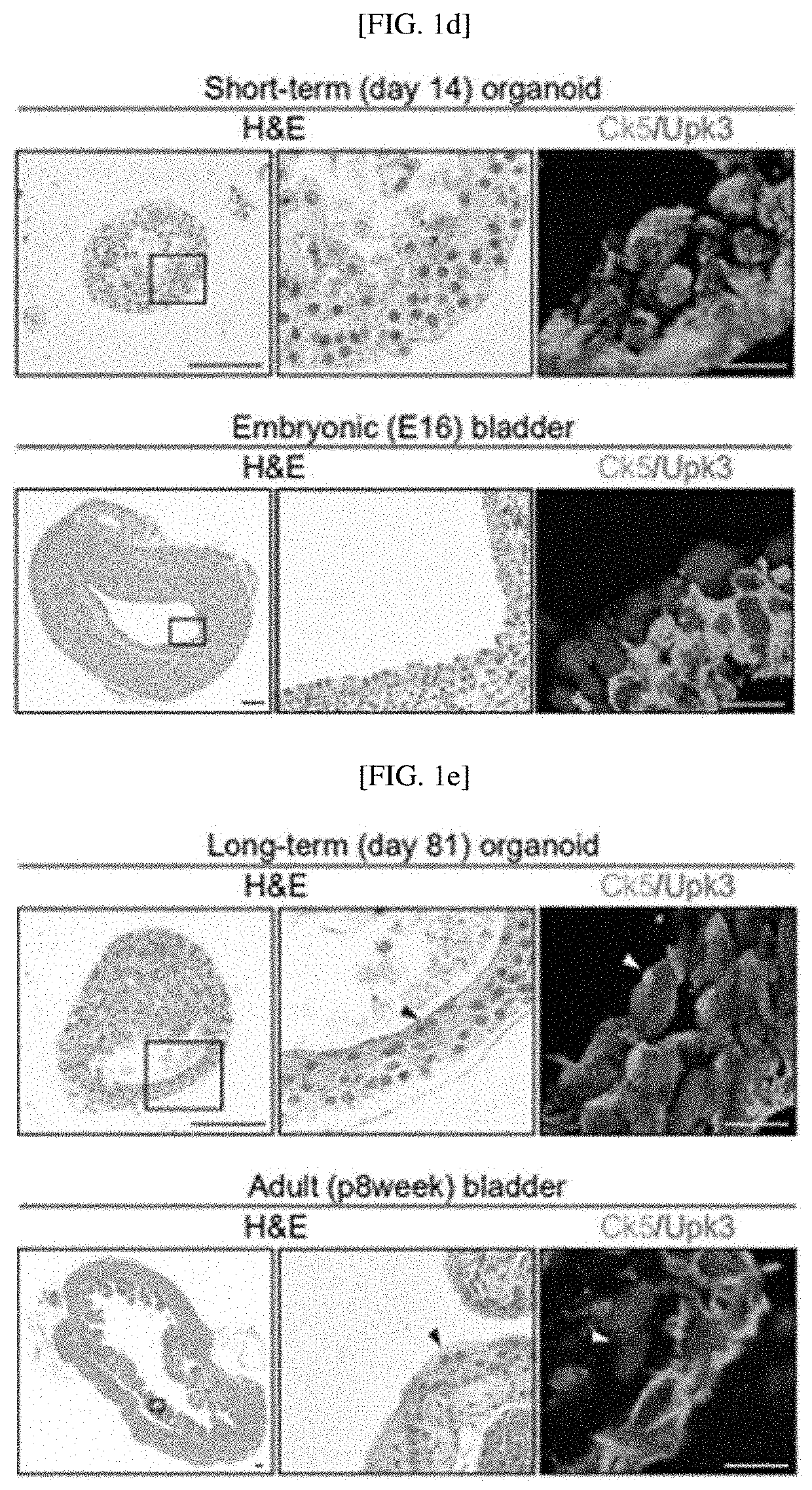
[0108]To confirm urothelial stem cells capable of forming a bladder tissue structure in vitro, and confirm whether organoids generated by the stem cells develop into mature bladder tissue including multiple layers of similarly differentiated epithelial cells as mature bladder tissue in vivo, as shown in FIG. 1A, two culture systems maintaining bladder organoids for a long time were established. For long-term serial passaging of short-term cultured organoids, as shown in FIG. 8A, primary bladder organoids generated by culturing single urothelial stem cells cultured in a 3D environment for 7 to 9 days were dissociated into single cells, and subcultured to form new bladder organoids. This process was repeated twenty times for 6 months, and as shown in FIG. 8B, it was confirmed that bladder tissue was su...

example 3

ion of In Vivo Bladder-Like Tissue Structure and Physiological Activity of In Vitro Reconstituted Three-Layered Miniature Bladder

[0117]3-1. Confirmation of In Vivo Bladder-Like Tissue Structure of In Vitro Reconstituted Three-Layered Miniature Bladder

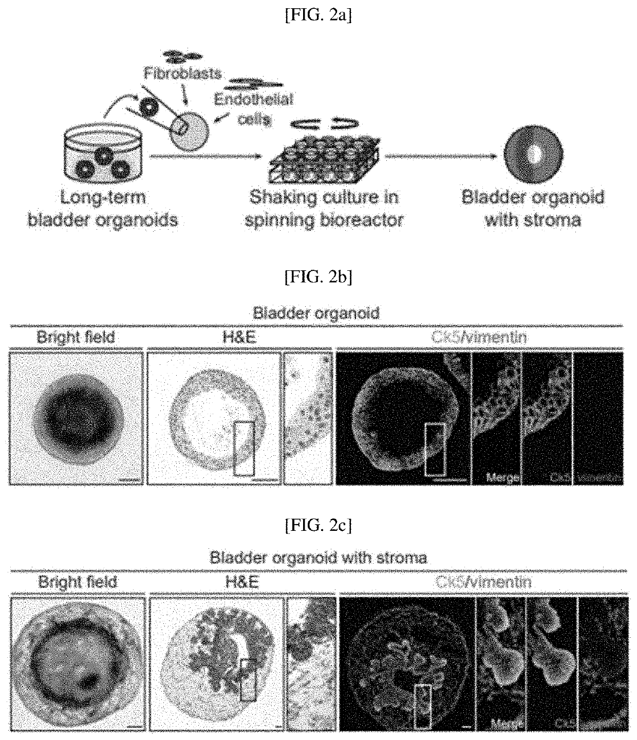
[0118]The inventors intended to determine that tissue stroma is a critical tissue component serving as a gap between stem cells for stimulating cell proliferation and differentiation and providing structural support, and to develop bladder organoids including tissue stroma. As shown in FIG. 2A, to mimic urothelial cells including mature stroma, bladder organoids cultured for a long period of over 200 days were reconstituted with two components of tissue stroma, such as fibroblasts and endothelial cells. In addition, as shown in FIG. 9A, to promote the growth and organization of bladder organoids with stroma, reconstituted organoids were cultured in a spinning bioreactor developed using 3D printing.

[0119]The cultured organoids were analy...


PUM

PropertyMeasurementUnit
pHaaaaaaaaaa
pressureaaaaaaaaaa
pressureaaaaaaaaaa

Abstract

The present invention relates to a 3 dimensional mimetic tissue structure—“Assembloid” based on patient-derived multiple cell types to develop next generation organoid technology serving as a novel platform for new drug development and a disease model and a method of manufacturing the same, and more particularly, to a stem cell- or tumor cell-based 3D multicellular mimetic tissue structure manufactured by reconstituting epithelial or tumor cells with various cellular components of a microenvironment such as stromal cells, vascular cells, immune cells or muscle cells based on three-dimensional (3D) bioprinting, and a method of manufacturing the same. As the stem cell- or tumor cell-based 3D multicellular mimetic tissue structure containing the major factors of a tissue microenvironment, such as stromal cells, vascular cells, immune cells and muscle cells, designed according to the present invention is confirmed to mimic physiological and pathological characteristics of tissue in the body better than conventional organoids, normal and tumor assembloids may be used as a new platform for new drug development and a disease model. More specifically, together with 3D bioprinting technology, it is expected that in vitro bladder tissue and bladder tumor tissue are effectively used as a platform to develop precise and personalized therapeutic options for bladder related diseases including bladder cancer.

Description

CROSS-REFERENCE TO RELATED APPLICATION[0001]This application claims priority to and the benefit of Korean Patent Application No. 10-2019-0113709, filed on Sep. 16, 2019, the disclosure of which is incorporated herein by reference in its entirety.BACKGROUND1. Field of the Invention[0002]The present invention relates to a 3 dimensional mimetic tissue structure (Assembloid) which is defined by ‘Organoids derived from tissue stem / tumor cells reconstituted with other components of the tissue stroma / microenvironment based on patient-derived multiple cell types and a method of manufacturing the same, and more particularly, to a multicellular mimetic tissue structure based on various normal cells, which is manufactured by constituting epithelial or tumor cells with various cellular components of a microenvironment such as stromal cells, vascular cells, immune cells or muscle cells based on three-dimensional (3D) bioprinting, and a method of manufacturing the same.2. Discussion of Related Ar...

Claims


Application Information

Patent Timeline
Patent Type & Authority Applications(United States)
IPC IPC(8): C12N5/071
CPCC12N5/0685C12N2513/00C12N2506/30C12N2502/1347C12N2502/02C12N5/0693C12N2501/11C12N5/0684C12N2502/253C12N2502/1114C12N2503/02C12N2506/02C12N2502/13C12N5/0697
Inventor SHIN, KUNYOOKIM, EUNJEEJUNG, SUNGJUNE
Owner POSTECH ACAD IND FOUND
Learn More