
Robotic device for a minimally invasive medical intervention on soft tissues

a robotic device and soft tissue technology, applied in the field of medical interventions, can solve the problems of avoiding sensitive anatomical structures, requiring a specific and restricting environment, and complicated procedures

Pending Publication Date: 2020-09-10
QUANTUM SURGICAL
View PDF1 Cites 12 Cited by
  • Summary
  • Abstract
  • Description
  • Claims
  • Application Information

AI Technical Summary

Benefits of technology

The present invention provides a solution to help medical operators position a medical instrument with precision and reproducibility in the body of a patient, regardless of the organ or anatomical structure's movement or deformation inside the patient. This is achieved through a robotic arm that improves the precision of positioning the medical instrument, resulting in improved clinical efficacy of the treatment and reduced risks of complications. The robotic device uses the trajectory, biomechanical model, and position information acquired during the medical intervention to determine the real position of a movable and deformable anatomical structure in the body of the patient. This avoids errors due to the operator compensating for the movements associated with respiration and with the internal deformations of the organs. The image capture system used during the intervention can be non-irradiating, reducing the irradiation dose for the patient and medical team and making its use less restricted.

Problems solved by technology

These procedures are sometimes lengthy and difficult since, in contrast to open surgery, the operator does not always have a direct view of the anatomy of the patient and of the organ that is to be treated.
This complicates the identification of the anatomical structures, the precise placement of the medical instrument and avoidance of sensitive anatomical structures (nerves, vessels, healthy organs, etc.).
However, they require a specific and restricting environment linked to the presence of a magnetic field formed by the magnetic field generator.
Although all of these known navigation systems are able to improve the precision of the medical procedure compared to the conventional manual method, by supplying in real time the position and the orientation of the medical instrument in images, they have important limitations in respect of minimally invasive medical interventions on deformable tissues.
A first limitation is that the final procedure of introducing the medical instrument as far as the targeted anatomical zone is performed manually by the operator, which means that the result depends on the skill of the operator and is unable to attain a high degree of precision.
A second limitation is that the functioning of these navigation systems assumes that the targeted organs or anatomical structures do not move and do not deform between the moment when the reference examination is carried out and the moment when the operator introduces the medical instrument.
In the case where the examination has been carried out several days before the medical intervention, and with the patient in a different position on the operating table compared to his position on the examination table, the targeted organs or anatomical structures may have moved or been deformed, and the offset between the displayed position and the real position of the targeted organ or anatomical structure may introduce a high degree of imprecision.
In addition, the targeted organs or anatomical structures may deform simply on account of the respiration of the patient, and the known navigation systems are based on controlling the respiration by the patient, which greatly limits the precision attainable by these navigation systems.

Method used


Image

  • Robotic device for a minimally invasive medical intervention on soft tissues

Examples


Embodiment Construction

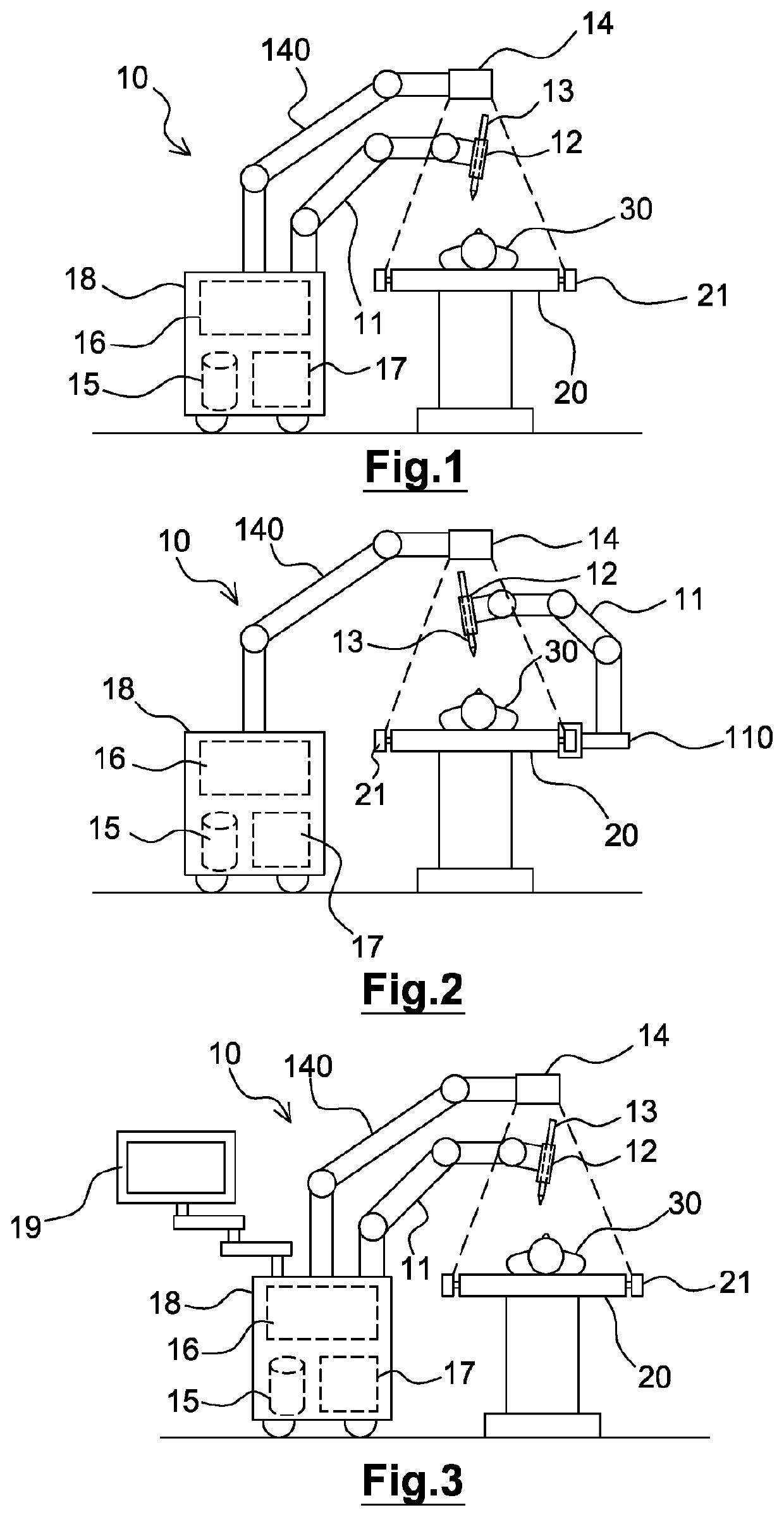
[0056]FIG. 1 shows schematically an embodiment of a robotic device 10 for assisting an operator in a medical intervention, for example a minimally invasive intervention on soft tissues.

[0057]As is illustrated in FIG. 1, the robotic device 10 has a robot arm 11 with several degrees of freedom. The robot arm 11 has an end suitable for receiving a medical instrument 13. In the example shown in FIG. 1, the medical instrument 13 is mounted on the end of the robot arm 11 by way of a guide tool 12 suitable for guiding said medical instrument 13. For this purpose, the robot arm 11 has, at said end, an interface suitable for receiving said guide tool 12.

[0058]The robot arm 11 preferably has at least 6 degrees of freedom in order to permit wide ranges for spatially monitoring the position and orientation of the guide tool 12 with respect to a patient 30, who is lying on an operating table 20 for example.

[0059]The guide tool 12 is suitable for guiding the medical instrument 13, that is to for ...


PUM


Abstract

The disclosure relates to a robotic device for performing a medical intervention on a patient using a medical instrument, comprising:a robot arm having several degrees of freedom and having an end suitable for receiving the medical instrument,an image capture system suitable for capturing position information concerning the anatomy of the patient,a storage medium having a biomechanical model of the human body,a processing circuit configured to determine a position setpoint and an orientation setpoint for said medical instrument on the basis of the biomechanical model, on the basis of the position information and on the basis of a trajectory to be followed by the medical instrument in order to perform the medical intervention, anda control circuit configured to control the robot arm in order to place the medical instrument in the position setpoint and the orientation setpoint.

Description

TECHNICAL FIELD[0001]The present invention belongs to the field of medical interventions and relates more particularly to a robotic device for minimally invasive medical interventions performed on the deformable tissues of a patient, for example for performing treatment or diagnosis on deformable organs or anatomical structures.PRIOR ART[0002]Medical interventions (for diagnosis, therapy and / or surgery) by a minimally invasive or percutaneous route are becoming increasingly important, especially in oncology for the local treatments of cancer, acting directly on the cells of the affected organ, such as the liver, the kidneys, the lungs, the pancreas, the breasts, the prostate, etc.[0003]Outside the field of oncology, there are many medical procedures and applications using a minimally invasive or percutaneous access route, for example by insertion of a needle: biopsies (collecting tissues for pathological analysis), placement of drains (aspiration of fluids), injection of therapeutic...

Claims


Application Information

Patent Timeline
Patent Type & Authority Applications(United States)
IPC IPC(8): A61B34/30A61B34/20A61B90/13A61B34/00A61B10/02A61N7/00A61B1/00A61B18/02A61B18/12A61N5/10A61B8/00
CPCA61B1/00149A61B2034/105A61B2034/742A61B2017/00199A61B2034/2055A61B8/4218A61B2090/502A61B17/3494A61B34/20A61B90/13A61B34/30A61B2017/00203A61B34/25A61B18/12A61B10/0233A61B2034/743A61B2034/2051A61N7/00A61B2034/107A61B2018/00613A61N5/10A61B18/02A61B2034/2046A61B34/32A61B90/361A61B90/37A61B34/10A61B2034/2065A61B2034/2057
Inventor BLONDEL, LUCIENBADANO, FERNANDNAHUM, BERTIN
Owner QUANTUM SURGICAL
Learn More