Methods of sample cycle multiplexing and in situ imaging

a sample cycle and multiplexing technology, applied in the field of in situ imaging of samples, can solve the problems of limited image-based sample analytical measurement techniques, inability to reveal essential spatial details, and inability to meet the needs of the sample, so as to achieve efficient, accurate and reliable results

Active Publication Date: 2019-11-14
LUNAPHORE TECH SA
View PDF1 Cites 1 Cited by
  • Summary
  • Abstract
  • Description
  • Claims
  • Application Information

AI Technical Summary

Benefits of technology

[0014]An object of this invention is to provide a method for in situ imaging of samples by cycle multiplexing that enables imaging of various molecular targets through multi-molecular read-outs on the same sample in an efficient, accurate and reliable manner.
[0015]It is advantageous to provide a method for in situ imaging of samples by cycle multiplexing that enables imaging of various molecular targets on the same sample in a rapid and sensitive manner.
[0016]It is advantageous to provide a method of in situ imaging of samples by cycle multiplexing where the sample integrity is maintained by the avoidance of de-mounting the sample from the sample support between each cycle of the multiplexing process.

Problems solved by technology

Image-based sample analytical measurement techniques have been limited by the number of molecular measurements that can be observed simultaneously (the extent of multiplexing) in a single sample, for example a tissue specimen.
As a consequence, essential spatial details, which currently only image-based approaches can reveal, are being missed.
Using immunofluorescence rather than classical immunohistochemistry can partly overcome this issue, but measurements are still limited to a maximum of 4-5 simultaneous molecular readouts.
The major limitation of image-based multiplexed sample analytical measurements is the separation of distinct signals in a single specimen without cross talk between signals.
In addition, fluorophores may exhibit self-quenching behavior at high labeling densities, further limiting the simultaneous application of multiple labels.
This could in principle be overcome by direct immunofluorescence, in other words labelling primary antibodies directly, but this approach gives rise to other problems, such as a decreased specificity and a lower signal output due to a lack of amplification.
While this method has the advantages of being image-based, applicable to tissue sections and able to be utilized in environments containing non-specific nucleases, the maximum number of simultaneous detections is limited.
The inclusion of quantifiable reference standards into the measurement process such as described in WO 2008 / 005464, while increasing the precision in quantification immunohistochemistry read-outs in certain applications such as semi-quantitative scoring of biomarker proteins, e.g. as applied to semi-quantitative scoring of Human Epidermal Growth Factor 2 (HER2) expression in breast cancer tissues, may further limit the possibility of multiple simultaneous readouts because of dividing the output signals into specific bands.
Although promising, translation of multi-cycle in situ imaging technologies to high-throughput, multiplexed molecular profiling of samples such as for example tumour sections is not straightforward.
First, long incubation and washing cycles (usually up to several hours) result in extremely long total protocol durations that cause degradation of tissue antigens under fluctuating ambient conditions.
Second, repeated mounting / demounting of imaging of sample coverslips steps further deteriorate tissue integrity.
Therefore, such manual handling of cycles affects reproducibility and practically impedes reliable molecular profiling of tissue specimens at high-throughput and renders the use of multi-cycle technologies impractical in applications like diagnostic purposes which require high throughput, reliability and relative low-cost implementation characteristics.
A further limitation of the existing methods available for in situ imaging of samples also originates from large-area imaging requirement for realizing multi-cycle assays.
This hinders the true localization of molecular signals, in particular those of subcellular features that can only be observed with a high resolution or super-resolution microscopy systems.
The issues of possible localization errors and analysis time increase resulting from the scanning process are also present in this method.
The method aims to overcome the several disadvantages of having to de-coverslip the sample between each successive run such as extra time consumption, tissue loss and slide-to-slide image variation by eliminating the need for this step, while it does not address the imaging area and process time requirements.
Finally, it has been observed that in situ imaging involving sequential and repeated fluorophore exposure of the sample leads to induced damages on the sample.
In particular, fluorescently labelled antibodies usually get cross-linked to the sample or tissue during imaging and cannot be removed from sample or tissue afterwards, which further limits the use of in situ imaging by cycle multiplexing.

Method used


Image

  • Methods of sample cycle multiplexing and in situ imaging
  • Methods of sample cycle multiplexing and in situ imaging
  • Methods of sample cycle multiplexing and in situ imaging

Examples


example 1

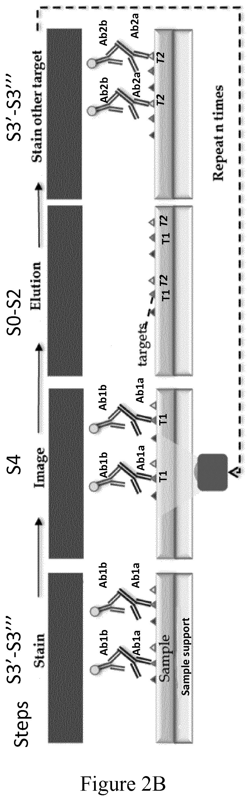
Example of Multiplexing Protocol to Carry out Successive Sample Labelling Cycles

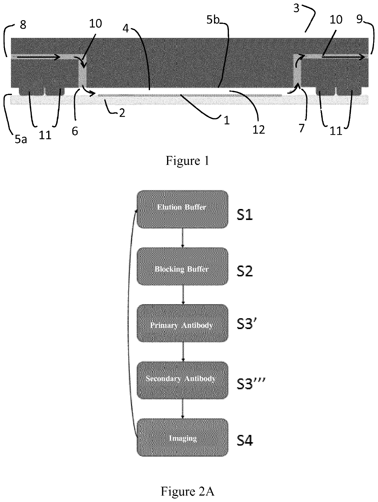
[0109]A method of the invention for in situ imaging of samples by cycle multiplexing is implemented in a device on a formalin-fixed paraffin-embedded (FFPE) tissue sample as illustrated on FIG. 1.

[0110]a) Example of Sample Preparation

[0111]Different types of sample preparation steps can be realized depending on the sample type and desired application. In the present example, the sample preparation steps for FFPE tissue samples which were carried out before multi-staining processes is described here as a typical example.

[0112]The biological samples are first dehydrated at 65° C. for 10 min. After 5 minutes of cool-down, the tissue sections are dewaxed for 10 minutes in a Histoclear solution (e.g. Xylol), followed by rehydration in 100%, 95%, 70% and 40% (vol. / vol.) ethanol, respectively. Finally, the heat-induced antigen-retrieval process is carried out with a sodium citrate buffer (about pH 6) or EDTA bu...

example 2

Example of Using an Imaging Buffer of the Invention in a Method of the Invention

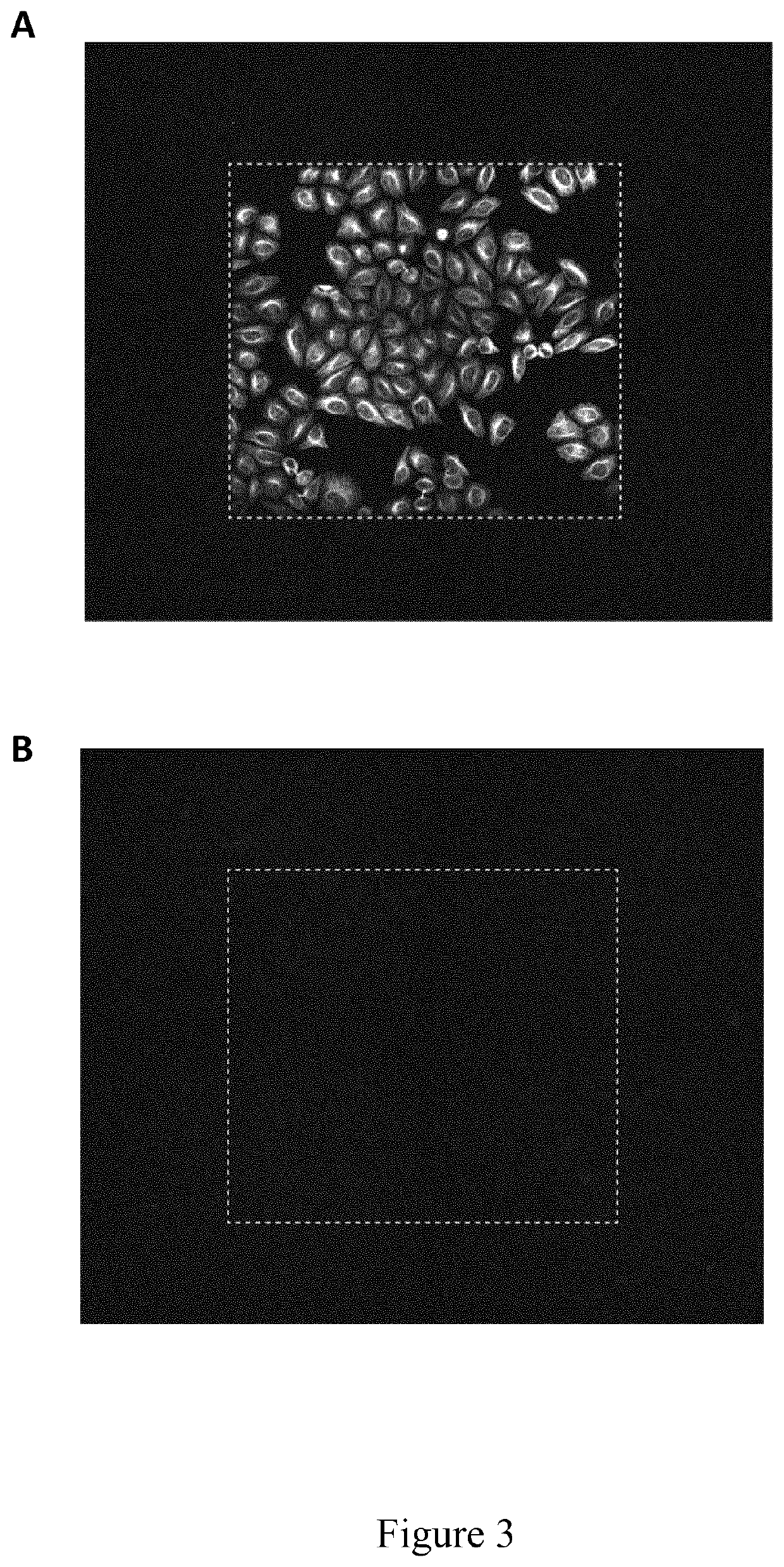
[0151]The present example illustrates the use of an imaging buffer according to the invention used to improve the efficiency of fluorescent molecule removal from samples under high intensity light, which is particularly useful in a method according to the invention.

[0152]Formalin fixed HeLa cells were applied to one sample labeling cycle according to the multiplexing protocol as described under Example 1. Then, imaging was performed using either phosphate buffered saline (PBS) alone or in combination with a radical-scavenging agent as imaging buffer. Afterwards, the elution step was performed as described under Example 1 and a second sample labeling cycle was performed, omitting the primary antibody binding step, and a larger imaging area was scanned when compared to the first imaging round, while leaving all other imaging parameters such as laser intensity and exposure time unchanged. In the shown image...

example 3

Multiplexed Colocalized Staining of ER, CK, PR and HER2 on a Breast Tumor Sample

[0156]Formalin-fixed paraffin-embedded (FFPE) tissue slides can be stained while also imaging the sample between each step using the methods disclosed in the present invention. As an illustrative example, an FFPE breast tumor section positive to estrogen receptor (ER), progesterone receptor (PR), cytokeratin (CK) and epidermal growth factor receptor 2 (HER2), has been stained using sequential multiplexing according to a method of the invention using in sequence a plurality of reagents and imaging steps determined according to data derived from preliminary test results. The sample has been first prepared for staining through the dewaxing, rehydration and antigen retrieval processes as described in the previous examples and then subjected to a method of in situ imaging of the invention are summarized in Table 2 below. Each marker has been detected in the imaging step using sandwich assays with fluorescent ...


PUM

PropertyMeasurementUnit
volumeaaaaaaaaaa
volumeaaaaaaaaaa
flow rateaaaaaaaaaa

Abstract

The invention relates to a method for in situ imaging of samples by cycle multiplexing that enables imaging of various molecular targets through multi-molecular read-outs on the same sample in a rapid, highly sensitive and reliable manner. The invention is further related to imaging buffers preventing the degradation of the sample and of the imaging reagents, which are particularly useful in a method of in situ imaging of samples by cycle multiplexing according to the invention.

Description

FIELD OF THE INVENTION[0001]The present invention pertains generally to the fields of in situ imaging of samples, in particular biological samples by cycle multiplexing.BACKGROUND OF THE INVENTION[0002]Image-based sample analytical measurement techniques have been limited by the number of molecular measurements that can be observed simultaneously (the extent of multiplexing) in a single sample, for example a tissue specimen. This has, so far, constrained this type of analytical approaches from large-scale ‘-omics’ use when compared to other highly multiplexed technologies, such as single-cell sequencing or mass cytometry. As a consequence, essential spatial details, which currently only image-based approaches can reveal, are being missed. Using immunofluorescence rather than classical immunohistochemistry can partly overcome this issue, but measurements are still limited to a maximum of 4-5 simultaneous molecular readouts. The major limitation of image-based multiplexed sample analy...

Claims


Application Information

Patent Timeline
Patent Type & Authority Applications(United States)
IPC IPC(8): G01N33/543C12Q1/6832C12Q1/6818G01N33/53
CPCC12Q1/6818C12Q1/6832G01N33/54366G01N33/5306G01N33/58C12Q2563/179C12Q1/6816
Inventor CIFTLIK, ATA TUNADUPOUY, DIEGO GABRIELJORIS, PIERREGIJS, MARTIN
Owner LUNAPHORE TECH SA
Why Eureka
Try Eureka