Methods for promoting t cells response
a t cell response and promoting technology, applied in the field of promoting t cell response, can solve the problems of colorectal cancer patients with increased risk of polymorphisms in the promoter of the dc-sign gene, and achieve the effect of improving the risk of colorectal cancer
- Summary
- Abstract
- Description
- Claims
- Application Information
AI Technical Summary
Benefits of technology
Problems solved by technology
Method used
Image
Examples
example 1
[0131]Material & Methods
Animals
[0132]Rats were purchased from the “Centre d′ElevageJanvier” (Genest, Saint-Isle, France) and experimental procedures were carried out in strict accordance with the protocols approved by the Committee on the Ethics of Animal Experiments of Pays de la Loire and authorized by the French Government's Ministry of Higher Education and Research. Clec-1 knock out (KO) were generated by the Transgenic Rats and Immunophenomics Platform facility (SFR-Sante-Nantes) with the zinc finger nucleases (ZFN) technology in the inbred RT1a Lewis background. Absence of CLEC-1 protein at the expected size of 32 kDa was confirmed by western blot.
Antibodies
[0133]Anti-human CLEC-1 monoclonal Abs (mAb) was generated by lymphocyte somatic hybridization (Biotem, Apprieu, France) by immunisation of Balb / c mice with a peptide encoding the extracellular domain of hCLEC-1 and selected by screening on recombinant human CLEC-1 protein (RD system) by ELISA and then purified by chromatog...
example 2
[0177]The inventors previously showed in rat that CLEC-1 blockade with CLEC-1 Fc fusion protein enhance T cell proliferation in a mixed leukocyte reaction (MLR) (see EXAMPLE 1). They have generated several mAbs directed against the extra-cellular part of human CLEC-1 and they show that one mAbs appears antagonist of CLEC-1 signalling and thus enhances T cell proliferation and IFN-γ production in mixte leukocyte reaction (MLR). MLR was consisted of purified T cells isolated from peripheral blood (5×10⁴) mixed with allogenic monocytes derived dendritic cells (12.5×10³) expressing high level of CLEC-1. Isotype control (IgG1) or anti-human CLEC-1 antibody were added at doses of 0.5 to 10 μg / ml for 5 days. Proliferation of T cells was then assessed by carboxyfluorescein succinimidyl ester dilution and IFNγ expression assessed by flow cytometry in T cells and by ELISA in supernatants (FIGS. 6A and 6B, 6C histograms and representative plots).
example 3
[0178]CLEC-1 is Highly Expressed in Tumors and Plays a Functional Role in Tumor Immunity
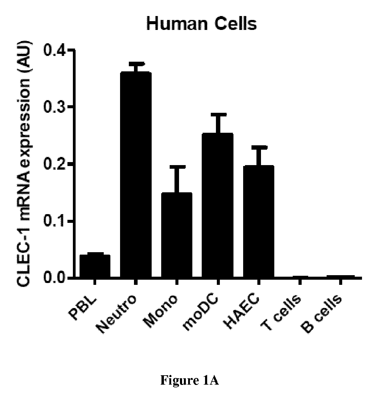
[0179]In a mouse hepatocarcinoma model (intraportal injection of hepa 1.6 tumor cells), the inventors observed an increased and long-lasting expression of CLEC-1 in tumors (FIG. 7a), Importantly, in this model, mice deficient in CLEC-1 (kindly gifted by Derrick J. Rossi, Harvard Stem Cell Institute, Cambridge) are better resistant to tumor growth and exhibit anincreased survival rate (median survival 28d in KO versus 21d in WT) (FIG. 7b). Similarly, in a subcutaneous (sc.) rat glioma model (C6), the inventors observed total regression of tumors in 3 out of 5 of CLEC-1 deficient rats (generated by ZFN technology in our lab) (FIG. 7c). Importantly, by Q-PCR in tumors harvested at day 18 after tumor cell inoculation, a higher mRNA expression of inos, tnfa and ifng were assessed in tumor from CLEC-1 deficient rats (FIG. 7d). In addition, a higher mRNA expression of ifng was detected in lymph nodes (F...
PUM
| Property | Measurement | Unit |
|---|---|---|
| Plasticity | aaaaa | aaaaa |
| immune resistance | aaaaa | aaaaa |
| cell-adhesion | aaaaa | aaaaa |
Abstract
Description
Claims
Application Information
 - R&D
- Intellectual Property
- Life Sciences
- Materials
- Tech Scout
- Unparalleled Data Quality
- Higher Quality Content
- 60% Fewer Hallucinations
Browse by: Latest US Patents, China's latest patents, Technical Efficacy Thesaurus, Application Domain, Technology Topic, Popular Technical Reports.
© 2025 PatSnap. All rights reserved.Legal|Privacy policy|Modern Slavery Act Transparency Statement|Sitemap|About US| Contact US: help@patsnap.com


