
A shunt system, shunt and method for treating an ocular disorder

a shunt and ocular disorder technology, applied in the field of shunt systems, can solve the problems of glaucoma damage, glaucoma damage, defect enlargement, etc., and achieve the effect of preventing the distal end from being pulled ou

Pending Publication Date: 2019-08-15
LIQID MEDICAL PTY LTD
View PDF9 Cites 8 Cited by
  • Summary
  • Abstract
  • Description
  • Claims
  • Application Information

AI Technical Summary

Benefits of technology

The invention is a shunt used to treat ocular disorders related to intraocular or intracranial pressure. The shunt is implanted in the eye and connected from the front to the spine, where it forms a lumen to prevent pressure buildup. The shunt has a distal stop formation to prevent it from moving backwards and a proximal stop formation to prevent it from migrating into other areas. The shunt is flexible to conform to the shape of the eye and is securely positioned to treat the disorder. The technical effect of this invention is to create a safe and effective device to prevent pressure buildup in the eye and maintain its health.

Problems solved by technology

If an imbalance occurs in the amount of aqueous produced or drained from the ocular globe, then the intraocular pressure becomes too high or too low.
This causes damage to the optic nerve head known as glaucoma.
These defects enlarge until a patient's field of view is severely restricted.
If the intraocular pressure remains very high, the eye can become persistently painful and may need to be removed.
1) The subtenon's spaceThis space is not adapted to the presence of pressurised aqueous fluid. The aqueous therefore incites an inflammatory response which results in the formation of a fibrovascular “bleb”. Consequently there is a high rate of complications, unpredictable outcomes and treatment failure.
2) The suprachoroidal spaceThis space is highly vascular and thus there is a high risk of bleeding. Outcomes are unpredictable and there is a high rate of failure.
3) The canal of schlemmOnly a small amount of fluid can drain through this space and thus the effect of these devices is minimal and treatment failure is common.
Currently, no medical or surgical treatment options exist which can effectively reverse chronically low intraocular pressure.
If more cerebrospinal fluid is produced than is drained from the subarachnoid space, then the cerebrospinal fluid pressure becomes too high.
This can cause damage to the optic nerve head due to reversed translaminar pressure.

Method used


Image

  • A shunt system, shunt and method for treating an ocular disorder
  • A shunt system, shunt and method for treating an ocular disorder
  • A shunt system, shunt and method for treating an ocular disorder

Examples


Embodiment Construction

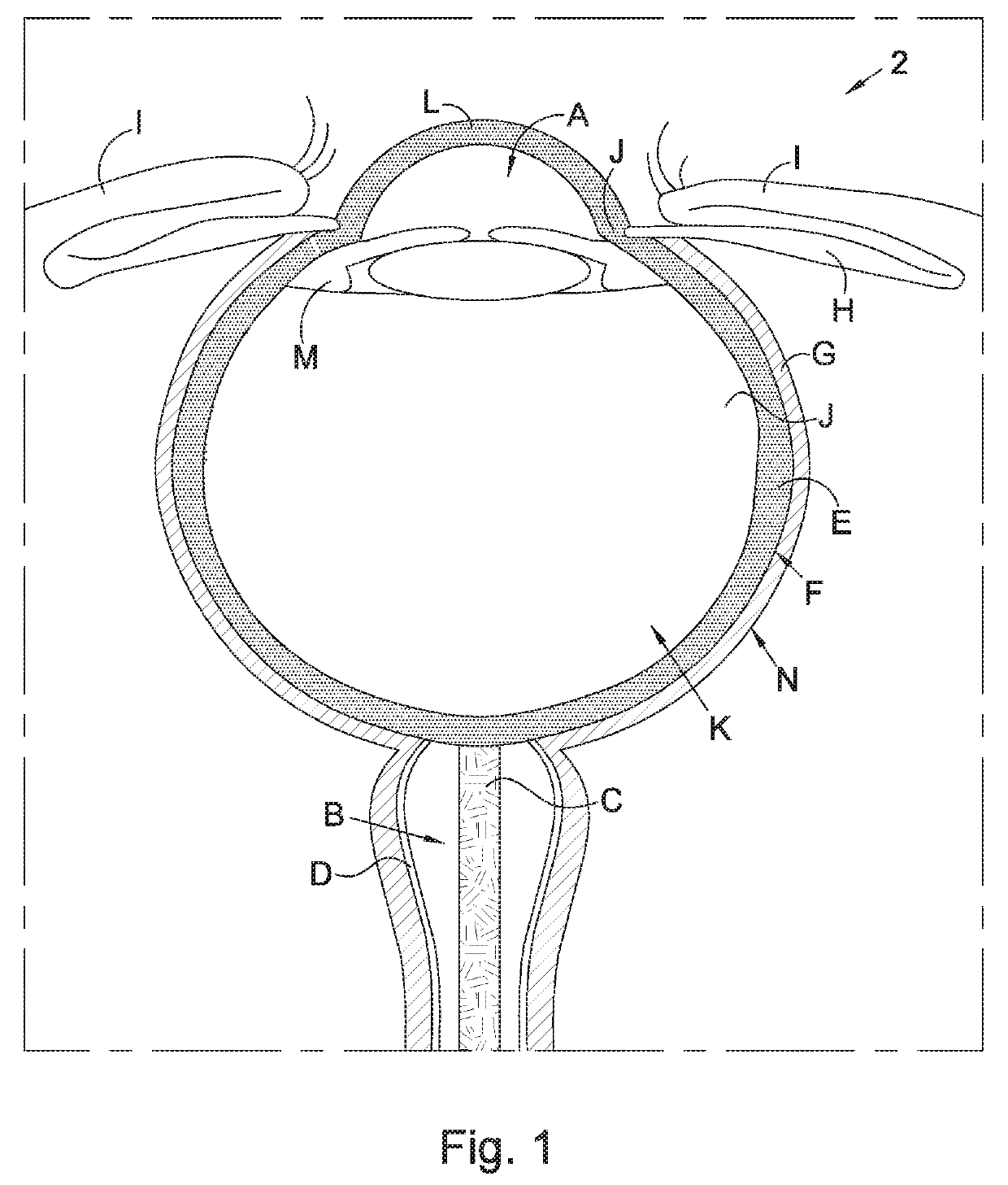
[0104]With reference to FIG. 1 of the drawings, a cross-sectional view illustrating anatomical parts of a human eye 2 which are required for use in the description which follows below, comprises:[0105]A: Anterior chamber filled with aqueous fluid[0106]B: Subarachnoid space filled with cerebrospinal fluid[0107]C: Optic nerve[0108]D: Optic nerve sheath[0109]E: Sclera[0110]F: Subtenon's space[0111]G: Tenon's capsule[0112]H: Conjunctiva[0113]I: Eyelids[0114]J: Limbus and trabecular meshwork[0115]K: Posterior segment filled with vitreous jelly[0116]L: Cornea[0117]M: Ciliary body[0118]N: Ocular globe

[0119]With reference to FIGS. 1 to 20 of the drawings, a shunt in accordance with the invention, is designated generally by the reference numeral 10. The shunt 10 is adapted for implantation in the human body so as to provide for flow communication between aqueous fluid in the anterior chamber A of the eye and cerebrospinal fluid in the subarachnoid space B surrounding the optic nerve C. The s...


PUM


Abstract

A shunt 10 for implantation in the human body for treating ocular disorders related to disorders of intraocular or intracranial pressure by providing for flow of aqueous fluid in the anterior chamber A of the eye and cerebrospinal fluid in the subarachnoid space B surrounding the optic nerve C. The shunt has a proximal end 12 which is implanted in the ocular anterior chamber and a distal end 14 which is implanted in the subarachnoid space. The shunt has a two-part construction, including a flexible distal tube 18 and a rigid proximal tube 20. The distal tube has a distal stop formation 26 near the distal end 14 which is located in the subarachnoid space upon implantation of the distal end, resisting withdrawal of the shunt. The proximal tube has a curved portion which conforms to the anatomical curvature of the ocular globe.

Description

FIELD OF INVENTION[0001]This invention relates to a shunt system for treating an ocular disorder related to an intraocular or intracranial disorder in a patient. The invention relates also to a method for treating an ocular disorder related to an ocular disorder of intraocular or intracranial pressure in a patient. More specifically, the invention relates to a shunt system and method for implanting a shunt between the eye and the subarachnoid space surrounding the optic nerve of a patient. The invention extends to the shunt of the shunt system.BACKGROUND TO INVENTION[0002]The implantation of a shunt connecting the anterior chamber of the ocular globe and the subarachnoid space is a procedure which, in order to be successful, needs to overcome the following difficulties: creating a clear passage from an external side of the optic nerve sheath to access the optic nerve sheath; penetrating the optic nerve sheath to access the subarachnoid space; ensuring a fluid-tight seal of the optic...

Claims


Application Information

Patent Timeline
IPC IPC(8): A61F9/007A61B17/02
CPCA61F9/00781A61B17/0231A61B3/00A61F2009/0052A61M2210/0612A61F9/007A61B1/00154
Inventor MCCLUNAN, DAEMON BRUCE
Owner LIQID MEDICAL PTY LTD
Learn More