Image Correction Design System and Method for Oral and Maxillofacial Surgery
a design system and technology for applied in the field of image correction design system and method for can solve the problems of prolonging the surgery time, misjudgment of the surgery portion, and inaccurate image data of the photographic equipment used in oral and maxillofacial surgery, so as to achieve the effect of reducing the time for performing dental, orthognathic and facial plastic surgery
- Summary
- Abstract
- Description
- Claims
- Application Information
AI Technical Summary
Benefits of technology
Problems solved by technology
Method used
Image
Examples
first embodiment
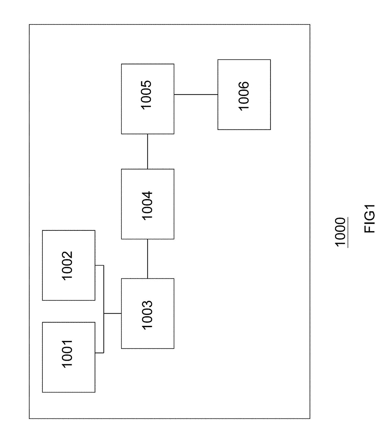
[0060]FIG. 1 shows a functional block diagram of an image correction design system for an oral and maxillofacial surgery of the invention, wherein the image correction design system for an oral and maxillofacial surgery 1000 comprises: a first image data scanning module 1001; a second image data scanning module 1002; a scanning image correction and comparison module 1003; an image block calculation module 1004; a surgery simulation image design module 1005; and an output module 1006.
second embodiment
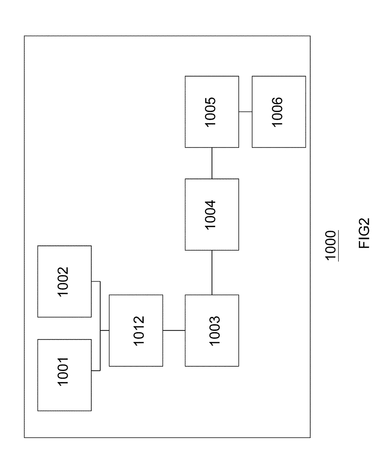
[0061]FIG. 2 shows a functional block diagram of an image correction design system for an oral and maxillofacial surgery of the invention. When an orthognathic surgery is performed by a physician, a clinic file of X-ray machine or computerized tomography (CT) scan is needed to confirm a predetermined operation site. An image correction design system for an oral and maxillofacial surgery 1000 can provide a correct and accurate image to a physician for reference. When an image correction design system for an oral and maxillofacial surgery 1000 is performed, starting the first image data scanning module 1001 is executed. The first image data scanning module 1001 is used for scanning to obtain image data of relative coordinate positions of maxillofacial bones, teeth, and soft tissues of a patient, wherein the first scanning image data is an image taken by X-ray machine or computerized tomography scan equipment, and the image is like a computerized tomography scan file (hereafter referre...
third embodiment
[0065]FIG. 3 shows a functional block diagram of an image correction design system for an oral and maxillofacial surgery of the invention, wherein the image correction design system for an oral and maxillofacial surgery 1000 comprises: a first image data scanning module 1001; an image data conversion module 1010; a second image data scanning module 1002; a scanning image correction and comparison module 1003; an image block calculation module 1004; a surgery simulation image design module 1005; and an output module 1006.
[0066]Following the above-mentioned third embodiment, for an image correction design system for an oral and maxillofacial surgery 1000, a first image data scanning module 1001 further comprises an image data conversion module 1010 for converting the first scanning image data such as Dicom file including bones, teeth and soft tissues that is stored in the first image data scanning module 1001 to a 3 dimension (3D) data file, wherein the format of the 3 dimension (3D) ...
PUM
 Abstract
Description
Claims
Application Information
 - R&D
- Intellectual Property
- Life Sciences
- Materials
- Tech Scout
- Unparalleled Data Quality
- Higher Quality Content
- 60% Fewer Hallucinations
Browse by: Latest US Patents, China's latest patents, Technical Efficacy Thesaurus, Application Domain, Technology Topic, Popular Technical Reports.
© 2025 PatSnap. All rights reserved.Legal|Privacy policy|Modern Slavery Act Transparency Statement|Sitemap|About US| Contact US: help@patsnap.com


