
Method for scoring pathology images using spatial analysis of tissues

a tissue and image analysis technology, applied in image enhancement, medical/anatomical pattern recognition, instruments, etc., can solve the problem that tissue-based assays only evaluate biomarker positivity or expression levels in tissue with limited granularity

Inactive Publication Date: 2018-03-29
FLAGSHIP BIOSCI
View PDF0 Cites 5 Cited by
  • Summary
  • Abstract
  • Description
  • Claims
  • Application Information

AI Technical Summary

Benefits of technology

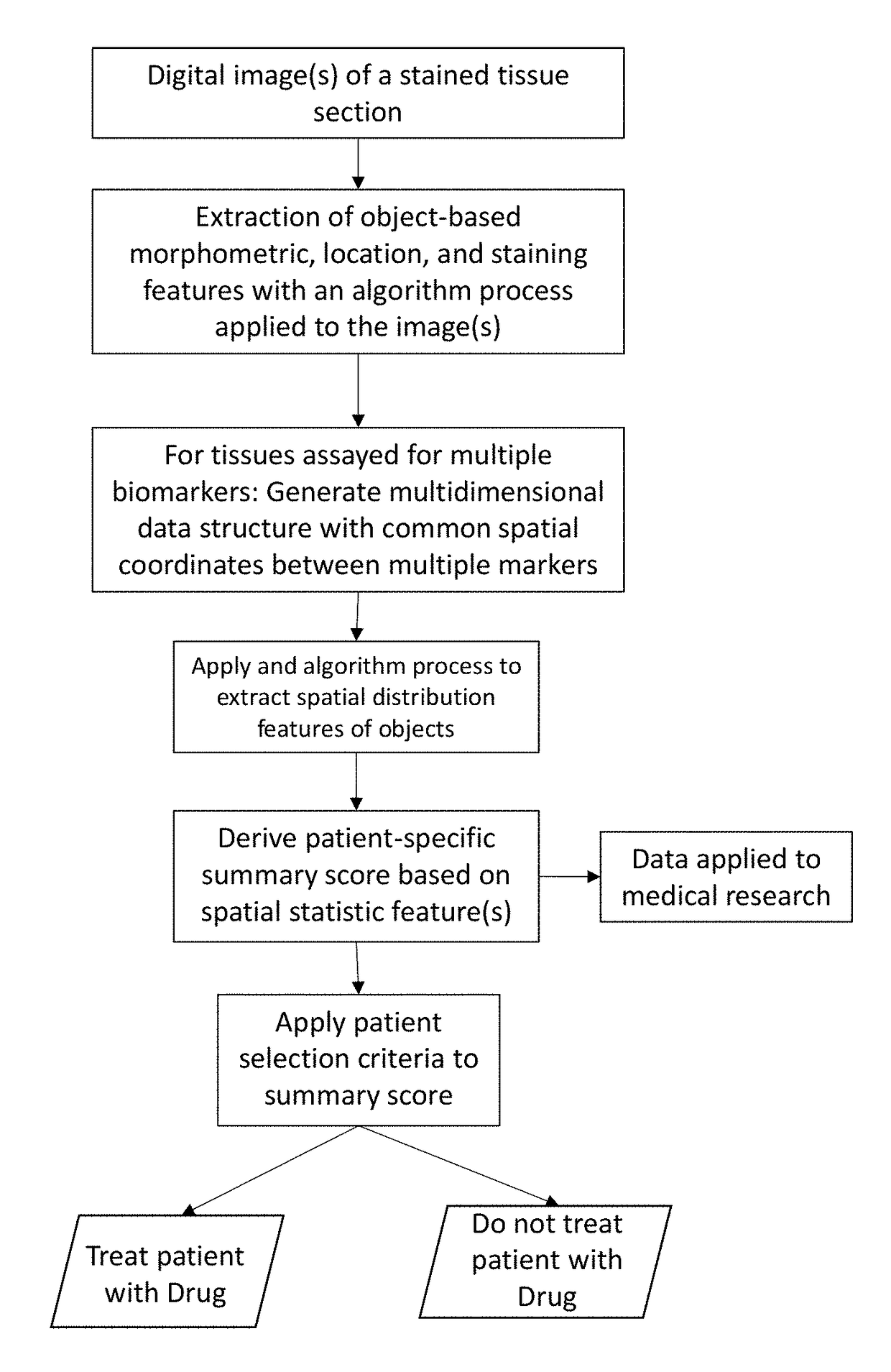
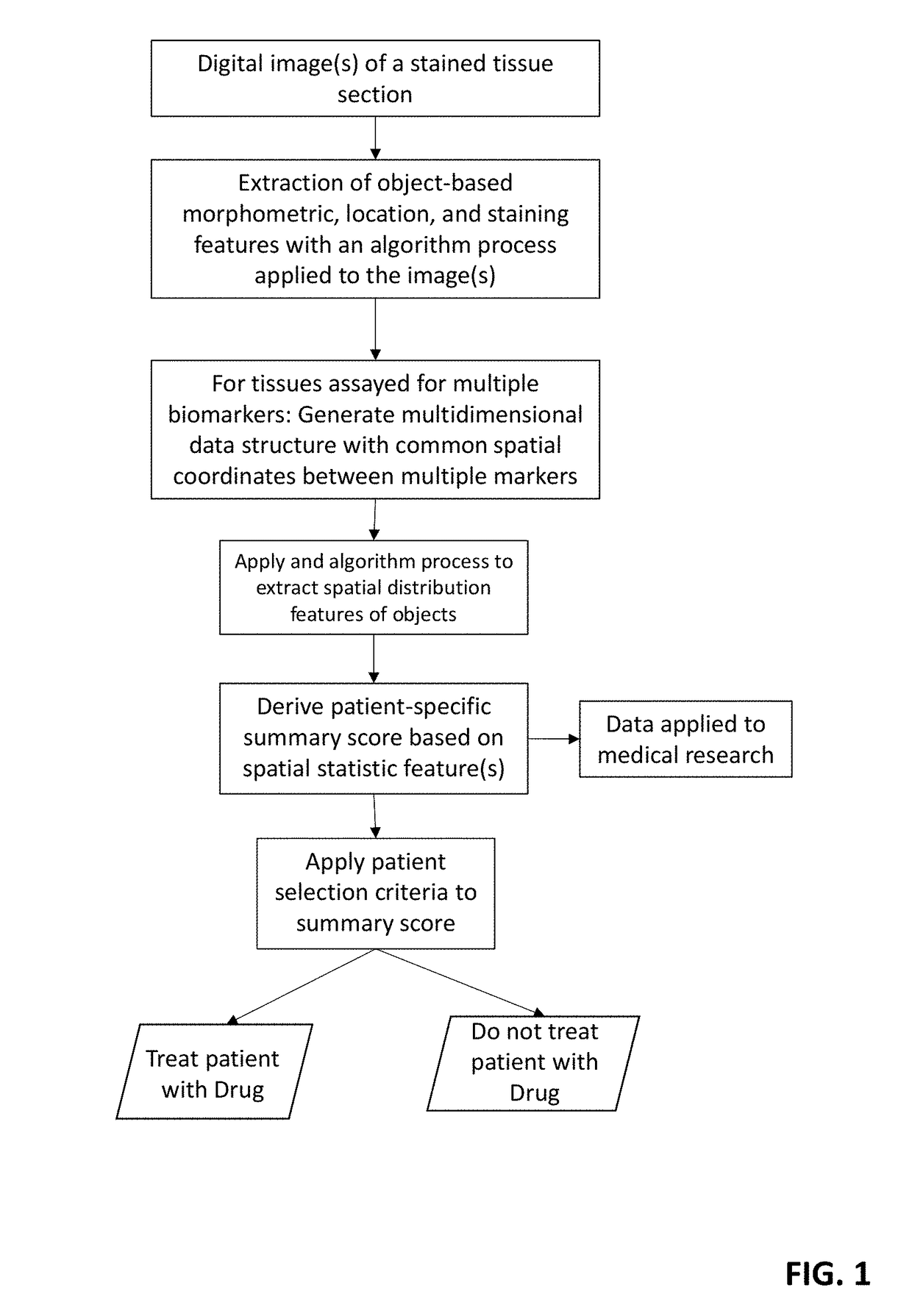
This patent describes a method for extracting detailed statistics about the distribution and patterns of cells in a tissue sample, using digital image analysis. These statistics can be used to score the sample and guide medical research or treatment options for the patient. The method can be implemented in medical research and practice to improve the diagnosis and treatment of various medical conditions.

Problems solved by technology

However, tissue-based assays only evaluate biomarker positivity or expression levels in tissue with limited granularity in regard to the spatial distribution or pattern of cells.

Method used


Image

  • Method for scoring pathology images using spatial analysis of tissues
  • Method for scoring pathology images using spatial analysis of tissues
  • Method for scoring pathology images using spatial analysis of tissues

Examples


Embodiment Construction

[0025]In the following description, for purposes of explanation and not limitation, details and descriptions are set forth in order to provide a thorough understanding of the present invention. However, it will be apparent to those skilled in the art that the present invention may be practiced in other embodiments that depart from these details and descriptions without departing from the spirit and scope of the invention.

[0026]Within this disclosure, a multitude of spatial analysis techniques are contemplated. For the purpose of example, and not limitation, some of these spatial analysis techniques include geographic distribution analysis, cluster analysis, topological analysis, spatial autocorrelation, network analysis, connectivity analysis, and spatial interaction. Other types of spatial analysis are contemplated.

[0027]For purpose of definition, a tissue object is one or more of a cell (e.g., immune cell), cell sub-compartment (e.g., nucleus, cytoplasm, membrane, organelle), cell...


PUM


Abstract

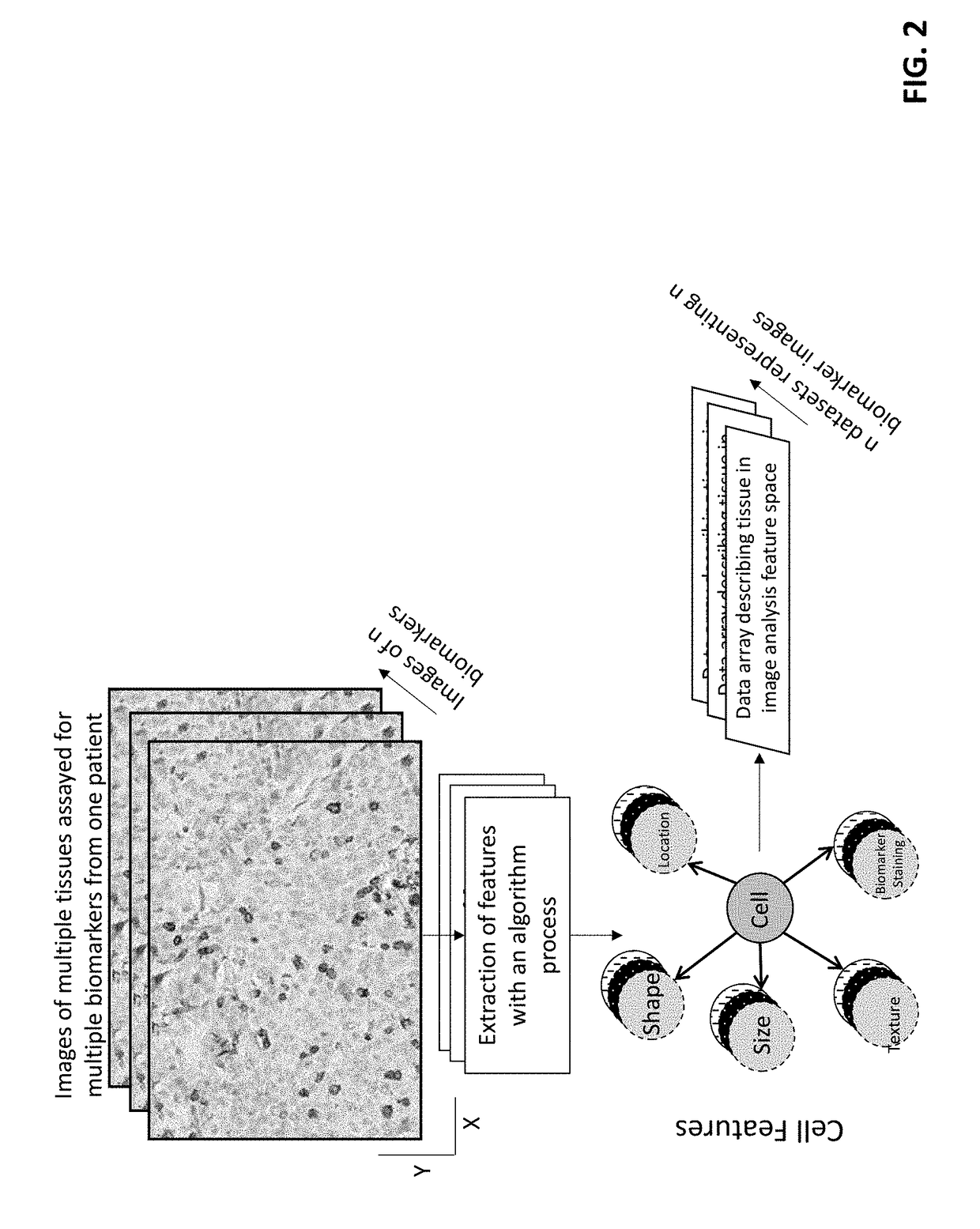
The disclosure concerns a method for extracting spatial distribution statistics from patient tissue samples assayed with a tissue-based test for the purpose of scoring the patient tissue samples and guiding treatment based on the score(s). The method described herein utilizes digital image analysis of an image of one or more tissue sections to extract object-based (e.g., cells) features. The object-based features within the image of the tissue section is further processed using one or more algorithm processes to extract sophisticated spatial distribution features of one or more object type or sub-type in the tissue. Statistics describing the spatial distribution features are summarized to generate a patient-specific diagnostic score, and this score can be evaluated to guide patient treatment decisions.

Description

CROSS-REFERENCE TO RELATED APPLICATIONS[0001]This application is a continuation-in-part (CIP) of U.S. Ser. No. 15 / 181,156, filed Jun. 13, 2016, and titled “METHODS FOR ASSESSMENT OF SPATIAL STATISTICS OF CELLS IN TISSUE SAMPLES”, which is a CIP of U.S. Ser. No. 14 / 189,833, filed Feb. 25, 2014, and titled “CELL-BASED IMAGE REGISTRATION”, which is a CIP of U.S. Ser. No. 13 / 247,991, filed Sep. 28, 2011, titled “METHODS FOR FEATURE ANALYSIS ON CONSECUTIVE TISSUE SECTIONS”, which issued as U.S. Pat. No. 8,787,651 on Jul. 22, 2014;[0002]the contents of each of which are hereby incorporated by reference.BACKGROUNDField of the Invention[0003]This application relates to methods for scoring the spatial relationships or patterns of cells within tissue biopsies. More specifically, the invention relates to the use of spatial analysis to determine distributions of cells in tissues.Description of the Related Art[0004]The majority of current tissue-based in vitro diagnostic assays (e.g., immunohist...

Claims


Application Information

Patent Timeline
IPC IPC(8): G06K9/00G06T7/00G06T7/11G06F19/26G06V10/40G06V10/42G16B45/00
CPCG06K9/00134G06K9/0014G06K9/00147G06T7/0012G06T7/11G06F19/26G06T2207/30024G06T2207/10056G06T2207/20076G06T2207/30096G06T2207/10024G06V20/695G06V20/698G06V10/40G06V10/42G06V2201/03G16B45/00G06V20/693
Inventor BLACK, JOSHUA C.CERKOVNIK, LOGAN JEROMEHARNEY, ALLISON S.RYALL, KARENKRUEGER, JOSEPH
Owner FLAGSHIP BIOSCI
Learn More