Anti-flt-1 antibodies for treating duchenne muscular dystrophy
a duchenne muscular dystrophy and antibody technology, applied in the field of anti-flt-1 antibodies for treating duchenne muscular dystrophy, can solve the problems of abnormal sarcolemal membrane function, dystrophin protein alteration or absence, shortage of oxygen and nutrients needed for cellular metabolism, etc., to promote angiogenesis, and increase the amount of vegf and/or other ligands
- Summary
- Abstract
- Description
- Claims
- Application Information
AI Technical Summary
Benefits of technology
Problems solved by technology
Method used
Image
Examples
example 1
n and Characterization of High Affinity Anti-Flt-1 Antibodies
[0232]Generation of Antibodies
[0233]Monoclonal antibodies were generated against soluble Flt-1 using llama monoclonal antibody methodology. Briefly, llamas were immunized with recombinant human soluble Flt-1 (purchased from ABCAM) and the serum was collected.
[0234]Antibody Characterization
[0235]Antibodies that bound to human and mouse Flt-1 were further characterized for 1) VH family; 2) affinity for Flt-1; 3) IC50; 4) off-rate screening by Biacore assay, 5) cross-reactivity to cynomolgus Flt-1 and 6) binding to VEGF R2 and VEGF R3. Candidate antibodies against human Flt-1 (hFl-1) and mouse Flt-1 (mFlt-1) were characterized as shown in Table 4.
TABLE 4IC50 by Bioassay Affinity ELISA(%Human(nM)(pM)rescue)identityAnti-VHhFlt-mFlt-hFlt-mFlt-hFlt-mFlt-(%)bodyfamily111111VHVL13B4150.6 1.4 13.3500>9072.294.393.710G12170.290.334003004487.590.88111A11200.2 33.3>9090.891.1
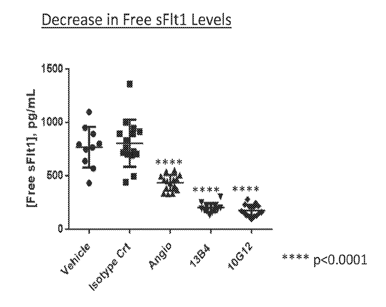
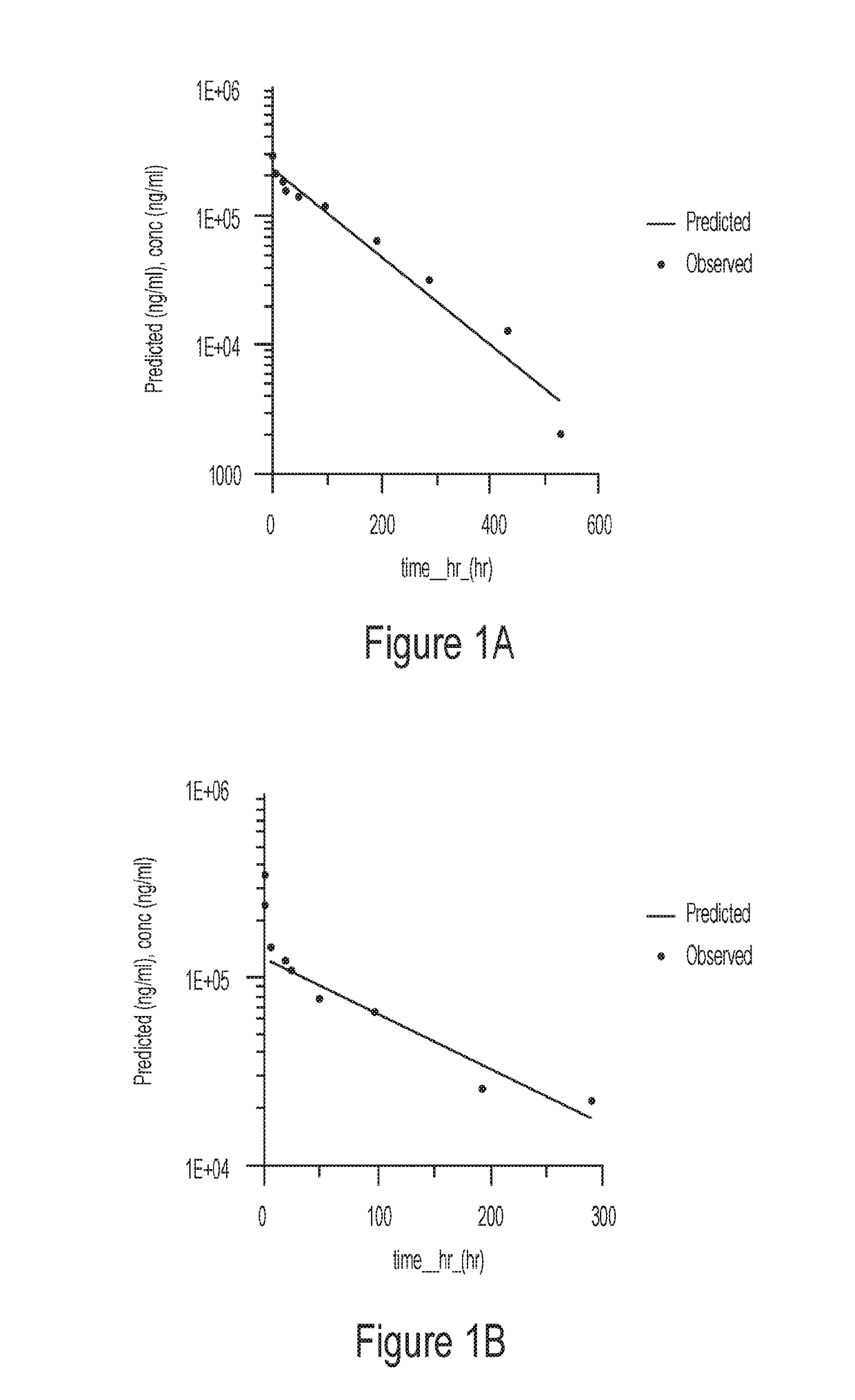
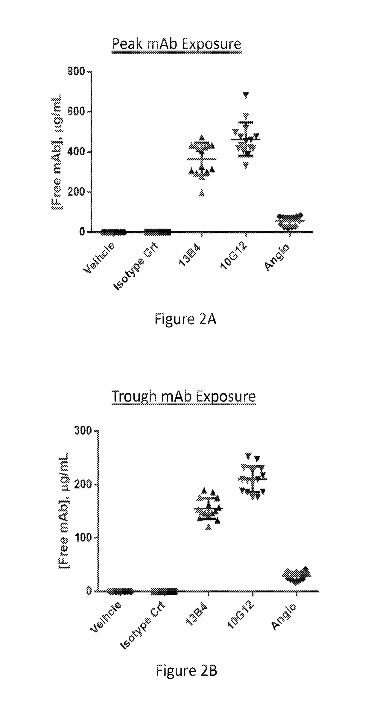
[0236]Pharmacokinetic properties of antibody 13B4 and 10G12 w...
example 2
n and Characterization of High Affinity Anti-Flt-1 Antibodies
[0243]Additional anti-Flt-1 monoclonal antibodies were generated as described above. These antibodies were further characterized for binding affinity to sFlt-1 antigen (by ELISA and Biacore), competition for VEGF in a sFlt-1:VEGF competition ELISA; and performance in a cell based assay.
[0244]Antibody Characterization—Binding to Target
[0245]The monoclonal anti-Flt-1 antibodies were assayed for binding to recombinant sFlt-1 antigen in an ELISA assay (FIG. 7). All antibodies demonstrated a dose-dependent increase in binding. Binding affinity of the anti-Flt-1 antibodies to mouse and human Flt-1 antigen was measured by surface plasmon resonance methodology (i.e., Biacore) (Table 6). The antibodies bound to human Flt-1 in the nanomolar range with antibody 11A11 demonstrating the highest binding affinity for human Flt-1. Biacore analysis also demonstrated that the antibodies did not cross-react with VEGF R2 or VEGF R3 (Table 6),...
example 3
ization of High Affinity Anti-Flt-1 Antibodies Generated by Light Chain Shuffling
[0250]Light chain shuffling of antibodies 18B6, 11A11 and 13B4 described in Example 2 was performed to increase the affinity and potency of the candidate antibodies.
[0251]Antibody Characterization—Binding to Target
[0252]The resulting antibodies displayed increased affinity for the Flt-1 antigen. For instance the KD of antibody 21C6 increased approximately 10-fold over that of the parent antibody, 11A11. Similarly, the KD of antibody 21B3 increased approximately 5-fold over that of the parent antibody, 13B4 (Table 7).
[0253]Antibody Characterization—Competition / Antagonism
[0254]To estimate the potency of the antibodies following the light chain shuffling, the antibodies were assay in a competition ELISA using human sFlt-1 and VEGF. Antibody concentrations tested ranged from 0.2 mg / mL to 200 ng / mL. The ability of parent antibody 13B4 to competitively bind sFlt-1 was compared to that of antibodies generated...
PUM
| Property | Measurement | Unit |
|---|---|---|
| concentration | aaaaa | aaaaa |
| concentration | aaaaa | aaaaa |
| concentration | aaaaa | aaaaa |
Abstract
Description
Claims
Application Information
 - R&D
- Intellectual Property
- Life Sciences
- Materials
- Tech Scout
- Unparalleled Data Quality
- Higher Quality Content
- 60% Fewer Hallucinations
Browse by: Latest US Patents, China's latest patents, Technical Efficacy Thesaurus, Application Domain, Technology Topic, Popular Technical Reports.
© 2025 PatSnap. All rights reserved.Legal|Privacy policy|Modern Slavery Act Transparency Statement|Sitemap|About US| Contact US: help@patsnap.com


