
Diffusion monitoring protocol for optimized tissue fixation

a tissue fixation and monitoring protocol technology, applied in the field of tissue specimen analysis, can solve the problems of poor tissue morphology, unbalanced samples, shrinking of cellular structures and condensing nuclei, etc., and achieves the effects of accurate prediction of the quality of fixation and subsequent staining, optimal fixation, and high quality staining

Active Publication Date: 2017-10-05
VENTANA MEDICAL SYST INC
View PDF0 Cites 16 Cited by
  • Summary
  • Abstract
  • Description
  • Claims
  • Application Information

AI Technical Summary

Benefits of technology

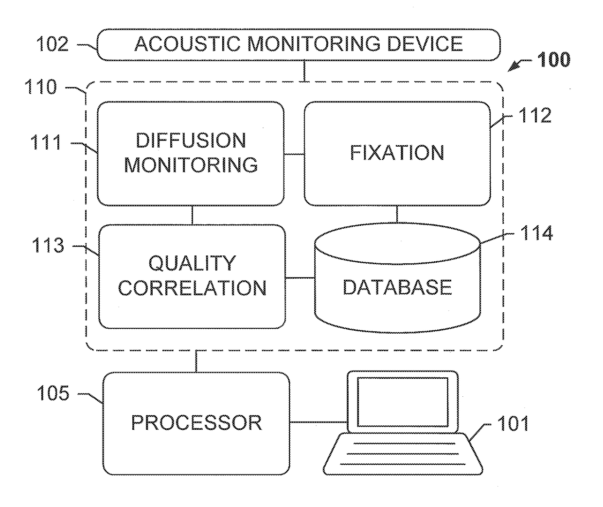
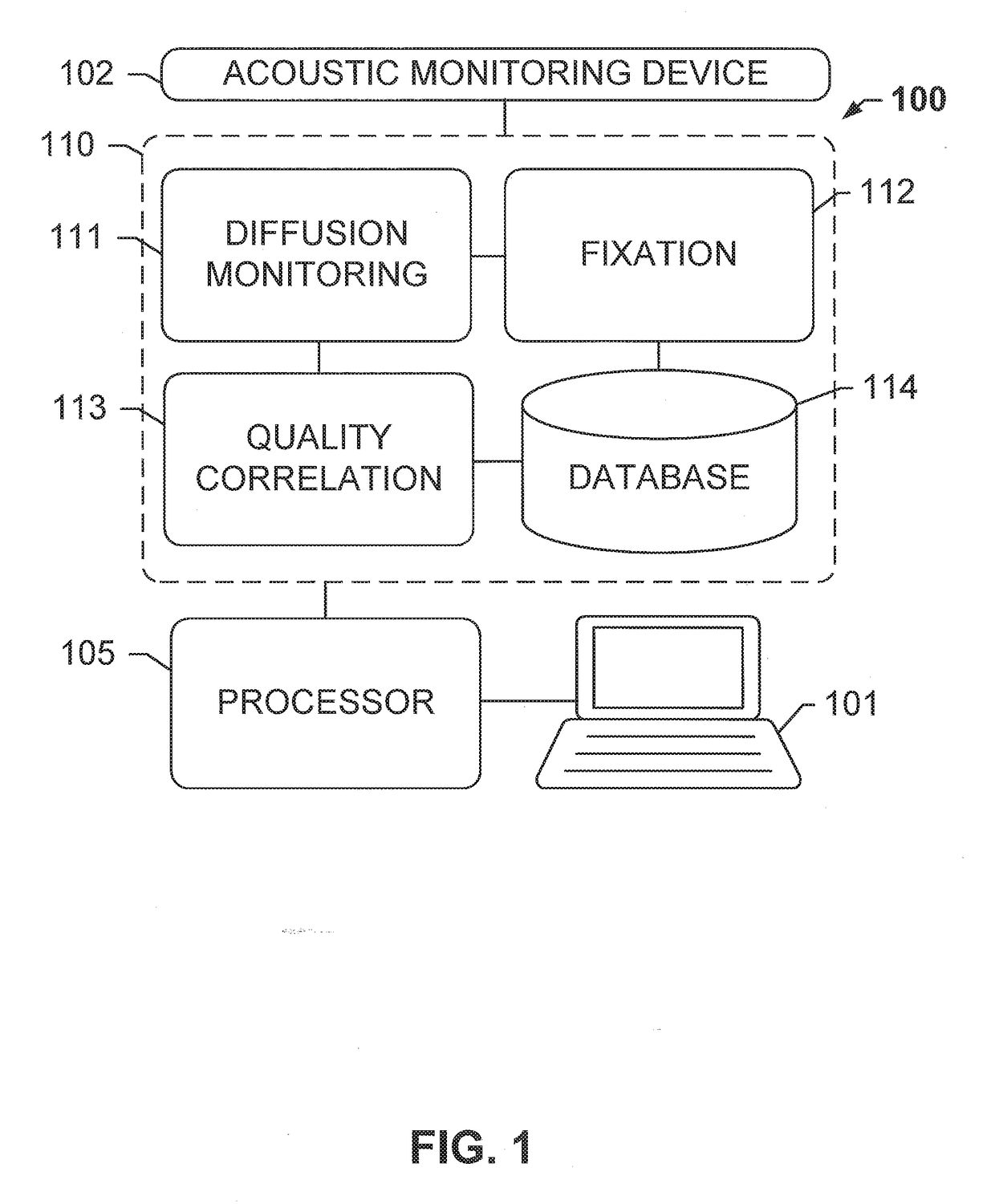
The patent presents a system and method for monitoring the diffusion of a fixative solution into a tissue sample to ensure optimal fixation and high quality staining for downstream assays. This is done by detecting changes in the speed of sound caused by the diffusion of fixative. By tracking and quantifying the fixative diffusion, the system can accurately predict the quality of fixation and subsequent stain, which in turn improves the efficiency of the tissue processing laboratory. The predictive metric is validated with results from a large tissue collection study. Overall, this technology helps ensures ideally-stained cross-sections while saving time and improving quality assurance.

Problems solved by technology

If formalin has not diffused properly through the tissue samples, outer regions of the tissue samples exposed to formalin may be over-fixed and interior regions of the tissue samples not exposed to formalin may be under-fixed, resulting in very poor tissue morphology.
In under-fixed tissue, subsequent exposure to ethanol often shrinks the cellular structures and condenses nuclei since the tissues will not have the chance to form a proper cross-linked lattice.
When under-fixed tissue is stained, such as with hematoxylin and eosin (H&E), many white spaces may be observed between the cells and tissue structures, nuclei may be condensed, and samples may appear pink and unbalanced with the hematoxylin stain.
Tissues that have been exposed to excess amounts of formalin or too long typically do not work well for subsequent immunohistochemical processes, presumably because of nucleic acid and / or protein denaturation and degradation.
As a result, the optimal antigen retrieval conditions for these tissues do not work properly and therefore the tissue samples appear to be under stained.
However, this is a broad and inefficient protocol, and the current standard of care is for laboratories to process tissues with a custom unverified protocol that is not standardized and sub-optimal.

Method used


Image

  • Diffusion monitoring protocol for optimized tissue fixation
  • Diffusion monitoring protocol for optimized tissue fixation
  • Diffusion monitoring protocol for optimized tissue fixation

Examples


embodiment 1

[0097] A method of fixing tissue sample, said method comprising: (a) immersing an unfixed tissue sample in a volume of a fixative solution at a temperature from 0 to 10° C.; (b) monitoring a rate of diffusion of the fixative solution into the tissue sample by: (b1) transmitting an ultrasonic acoustic signal through the tissue sample and detecting the ultrasonic acoustic signal after the ultrasonic signal has passed through the tissue sample; (b2) calculating the time of flight (TOF) of the ultrasonic acoustic signal; (b3) repeating (b1) and (b2) at a plurality of subsequent time points; (b4) calculating a rate of diffusion at least at each of the subsequent time points by analyzing a slope of a curve of the TOFs calculated from the ultrasonic acoustic signals measured at each of the time points; (b5) determining when the rate of diffusion falls below a predetermined threshold; and (c) after the rate of diffusion falls below the predetermined threshold, allowing the tissue sample to ...

embodiment 4

[0100] The method of embodiment 2 or 3, wherein the single exponential curve is a curve according to Equation I:

TOF(t, r)=C(r)+Ae−t / τ(r)   (I), and

wherein the rate of diffusion is calculated according to Equation IIIa:

dTOF(t)dt(t=to)=-Aτe-to / τ,(IIIa)

wherein C is a constant offset, A is amplitude of decay, τ is a decay constant, t is diffusion time, and r is the spatial dependence, and t0 is diffusion time at the time point at which the rate of diffusion is calculated.

[0101]Embodiment 5: The method of any one of the previous embodiments 2-4, wherein the single exponential curve is a curve according to Equation I:

TOF(t, r)=C(r)+Ae−t / τ(r)   (I), and

wherein the rate of diffusion is an amplitude-normalized rate of diffusion is calculated according to Equation IVa:

m~(t=to)=100(-1τe-to / τ),[%time],(IVa)

wherein C is a constant offset, A is amplitude of decay, τ is a decay constant, t is diffusion time, r is the spatial dependence, {tilde over (m)} is the amplitude-normalized rate of diffusio...

embodiment 9

[0105] The method of any of the foregoing embodiments, wherein the threshold rate of diffusion has been met when the rate of diffusion at a discrete time point calculated according to (b4) meets or exceeds the threshold rate of diffusion.


PUM

PropertyMeasurementUnit
temperatureaaaaaaaaaa
fixation timeaaaaaaaaaa

Abstract

The subject disclosure presents systems and computer-implemented methods for evaluating a tissue sample that has been removed from a subject. A change in speed of the energy traveling through the sample is evaluated to monitor changes in the biological sample during processing. The rate of change in the speed of the energy is correlated with the extent of diffusion. A system for performing the method can include a transmitter that outputs the energy and a receiver configured to detect the transmitted energy. A time-of-flight of acoustic waves and rate of change thereof is monitored to determine an optimal time for soaking the tissue sample in a fixative.

Description

CROSS-REFERENCE TO RELATED APPLICATIONS[0001]This is a continuation of International Patent Application No. PCT / EP2015 / 080254 filed Dec. 17, 2015, which claims priority to and the benefit of U.S. Provisional Patent Application No. 62 / 093,151, filed Dec. 17, 2014, U.S. Provisional Patent Application No. 62 / 093,173, filed Dec. 17, 2014, and U.S. Provisional Patent Application No. 62 / 097,520, filed Dec. 29, 2014, all of which prior patent applications are incorporated by reference herein.BACKGROUND OF THE SUBJECT DISCLOSUREField of the Subject Disclosure[0002]The present subject disclosure relates to analysis of tissue specimens. More particularly, the present subject disclosure relates to monitoring processing of tissue samples.Background of the Subject Disclosure[0003]In the analysis of biological specimens such as tissue sections, blood, cell cultures and the like, biological specimens are stained with one or more combinations of stain and biomarkers, and the resulting assay is view...

Claims


Application Information

Patent Timeline
Patent Type & Authority Applications(United States)
IPC IPC(8): G01N13/00G01N29/44G01N1/30G01N29/07
CPCG01N13/00G01N29/07G01N29/44G01N2013/003G01N2291/02475G01N2291/011G01N1/30G01N29/024G01N2291/0245
Inventor BAUER, DANIELCHAFIN, DAVIDOTTER, MICHAEL
Owner VENTANA MEDICAL SYST INC
Learn More