
3D tissue-engineered bone marrow for personalized therapy and drug development

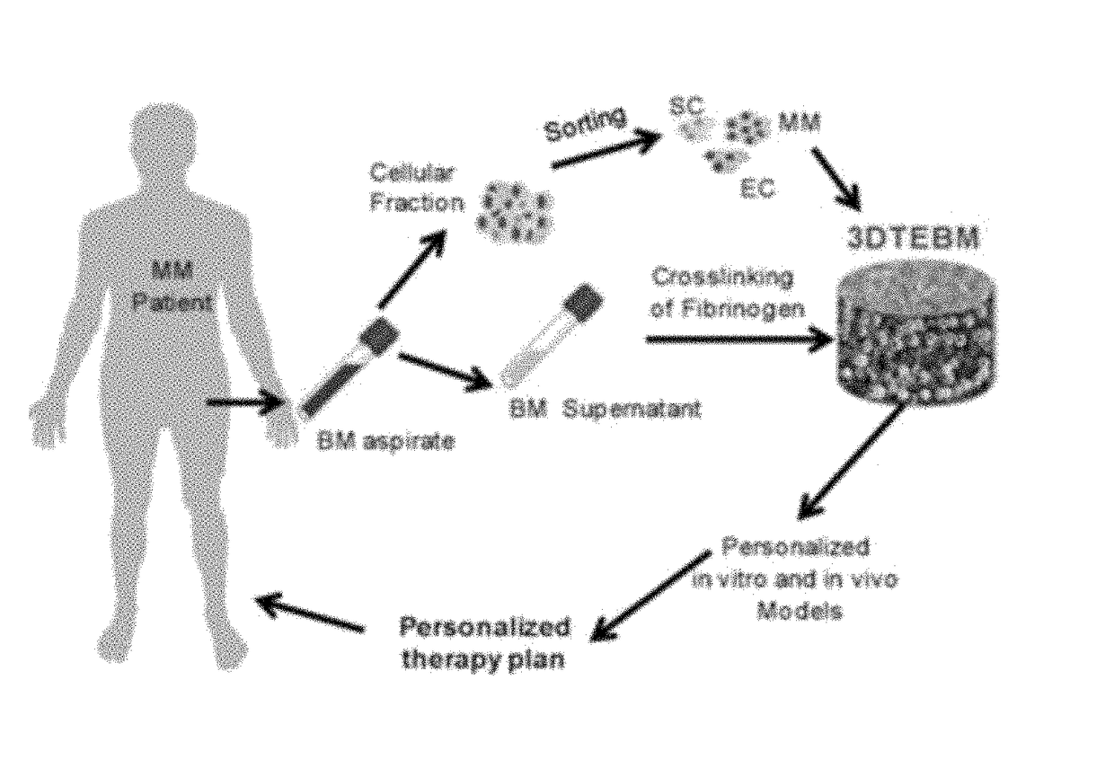
a bone marrow and personalized therapy technology, applied in the field of tissue-engineered bone marrow, can solve the problems of inability to accurately predict, each has its limitations, and the in vitro efficacy is not clear, so as to increase the stability of 3dtebm, the effect of maximizing the scaffold and increasing the cell density

Active Publication Date: 2017-08-17
WASHINGTON UNIV IN SAINT LOUIS
View PDF0 Cites 1 Cited by
  • Summary
  • Abstract
  • Description
  • Claims
  • Application Information

AI Technical Summary

Benefits of technology

This model provides a more accurate prediction of drug responses and resistance by recreating the 3D bone marrow microenvironment, enabling personalized therapeutic strategies and improving the prediction of therapeutic efficacy for multiple myeloma and other hematologic malignancies.

Problems solved by technology

The discrepancy between in vitro efficacy and clinical outcomes can be attributed to several limitations of the classic tissue culture drug screening models including: (1) Most of the in vitro models use MM cell line cultures and neglect the vital role of the bone marrow (BM) microenvironment in MM progression, which promotes drug resistance; (2) The BM niche is a three-dimensional (3D) structure with a gradient of both oxygen and drug concentration as a function of distance from blood vessels.
The classic two-dimensional (2D) in vitro tissue culture system cannot mimic oxygen and drug gradients in culture wells, making all cells highly oxygenated; therefore, 2D cultures cannot accurately predict drug sensitivity in different parts of the BM niche due to lack of accurate effects throughout various tissue depths; and (3) The MM patient population is highly variable, both genetically and epigenetically, and the biological characteristics of patients are widely different, which demonstrates sensitivity of individual patients to different therapies.
These provide a better alternative compared to 2D cultures; however, each has its limitations.
The Matrigel and synthetic polymer-based models are made from materials which are not physiologically found in the BM and may cause significant changes in interactions between different culture components and the matrix.
The bone-chip-based model is closer to BM physiological conditions; however, it is technically challenging, as it needs about eight weeks before it is ready for drug testing.

Method used


Image

  • 3D tissue-engineered bone marrow for personalized therapy and drug development
  • 3D tissue-engineered bone marrow for personalized therapy and drug development
  • 3D tissue-engineered bone marrow for personalized therapy and drug development

Examples


example 1

Cells for the 3DTEBM

[0072]The MM cell line (MM1s, and MM1s-GFP-Luc) and BM stromal cell (BMSC) line Hs5 were a kind gift from Dr. Irene Ghobrial, Dana-Farber Cancer Institute, Harvard Medical School, Boston, Mass. Human umbilical vein endothelial cells (HUVECs) were purchased from Lonza. All cells were cultured at 37° C., 5% CO2; MM1s cells in RPMI1460 media (Corning CellGro, Mediatech, Manassas, Va.) supplemented with 10% fetal bovine serum (FBS, Gibco, Life technologies, Grand island, N.Y.), 2 mmol / L of L-glutamine, 100 U / mL Penicillin and 100 μg / mL Streptomycin (CellGro, Mediatech, Manassas, Va.), HUVECs in EGM-2 completed media (Lonza, Walkersville, Md.), and stromal cells (Hs5) in Dulbecco's Modified Eagle's Medium (DMEM, Corning CellGro, Mediatech, Manassas, Va.) supplemented with 20% FBS, L-glutamine, and penicillin and streptomycin. Before experiments MM1s, Hs5 stromal cells, and HUVECs (1×10⁶ cells / ml) were pre-labeled with calcein violet (1 μg / mL), DiD (10 μg / mL), or DiI (...

example 2

Three-Dimensional (3D) Tissue-Engineered Bone Marrow (3DTEBM) Cultures

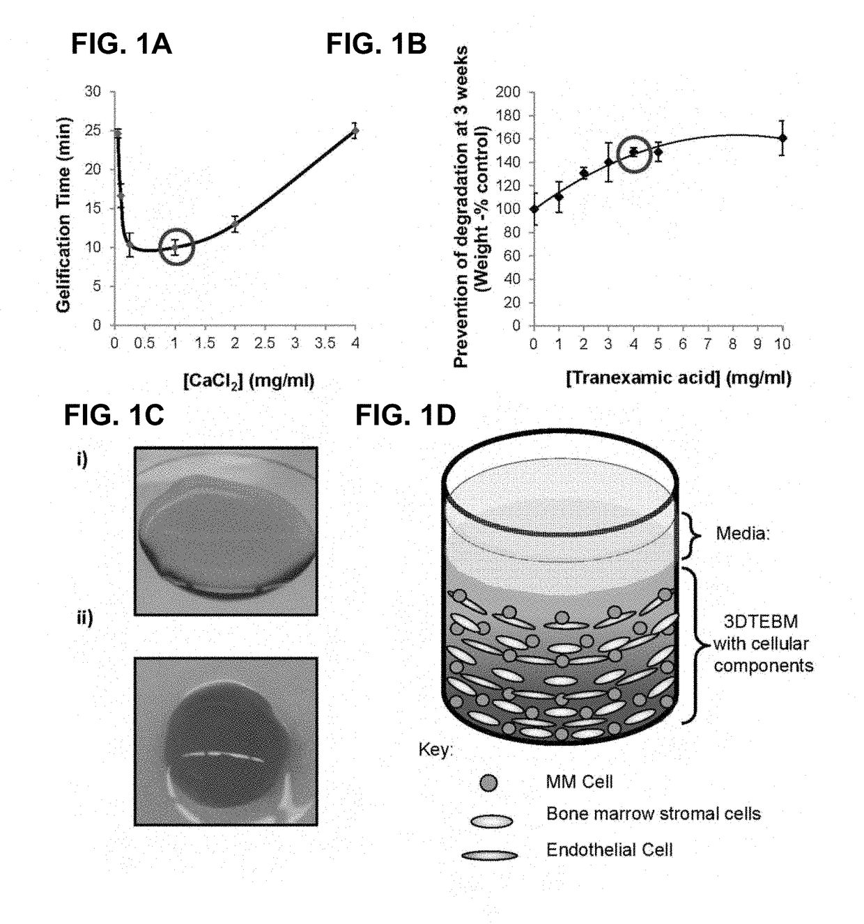
[0075]The three-dimensional (3D) tissue-engineered bone marrow was formed through cross-linking of fibrinogen (naturally found in the plasma of the BM supernatant) with calcium chloride concentrations 0-4 mg / ml. Gelification time was measured and the calcium chloride concentration that induced the fastest gelification time was selected for further studies. Prevention of degradation of the fibrin fibers and stabilization of the scaffolds were achieved by addition of tranexamic acid 0-10 mg / ml. Stability of the scaffold was measured at 3 weeks by weight of scaffolds compared to the weight of non-stabilized scaffolds, and the tranexamic acid concentration that induced the maximal stabilization was selected for further studies. Briefly, 3DTEBM was formed by mixing the following components to achieve final concentrations of 400 μl / ml plasma, 1 mg / ml calcium chloride, and 4 mg / ml tranexamic acid diluted in RPMI complete...

example 3

Flow Cytometry and Cell Proliferation

[0076]For flow cytometry analysis, an internal control (MM1s-calcein violet) was added and a minimum of 5,000 events were acquired using BD FACS Aria (BD Biosciences) and DiVa v6.1.2 software. The data was analyzed using FlowJo program v10 (Ashland, Oreg.). MM cells or CD138+ cells were identified by gating cells with a high GFP / DiO signal (excitement, 488 nm; filter, 530 / 30 nm); Hs5 stroma or CD138-stromal cells were identified by gating cells with a high DiD signal (excitement, 638 nm; filter, 660 / 20 nm); HUVECs were identified by gating cells with a high DiI signal (excitement, 488 nm; filter, 585 / 15 nm); and MM1s cells control were identified by gating cells with a high UV signal (excitement, 355 nm; filter, 450 / 50 nm). A 3DTEBM was digested with type I collagenase at 25 mg / ml for 2 h at 37° C. Classic bi-dimensional (2D) cultures were washed with PBS and removed by pipetting. The effect of cell density (5×10³-50×103 cells / well) of MM1s-GFP, ...


PUM


Abstract

A tissue-engineered bone marrow for personalized therapy of a patient is described. The tissue-engineered bone marrow includes an autologous fibrin scaffold and a plurality of patient-derived cells isolated from the patient's bone marrow. The autologous fibrin scaffold is made using fibrinogen isolated from the patient's bone marrow. The plurality of patient-derived cells may include cells associated with a hematological or metastatic malignancy, bone marrow stromal cells, and endothelial cells. The patient-derived cells are cultured on the autologous fibrin scaffold to create the tissue-engineered bone marrow. The tissue-engineered bone marrow may be used for personalized drug screening.

Description

CROSS REFERENCE TO RELATED APPLICATIONS[0001]This application is a divisional application of U.S. application Ser. No. 14 / 942,543 filed on Nov. 16, 2015, which claims priority from U.S. provisional patent application Ser. No. 62 / 079,868 entitled “3D Tissue Engineered Bone Marrow for Personalized Therapy and Drug Development” filed on Nov. 14, 2014, both of which are hereby incorporated by reference herein in their entirety.FIELD OF THE INVENTION[0002]This application relates to a tissue-engineered bone marrow and methods of preparing and using the tissue-engineered bone marrow for personalized therapy of a patient. In particular, this application relates to a tissue-engineered bone marrow including an autologous fibrin scaffold with fibrinogen isolated from the patient's bone marrow and a plurality of patient-derived cells isolated from the patient's bone marrow, such as, cells associated with a hematological or metastatic malignancy, bone marrow stromal cells, and endothelial cells...

Claims


Application Information

Patent Timeline
Patent Type & Authority Applications(United States)
IPC IPC(8): A61L27/38G01N33/50C12N5/077A61L27/22A61L27/54
CPCA61L27/3834A61L27/225A61L27/54A61L27/3847C12N5/0669G01N33/5011C12N2533/56A61L2300/64A61L2300/418A61L2430/02A61L2430/40C12N2513/00G01N33/5088A61L27/3608A61L27/3804A61L27/3808A61L2300/414A61L2300/42A61L2300/426C12N5/0694C12N2502/30G01N33/5082
Inventor AZAB, ABDEL KAREEMDE LA PUENTE, PILAR
Owner WASHINGTON UNIV IN SAINT LOUIS
Learn More