Electrophoretic tissue clearing chamber and uses thereof

a tissue clearing and electrophoretic technology, applied in the field of electrophoretic chambers, can solve the problems of ineffectiveness, fluorophores become very unstable or sanctioned in the clarification process, and the application of light microscopy remains limited for imaging through intact nervous systems, so as to increase the mechanical strength or stability of the treated tissues, the effect of preventing autolysis or putrefaction

Inactive Publication Date: 2017-06-22
UNIV DE MONTREAL
View PDF2 Cites 4 Cited by
  • Summary
  • Abstract
  • Description
  • Claims
  • Application Information

AI Technical Summary

Benefits of technology

[0057]The term “tissue sample” is intended to mean an entire organ or pieces, slices, segments or fragments thereof, of animal or human origin, that may be treated with the tissue clarification chamber of the present invention. According to an embodiment, the tissue sample may be fixed. In the fields of histology, pathology, and cell biology, fixation is a critical step in the preparation of histological sections by which biological tissues are preserved from decay, thereby preventing autolysis or putrefaction. The structure of a tissue is determined by the shapes and sizes of macromolecules in and around cells. The principal macromolecules inside a cell are proteins and nucleic acids. Fixation terminates any ongoing biochemical reactions, and may also increase the mechanical strength or stability of the treated tissues. The broad objective of tissue fixation is to preserve cells and tissue components and to do this in such a way as to allow for the preparation of thin, stained sections. The fixation solution may be for example a hydrogel solution such as described in US Patent Application No. US2015 / 2015 / 0144490, which is incorporated herein by reference in its entirety. For example, the hydrogel solution may comprise acrylamide (40% wt / wt), nis-acrylamide (0.025% wt / wt), VA-044 initiator (i.e. 2,2′-Azobis[2-(2-imidazolin-2-yl)propane]dihydrochloride—0.25% wt / wt), paraformaldehyde (4% wt / wt), optionally saponin (0.05% wt / wt), and phosphate buffered saline (PBS 1x), and water to in quantity sufficient to arrive at the indicated concentrations.

Problems solved by technology

However, the applicability of light microscopy remains limited for imaging through intact nervous systems, as mouse brains cover many millimeters even in the shortest dimension, and they are opaque at this scale mainly because of the fat which prevents the dispersion of light.
Although these approaches are feasible, they are not effective because that they cannot render oligonucleotides transparent.
When soft tissues, such as the mammary glands, are probed by immunohistochemistry using hydrophobic solutions that reduce lipid barriers, fluorophores become very unstable or sanctioned in the clarification process (a step which must be followed during antibody staining, because transparency will nevertheless be lost).
The first involves passive diffusion of clarifying solution through the tissue or organ, which lasts approximately 28 days, and the second involves an electrophoresis based approach, which is much faster, but is riskier as it may damage the tissue sample or simply fail to clarify the sample.
The second approach makes use of an electrophoretic tissue clarification chamber that appears to be less then optimal for the desired use.

Method used


Image

  • Electrophoretic tissue clearing chamber and uses thereof
  • Electrophoretic tissue clearing chamber and uses thereof
  • Electrophoretic tissue clearing chamber and uses thereof

Examples


example 1

Test of Electrophoretic Clearing Chambers

[0093]The electrophoretic tissue clearing chambers of the present invention were tested to compare their performance with the original tissue clearing chamber employed with the CLARITY technique. The results are presented in Table 1 below.

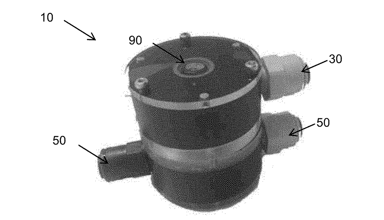
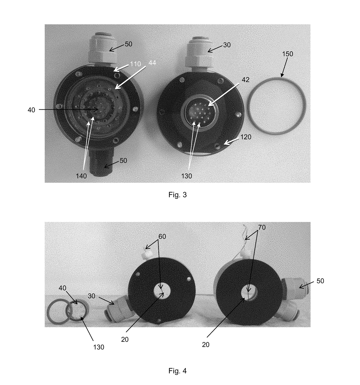
TABLE 1comparison of electrophoretic tissue clearing chambersInterval forBrainreplacementsampleDuration ofElectricConstantof CLARITYsizeprocedurecurrentVoltagesolutionETC(mm)(days)(mA)(V)(days)TransparencyOriginalComplete835025N / AVery obscurebrain−None -Slice -28N / AN / AEvery twoGoodPassive600 μmdays++diffusionPresentSlice -21620N / AGoodinvention -600 μm++FIG. 1PresentSlice -55027N / AExcellentinvention -2.5 mm+++FIG. 4

[0094]The electrophoretic tissue clearing chambers were capable of decreasing the duration of the procedure as well as dramatically improve the quality (i.e. transparency) of the tissue sample treated therein. FIG. 8 illustrates the slice of brain before and after the 5 day treatment in the chamber...


PUM

PropertyMeasurementUnit
pHaaaaaaaaaa
diameteraaaaaaaaaa
diameteraaaaaaaaaa

Abstract

The present document describes an electrophoretic tissue clearing chamber comprising an electrophoresis channel, configured to receive a clarification fluid therethrough; a first clarification fluid inlet, in fluid communication with the electrophoresis channel, configured to be connected to a source of the clarification fluid; a tissue sample holder in fluid communication with the electrophoresis channel, configured to receive a tissue sample to be clarified, and pressurize and homogenously apply the clarification fluid onto the tissue sample; a clarification fluid outlet, in fluid communication with the tissue sample holder, for exit of the clarification fluid from the electrophoretic tissue clearing chamber; and first and a second electrode, opposite one another in the electrophoresis channel, for transmission of an electric field therethrough.

Description

CROSS-REFERENCE TO RELATED APPLICATIONS[0001]This application claims priority of U.S. provisional patent application 62 / 268,189 filed on Dec. 16, 2015, the specification of which is hereby incorporated by reference in its entirety.BACKGROUND[0002](A) Field[0003]The subject matter disclosed generally relates to electrophoretic chambers, and more particularly to electrophoretic chambers for tissue clearing.[0004](b) Related Prior Art[0005]Confocal microscopy methods have revolutionized light microscopy by initiating the optical sectioning of different thicknesses (tens of micrometers) through a tissue sample and have allowed the visualization of the fluorescence of samples after laser excitation, enabling 3D reconstruction without the need to make physical isolation. However, the applicability of light microscopy remains limited for imaging through intact nervous systems, as mouse brains cover many millimeters even in the shortest dimension, and they are opaque at this scale mainly be...

Claims


Application Information

Patent Timeline
Patent Type & Authority Applications(United States)
IPC IPC(8): G01N1/40G01N27/453G01N1/30
CPCG01N1/40G01N1/30G01N1/31G01N2001/4038G01N27/453G01N1/34
Inventor WAJCER-LESSARD, JUSTIN
Owner UNIV DE MONTREAL
Why Eureka
Try Eureka