Method for diagnosing colorectal cancer from a human feces sample by quantitive pcr, primers and kit

Active Publication Date: 2017-04-06
FUNDACIO INST DINVESTIGACIO BIOMEDICA DE GIRONA DR JOSEP TRUETA +1
View PDF2 Cites 1 Cited by
  • Summary
  • Abstract
  • Description
  • Claims
  • Application Information

AI Technical Summary

Benefits of technology

The invention is about a method for screening for colorectal cancer (CRC) and polyps in humans using a specific set of bacterial sequences from fecal samples. These sequences are quantified from the sample and used as a biomarker for diagnosis, early detection, recurrence, risk assessment, and guiding therapy. The method can also be used with a set of nucleic acid probes or primers to amplify the bacterial sequences. Overall, the invention provides a non-invasive and effective way to screen for CRC and polyps.

Problems solved by technology

While fecal blood tests may detect some early stage tumors in the lower colon, they are unable to detect (i) CRC in the upper colon because any blood will be metabolized and / or (ii) smaller adenomatous polyps, thus creating false negatives.
While some guidelines recommend sDNA testing other guidelines are more conservative and do not recommend it.
The main disadvantages associated with this technique are however: high cost because it requires trained personnel, the risk of death associated with anesthesia and risk of intestinal perforation (Garborg K. et al., Ann Oncol., 2013; 24(8):1963-72).
Colonoscopy in the hands of an experienced professional is 100% sensitive and 100% specific, but because of colonoscopy's risk and cost, previous to colonoscopy it is required to carry out a screening of risk population using non-invasive tests such as the above mentioned fecal tests for detection of blood in feces, i.e., gFOBT or FIT, which as above mentioned lead to an important number of false positive results, thus to an equal number of unnecessary colonoscopies.
The current lack of an efficient CRC screening tool results in long waiting lists (>6 weeks) for colonoscopy and high amount of economic resources allocated for colonoscopies that were wrongly indicated.
It is well known in the art, however, that there are differences between the microbiota composition in mucosal biopsies and feces samples.

Method used


Image

  • Method for diagnosing colorectal cancer from a human feces sample by quantitive pcr, primers and kit
  • Method for diagnosing colorectal cancer from a human feces sample by quantitive pcr, primers and kit
  • Method for diagnosing colorectal cancer from a human feces sample by quantitive pcr, primers and kit

Examples


example 1

and Methods

1. Biological Samples

[0170]Fecal samples were obtained from 27 patients recently diagnosed of a colorectal cancer (CRC) (phase 0-I), 24 patients Lynch syndrome carriers (L) and 19 healthy individuals (C). All patients had a colonoscopy at least 15 days before sample collection.

[0171]All of them signed up the corresponding informed consent. Exclusion criteria included antibiotic treatment within the 1 month before the study and age <18 years.

2. 16S rDNA Bacterial Sequences

[0172]B3, B10, B41, B46, B48 and B50 sequences correspond to denaturating gradient gel electrophoresis (DGGE) gel bands previously isolated from uncultured bacterium isolates.[0173]16S rDNA bacterial sequence SEQ ID NO: 1 (B3), published in the EMBL-EBI European Nucleotide Archive (ENA) database under GQ411111.1;[0174]16S rDNA bacterial sequence SEQ ID NO: 4 (B10), published in the EMBL-EBI European Nucleotide Archive (ENA) database under GQ411118.1;[0175]16S rDNA bacterial sequence SEQ ID NO: 7 (B46), pu...

example 2

sign and Validation

[0196]Primer for the quantification of the bacterial markers B3, B10, B46, B48, B41 and B50 in biopsy were designed from the comparative analysis with sequences previously obtained from phylogenetic groups using bioinformatics tools: ClustalX from European Bioinformatics Institute (www.ebi.ac.uk), Netprimer (Premier Biosoft) and PrimerExpress (Life Tecnologies—Thermo Fischer).

[0197]The detection system chosen was SybrGreen®. Set of primers with less than 3 positions different with respect to groups nearby were discarded. Set of primers with Tm values in dissociation curves different of expected ones were also discarded. Primer validation was done in biopsy sample and fecal sample. Dissociation curve was determined in order to analysis primer performance.

[0198]In FIGS. 8a to 13b are shown the amplification plots and dissociation curves for the validation of the designed primers for the amplification of B3, B10, B46, B48, B41 and B50, respectively. Primers showing a...

example 3

16S rDNA Biomarkers Analysis in Colorectal Cancer Patients Vs Healthy Subjects

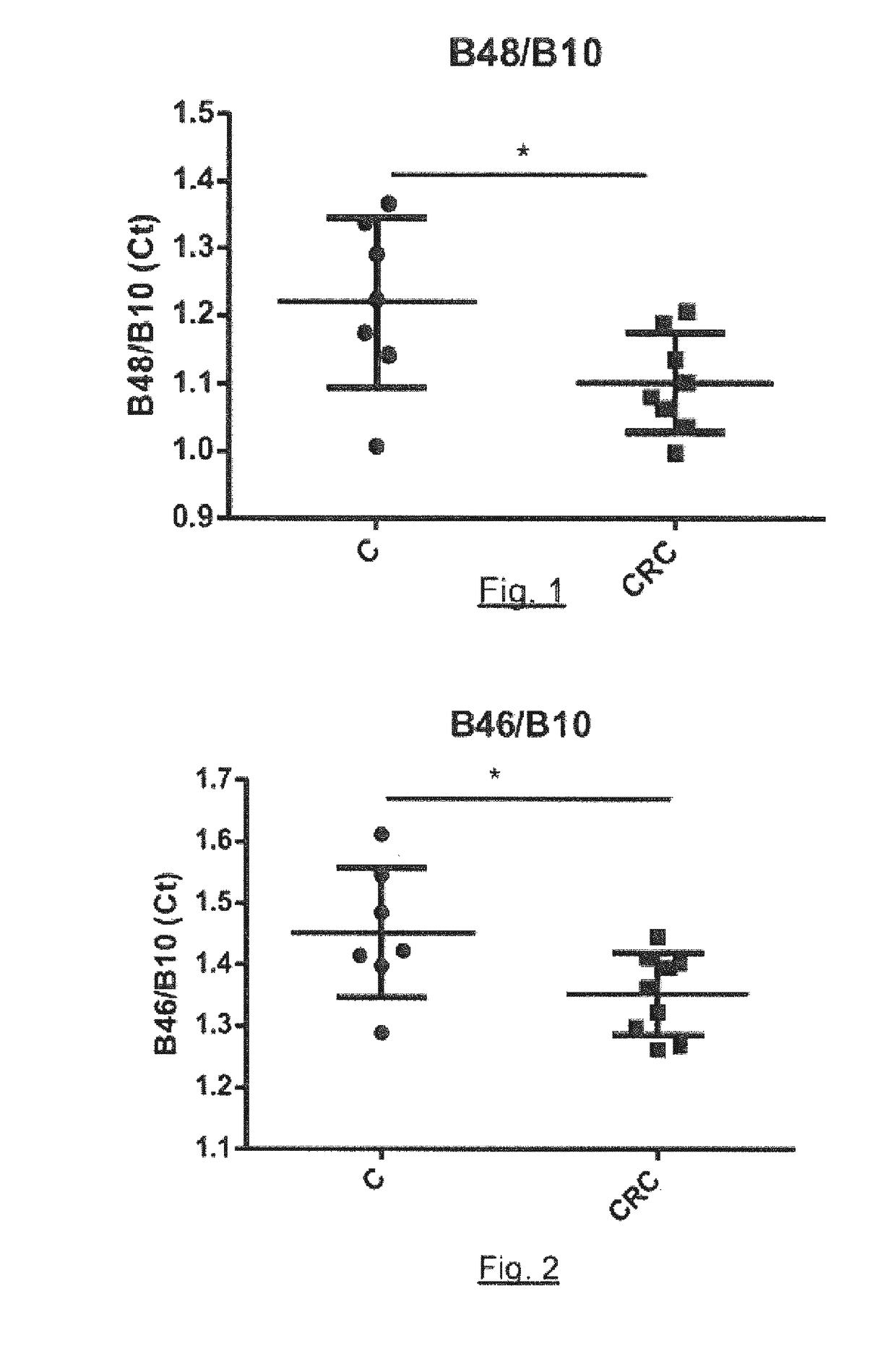
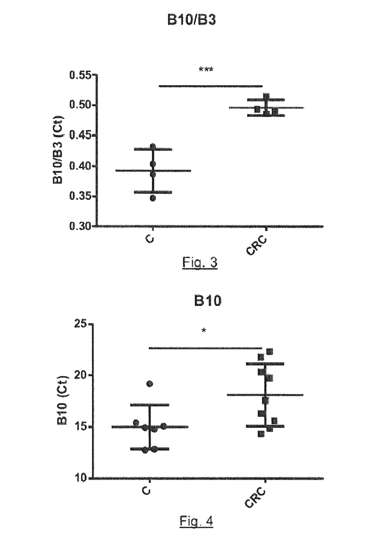
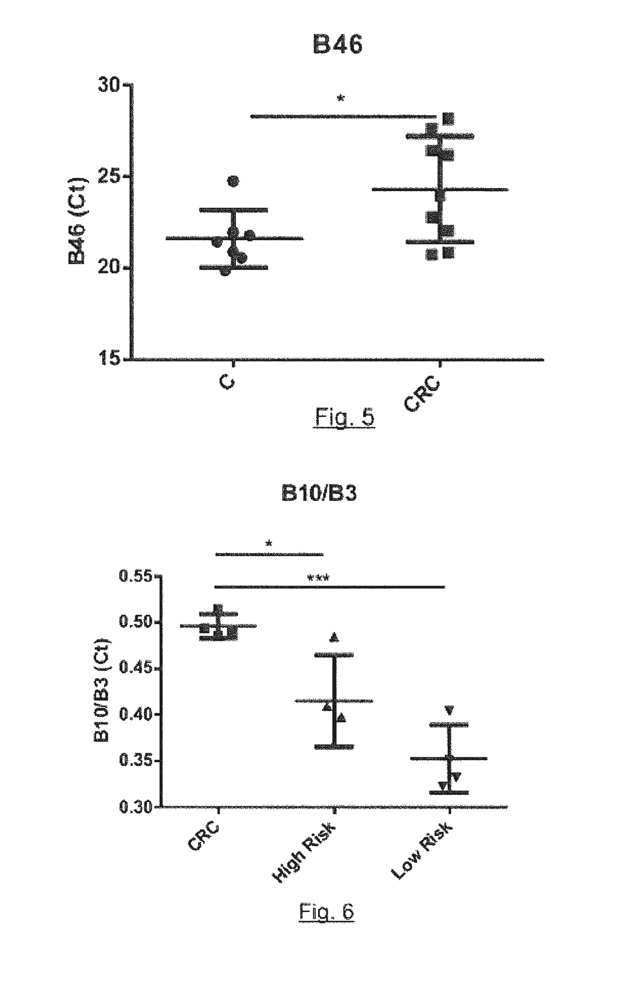
[0199]A total of 16 samples of feces in 7 controls and 9 colorectal cancer patients have been analyzed. The quantification of each of the bacteria markers cited in Example 1 in feces samples analyzed is expressed in Ct values. The Ct (cyclethreshold) is defined as the number of q-PCR cycles required for the fluorescent signal to cross the threshold. Ct levels are inversely proportional to the logarithm of target nucleic acid concentration in the sample (ie, the lower the Ct level the greater the amount of target nucleic acid in the sample). The real time assays undergo 40 cycles of amplification. The obtained results are shown below, in Tables 1 and 2. Table 1 represents Ct absolute values and Table 2 represents Ct ratios. Two sample t-test was applied.

TABLE 1Ct absolute valuesIDGroupConditionB48B46B10B3F10CRC118.5922.7816.3333.64F11CRC120.3326.4520.3741.57F3CRC123.5226.219.838.45F4CRC116.4120.8714.88F5CRC...


PUM

PropertyMeasurementUnit
Fractionaaaaaaaaaa
Electrical conductanceaaaaaaaaaa
Ratioaaaaaaaaaa

Abstract

The present invention relates to the field of detection of colorectal cancer (CRC). Specifically it relates to methods for the early detection, risk screening and monitoring of CRC and / or adenomatous polyps in a human subject based on the quantification of one or more 16S rDNA bacterial sequences in feces. It further relates to the use of said bacterial sequences as biomarkers of colorectal cancer and / or adenomatous polyps; to a kit comprising a reagent and instructions for the quantification of said bacterial sequences; and to nucleic acids for the quantification of said 16S rDNA bacterial sequences.

Description

FIELD OF INVENTION[0001]The present invention relates to the field of detection of colorectal cancer (CRC). Specifically it relates to methods for the early detection, risk screening and monitoring of CRC and / or adenomatous polyps in a human subject based on the quantification of one or more 16S rDNA bacterial sequences in feces. It further relates to the use of said bacterial sequences as biomarkers of colorectal cancer and / or adenomatous polyps; to a kit comprising a reagent and instructions for the quantification of said bacterial sequences; and to nucleic acids for the quantification of said 16S rDNA bacterial sequences.BACKGROUND OF THE INVENTION[0002]Colorectal cancer (CRC) is the second leading cause of cancer death in Europe and in the United States, and is the most frequently diagnosed cancer in Europe, with over 400,000 new cases and 200,000 deaths in 2008. These data reflect that preventive measures need to be taken.[0003]The most effective and economic measure to reduce ...

Claims


Application Information

Patent Timeline
IPC IPC(8): C12Q1/68
CPCC12Q2600/118C12Q1/6886C12Q1/689C12Q2600/158C12Q1/6806
Inventor SERRA PAG S, MARIONAGARC A-GIL, LIBRADO JES SMAS DE XAXARS, TERESAALDEGUER MANTE, XAVIER
Owner FUNDACIO INST DINVESTIGACIO BIOMEDICA DE GIRONA DR JOSEP TRUETA
Why Eureka
Try Eureka