Systems and methods for identifying coagulopathies
a technology of coagulopathy and coagulopathy, applied in the field of systems and methods for identifying coagulopathies, can solve the problems of lack of sensitivity to measuring fibrinolytic activity, difficult laboratory assessment of hemostasis, and lack of insight into the risk of thrombosis by existing methods, etc., to achieve simple practice, rapid and reliable
- Summary
- Abstract
- Description
- Claims
- Application Information
AI Technical Summary
Benefits of technology
Problems solved by technology
Method used
Image


Examples
example 1
Blood Clotting, Retraction, and Lysis with Thrombin and Tissue Plasminogen Activator
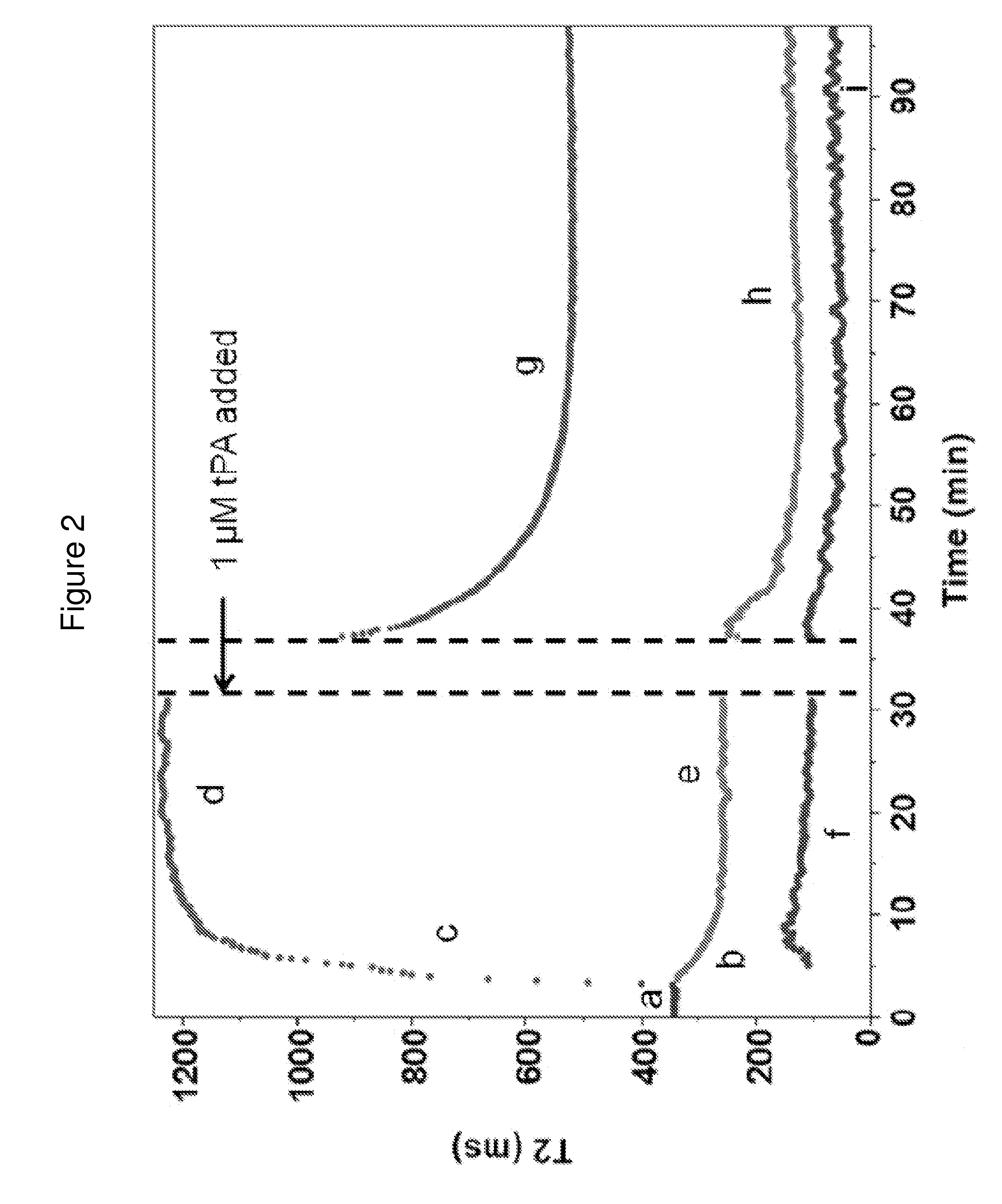
[0167]Blood clotting was initiated by addition of 2 μL of a 0.2 M CaCl2 solution and 2 μL of thrombin (Sigma-Aldrich, St. Louis, Mo., final concentration 0.1-3.0 U / ml) to 34 μL of blood in a 200 μL PCR tube (Eppendorf, Hauppauge, N.Y.). All components were pre-warmed for 1 min at 37° C. before mixing. Samples were mixed by three aspiration and dispersion cycles using a pipette, then put into the T2MR reader for measurement. Typical run length was 30 min with a 10 s sampling rate. For some experiments, data collection time was extended to 1 hr.
[0168]To establish T2MR signatures for fibrinolysis, tissue plasminogen activator (tPA, Alteplase, Genentech, South San Francisco, Calif.) was added to samples clotted by thrombin. Blood clotting was initiated as described above, and the sample was incubated for 60 min to allow for complete clot contraction. Then, 0.5-1 μM tPA was added to clotted and contracted...
example 2
Real-Time Monitoring of Clot Formation, Contraction and Fibrinolysis
[0169]We measured the dependencies of the T2MR signals during clotting of re-calcified citrated blood samples from healthy donors initiated by adding 3 U / ml thrombin. Thrombin activates platelets and cleaves fibrinogen to form a three-dimensional fibrin network stabilized by factor XIIIa. Addition of thrombin led to rapid formation of a gelatinous meshwork that filled the sample volume accompanied by a small, rapid decrease in the T2MR signal over tens of seconds due to the sample transitioning from a liquid to gel state. In the initial gel state, only one relaxation rate was observed (FIG. 2, part a), reflecting uniform distribution of erythrocytes and other blood components. Approximately four minutes after thrombin addition, the T2MR signal split into two peaks representing distinct water populations in slow exchange with each other. One peak decreased in T2 value (FIG. 2, part b), indicating increasing erythrocy...
example 3
Analyzing Isolated Sample Components
[0171]The T2 values of individual components of blood were determined using samples fractionated as described above or using clotted whole blood components. All samples were pre-warmed at 37° C. for 1 min before transferring to a T2MR reader for measurement. For plasma, 40 μL of PPP was measured. For serum, 200 μL of whole blood was clotted by addition of 2 U / ml thrombin to re-calcified blood. After a 30 min incubation at 37° C., the tube was centrifuged for 1 min at 10,000 g and 40 μL of the upper (serum) fraction was measured. To measure isolated retracted clots, re-calcified blood was allowed to clot for 1 hr following addition of 2 U / ml thrombin at 37° C. Then, erythrocytes excluded from clot were removed by washing the clot with 100 μL of PPP by gentle pipetting. The liquid was aspirated and disposed. This washing protocol was repeated two more times. To measure the isolated clot, all liquid was aspirated after the washing steps.
[0172]To inte...
PUM
 Abstract
Description
Claims
Application Information
 - R&D
- Intellectual Property
- Life Sciences
- Materials
- Tech Scout
- Unparalleled Data Quality
- Higher Quality Content
- 60% Fewer Hallucinations
Browse by: Latest US Patents, China's latest patents, Technical Efficacy Thesaurus, Application Domain, Technology Topic, Popular Technical Reports.
© 2025 PatSnap. All rights reserved.Legal|Privacy policy|Modern Slavery Act Transparency Statement|Sitemap|About US| Contact US: help@patsnap.com