Isolation of human neural stem cells from amniotic fluid of patients with neural tube defects
a technology of amniotic fluid and which is applied in the field of human neural stem cells, can solve the problems of limited availability, ethical concerns, and difficulty in harvesting, and achieve the effects of reducing the risk of infection, and improving the quality of li
- Summary
- Abstract
- Description
- Claims
- Application Information
AI Technical Summary
Benefits of technology
Problems solved by technology
Method used
Image


Examples
example 1
Isolation of NSCs from Amniotic Fluid
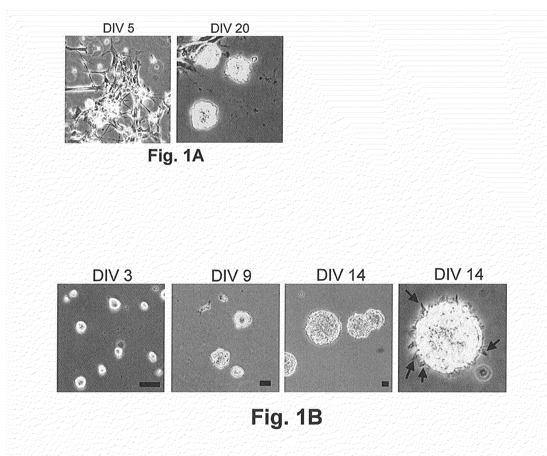
[0078]To isolate NSCs from amniotic fluid, normal (n=7) and NTD (anencephaly n=6, non-anencephaly n=6)-derived amniotic fluid specimens were collected by amniocentesis. When the cells isolated from these samples were cultured in NeuroCult™ NS-A proliferation medium, only a subset of the anencephaly samples produced some slightly attached neural-like cells and colonies during the first 3-5 days (FIG. 1A). After careful removal of the unattached cells and cellular debris, these neural-like cells proliferated and rounded-up to form primary neuroshperes that grew in suspension after 3 weeks (FIG. 1A). Upon passaging, the neurospheres were trypsinized into single cells, and cells proliferated and reformed neurospheres in suspension with each passage (FIG. 1B). The diameter of the neurospheres ranged from approximately 50 to 100 nm, and they possessed the classic microspikes on their outer surface (FIG. 1B). These AF-NSCs could be expanded by continual...
example 2
Characterization of the AF-NSCs
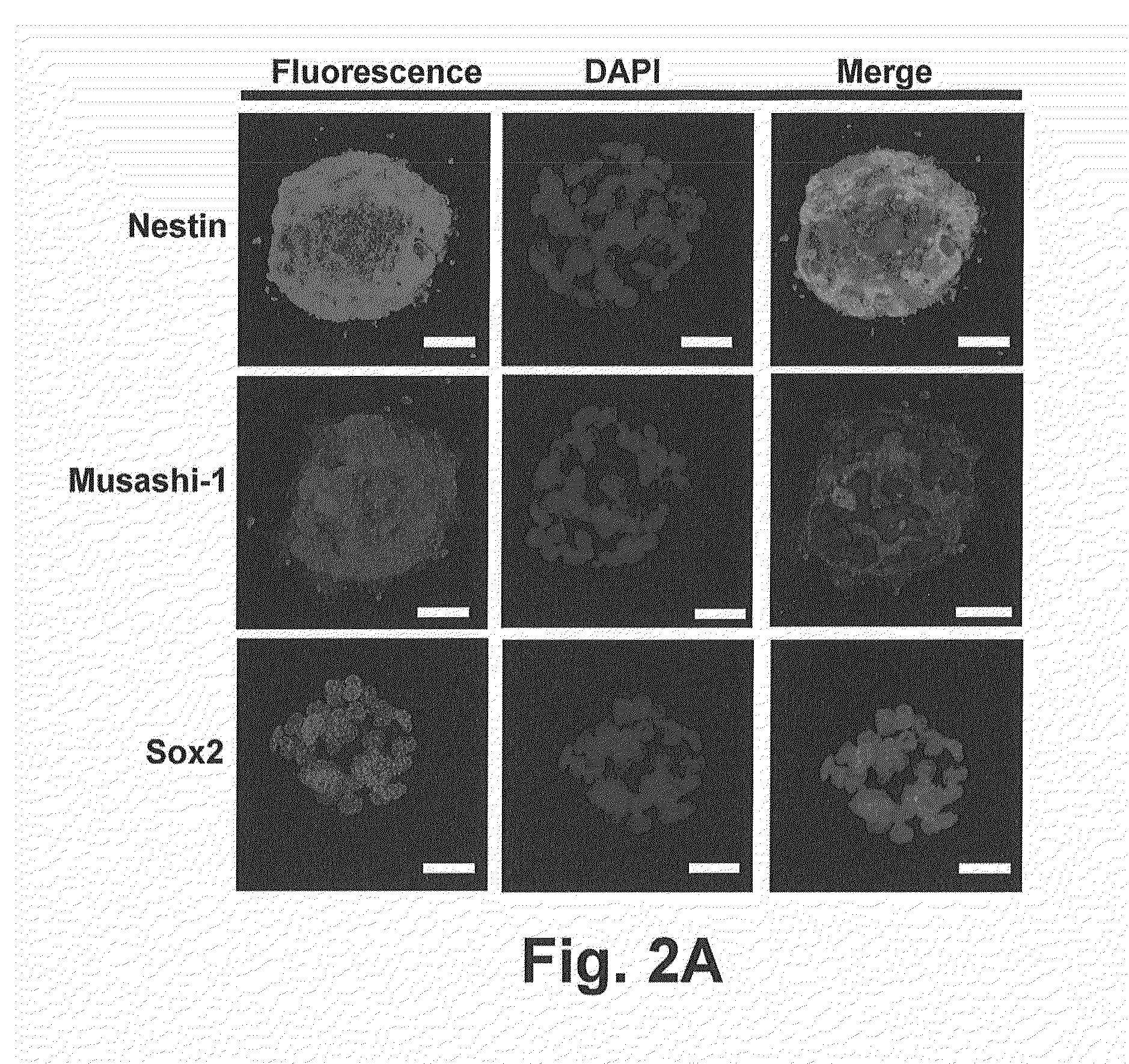
[0081]To characterize the AF-NSCs, immunocytochemistry and flow cytometry were used to detect the expression NSC-specific markers. Confocal microscopy revealed that AF-NSC-derived neurospheres strongly expressed both Nestin and Musashi-1 within cytoplasm. Sox2, a nucleus protein, could also be observed in the neurospheres (FIG. 2A).
[0082]Flow cytometry revealed that AF-NSCs express NSC-specific markers Nestin, Sox2, Musashi-1 and ABCG2 (FIG. 2B). CD133 was initially expressed at a low level, but the intensity of the signal increased with subsequent passages. We also analyzed the expression of embryonic stem (ES) cell-specific markers and determined that our AF-NSCs expressed a similar set of the transcription factors to ES cells, including Nanog, Oct-4, and Sox2, and low levels of SSEA-1; however, they did not express SSEA-3, SSEA-4, TRA-1-60, and TRA-81. Furthermore, the pattern of human leukocyte antigen (HLA) expression in AF-NSCs was similar to mos...
example 3
In Vitro Neural Differentiation of AF-NSCs
[0084]To determine whether AF-NSCs could differentiate into neurons, the cells were dissociated into a single cell suspension, cultured in NeuroCult™ differentiation medium and subsequently analyzed by immunocytochemistry. After 2 days of induction, the AF-NSCs began to undergo morphological changes, and only Nestin and Tuj-1 were expressed within cells at this stage (FIG. 3A). Additional neuron-specific markers, including Nestin, Tuj-1, MAP2, hNeuN and NFH, could be detected after 7 days of induction (FIG. 3B). We also used qPCR to determine the transcription levels of these neuron-specific genes and found that Tuj-1 and MAP2 were upregulated by 5.2- and 6.2-fold, respectively, after 7 days of differentiation. In contrast, the expression of Nestin, a NSC-specific gene, significantly decreased during this same time period (FIG. 3C).
[0085]Next, we induced our AF-NSCs into astrocytes, oligodendrocytes and dopaminergic neurons by exposing them ...
PUM
| Property | Measurement | Unit |
|---|---|---|
| diameter | aaaaa | aaaaa |
| density | aaaaa | aaaaa |
| density | aaaaa | aaaaa |
Abstract
Description
Claims
Application Information
 - R&D
- Intellectual Property
- Life Sciences
- Materials
- Tech Scout
- Unparalleled Data Quality
- Higher Quality Content
- 60% Fewer Hallucinations
Browse by: Latest US Patents, China's latest patents, Technical Efficacy Thesaurus, Application Domain, Technology Topic, Popular Technical Reports.
© 2025 PatSnap. All rights reserved.Legal|Privacy policy|Modern Slavery Act Transparency Statement|Sitemap|About US| Contact US: help@patsnap.com