Composition for Improving Skin Conditions Using Fetal Mesenchymal Stem Cells from Amniotic Fluid
a technology of amniotic fluid and mesenchymal stem cells, which is applied in the field of composition for improving skin conditions, can solve the problems of urgent need for sources and difficulty in obtaining a large amount of mesenchymal stem cells, and achieve the effect of improving skin conditions
- Summary
- Abstract
- Description
- Claims
- Application Information
AI Technical Summary
Benefits of technology
Problems solved by technology
Method used
Image
Examples
example 1
Culture of Fetus-Derived Cell Line in Amniotic Fluid Obtained from Pregnant Woman and Morphology Identification of Homogeneous Mesenchymal Stem Cells under Culture Conditions
[0060]There are many floating particles in the amniotic fluid obtained from a pregnant woman. In order to separate them, the amniotic fluid was put in a T-flask and incubated at 37° C. The next day, all the cells, with the exception of cells attached to the bottom, were removed. The cells attached to the bottom were detached from the bottom using trypsin, and then collected by centrifugation. Then, the cells were suspended in a basal medium of low glucose DMEM containing 10% FBS, 1% L-glutamine and 1% penicillin-streptomycin and 4 ng / ml bFGF, and the suspended cells were seeded in a 100 mm cell culture plate. After 12˜24 hours, the medium was replaced with fresh medium. It was found that the fetus-derived cell line in amniotic fluid showed heterogeneous populations (FIG. 1A: a-d), and had only a fibroblast-like ...
example 2
Identification of Fetus-Derived Cells in Amniotic Fluid via Karyotyping
[0061]In order to examine whether the cells are derived from the fetus in amniotic fluid, karyotyping was performed.
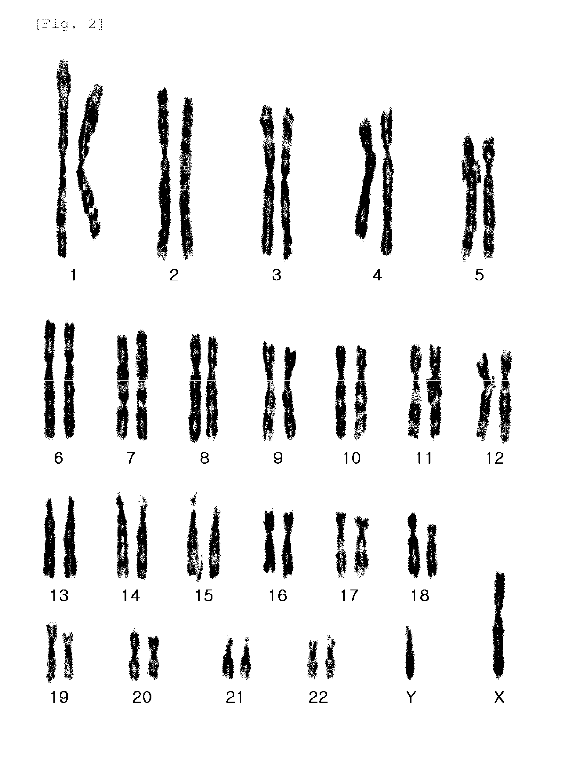
[0062]A karyotype shows the number, size, and shape of each chromosome type, and chromosome analysis is performed to detect mutations and to determine fetal sex. In order to perform karyotyping, the cell division was arrested at metaphase using colcemid for 1˜2 hrs, and chromosome analysis was performed by G-banding staining. The karyotyping results showed that the fetus-derived cells in amniotic fluid had normal chromosomes and sex chromosomes of XY, indicating that the cells were derived from the fetus, not the mother (FIG. 2).
example 3
Analysis on Similarity between Fetus-Derived Cells in Amniotic Fluid and Mesenchymal Stem Cells
[0063]It was examined whether the fetus-derived cells in amniotic fluid have the characteristics of pluripotent mesenchymal stem cells.
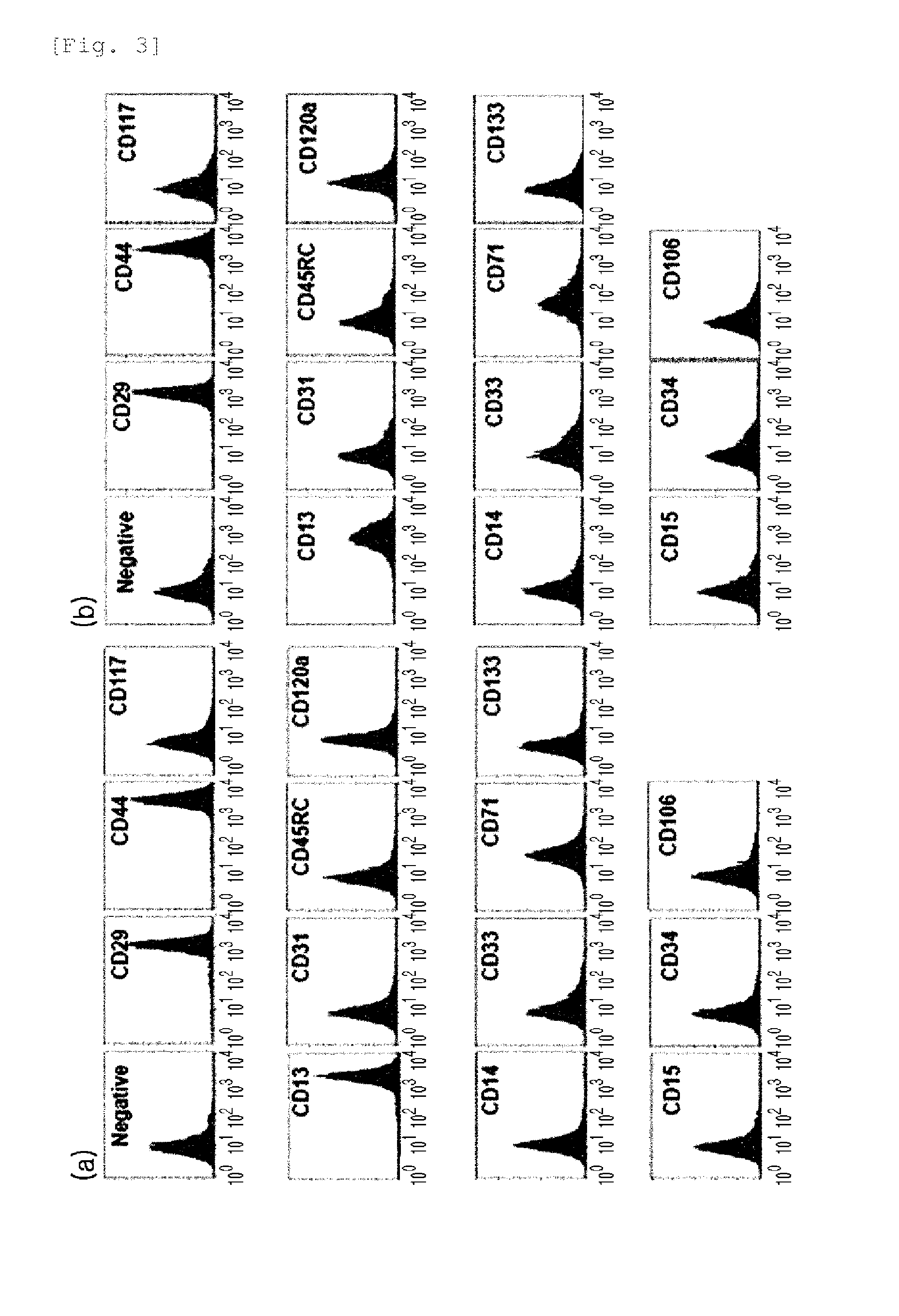
[0064]First, when immune phenotyping was performed using a flow cytometry, expression of CD13, CD29, and CD44 were observed in the fetus-derived cells in amniotic fluid, compared to the human bone marrow-derived mesenchymal stem cells (FIG. 3). Although their expression is specific to the mesenchymal stem cells, each cell may show different characteristics. Thus, the capability to differentiate into osteoblasts, adipocytes, and chondrocytes, which is a representative characteristic of the mesenchymal stem cells, was examined.
[0065]The fetus-derived cells in amniotic fluid were differentiated like the human bone marrow-derived mesenchymal stem cells, and then osteogenic differentiation was demonstrated by the expression of osteopontin and osteocalcin, adipog...
PUM
| Property | Measurement | Unit |
|---|---|---|
| Time | aaaaa | aaaaa |
Abstract
Description
Claims
Application Information
 - R&D
- Intellectual Property
- Life Sciences
- Materials
- Tech Scout
- Unparalleled Data Quality
- Higher Quality Content
- 60% Fewer Hallucinations
Browse by: Latest US Patents, China's latest patents, Technical Efficacy Thesaurus, Application Domain, Technology Topic, Popular Technical Reports.
© 2025 PatSnap. All rights reserved.Legal|Privacy policy|Modern Slavery Act Transparency Statement|Sitemap|About US| Contact US: help@patsnap.com


