
Laparoscopic Cholecystectomy With Fluorescence Cholangiography

Inactive Publication Date: 2016-09-15
YU STEVEN SOUNYOUNG
View PDF2 Cites 17 Cited by
  • Summary
  • Abstract
  • Description
  • Claims
  • Application Information

AI Technical Summary

Benefits of technology

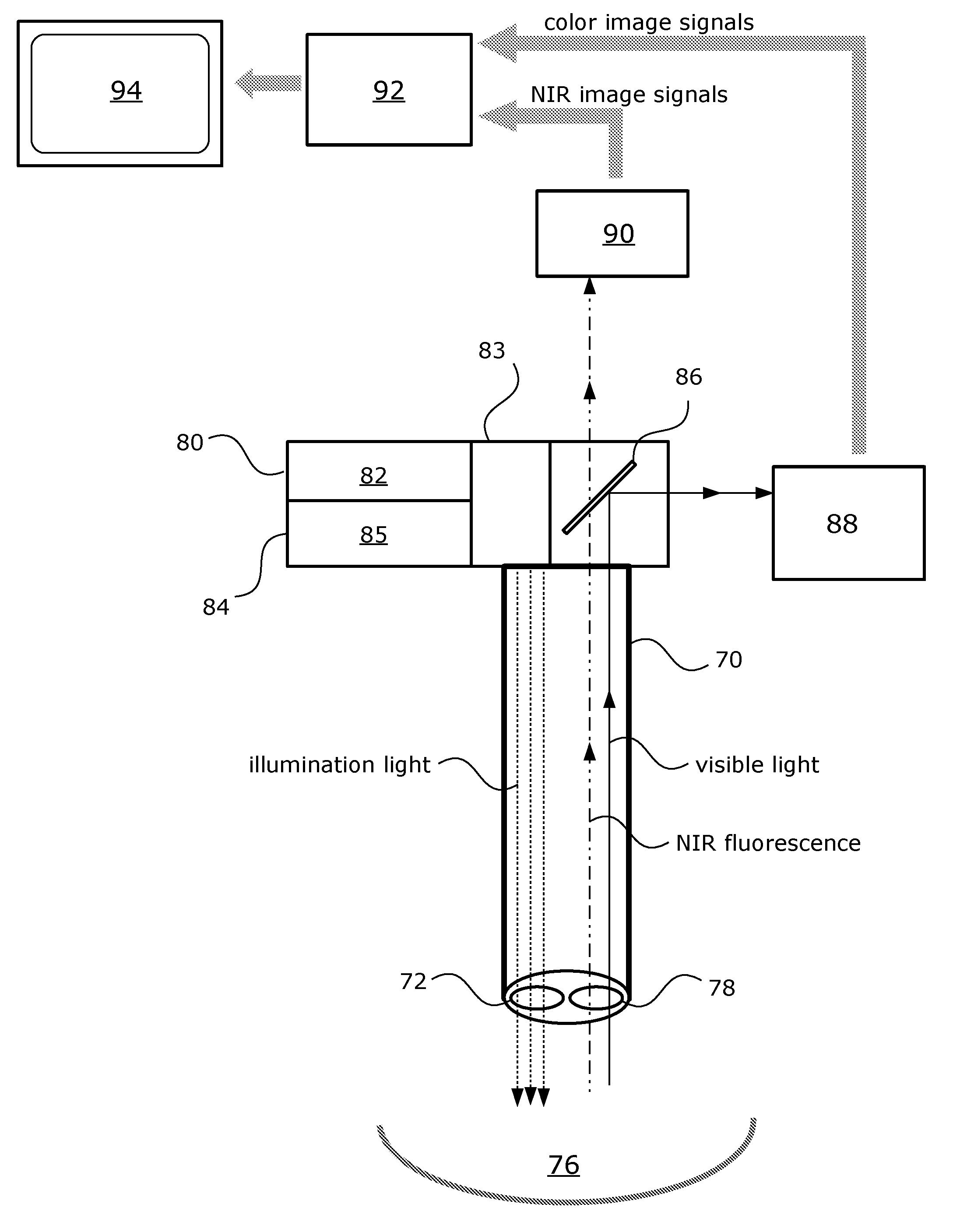
The patent describes a method of using near-infrared fluorescence imaging in endoscopic surgeries, such as laparoscopic cholecystectomy, to improve surgical precision. The method involves administering a near-infrared fluorescent dye to the patient and using an endoscope to view the surgical field. The fluorescence image is then combined with a color image to create a composite video image. This composite video image can be used to guide the surgery and ensure better visualization of the anatomy. The method can also involve selectively imaging specific areas of interest in the surgical field, such as the biliary ducts. The use of near-infrared fluorescence imaging has been shown to improve the accuracy of surgical procedures and reduce the risk of complications.

Problems solved by technology

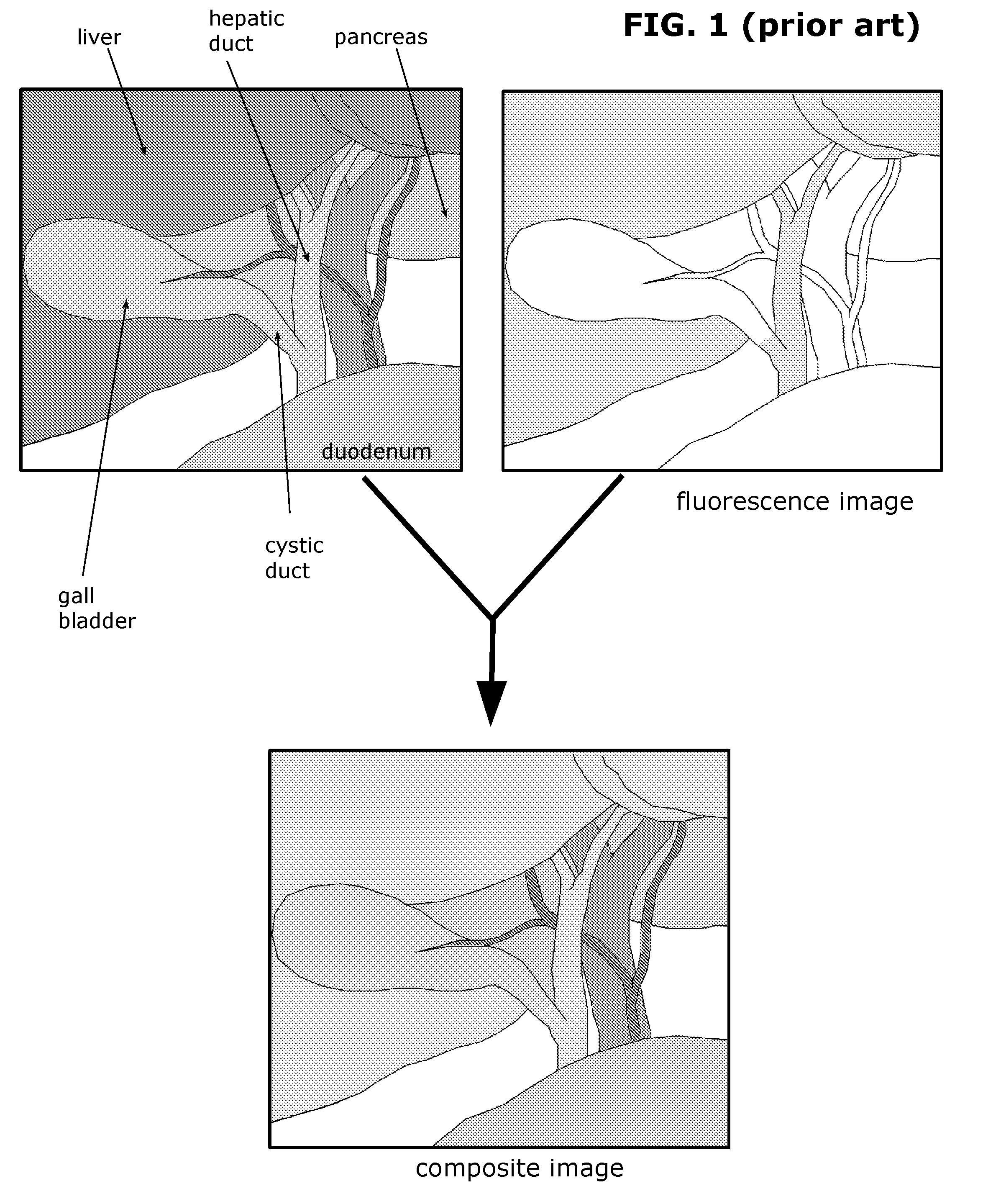
As seen here, because much of the fluorescent dye is still remaining in the liver, the fluorescence emission from the liver dominates the view and makes it more difficult to contrast the biliary ducts from the liver.

Method used


Image

  • Laparoscopic Cholecystectomy With Fluorescence Cholangiography
  • Laparoscopic Cholecystectomy With Fluorescence Cholangiography
  • Laparoscopic Cholecystectomy With Fluorescence Cholangiography

Examples


Embodiment Construction

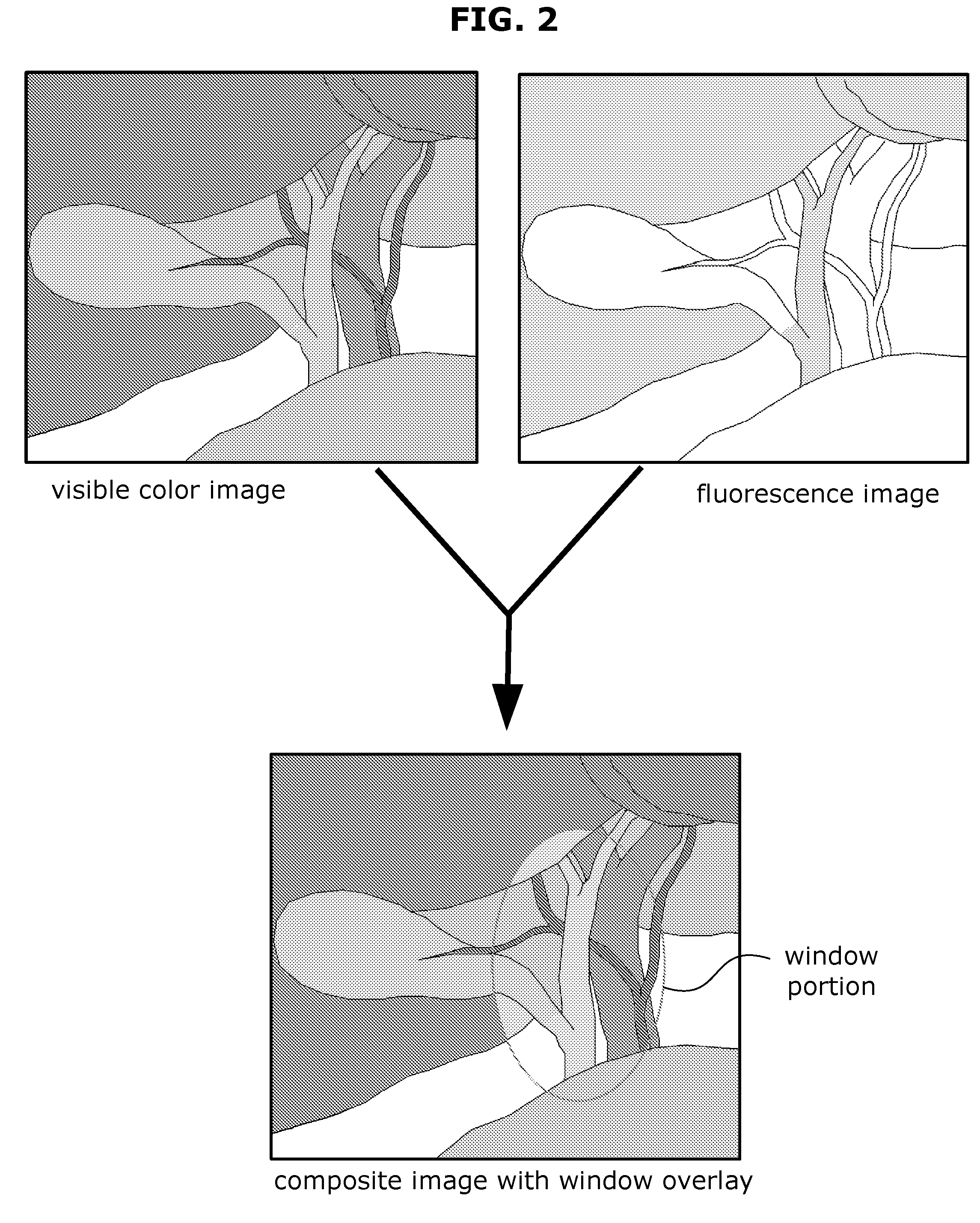
[0034]In my invention, a fluorescent video image of the surgical field is composited with a color video image of the surgical field in real-time to produce a composited video image. In the composited video image, the fluorescence video image (which is in greyscale) is shown in a pseudo-color. Other terms of the art that are equivalent or encompassed by the term “compositing” include blending, overlaying, superimposing, or merging the images. The expression stating that the fluorescence image is overlaid or superimposed over the color image does not necessarily indicate any particular order of layering. Whether the fluorescence image is treated as a layer over the color image or the reverse arrangement, the same desired effect can be rendered, i.e. that the fluorescence image appears to the human user as being placed over the color image.

[0035]The image processing required to composite the color video image with the NIR fluorescence video image may be performed by any suitable image ...


PUM


Abstract

Use of near-infrared fluorescence imaging in performing an endoscopic surgery, such as laparoscopic cholecystectomy. A composite video image of the surgical field (e.g. the biliary anatomy) is shown on a display screen. The composite video image combines a visible color image and a fluorescence image of the surgical field. Using various techniques, selective fluorescence imaging of a particular area of interest in the surgical field is made possible. For example, in a cholecystectomy procedure, the biliary ducts may be the particular area of interest for the selective fluorescence imaging.

Description

FIELD OF THE INVENTION[0001]My invention relates to fluorescence imaging in surgery.BACKGROUND[0002]Inadvertent injury to the bile ducts from anatomic misidentification is among the more serious complications of laparoscopic cholecystectomy. Real-time intraoperative imaging of the biliary anatomy by fluorescence cholangiography is a recently introduced technique that helps the surgeon to delineate the bile ducts during the laparoscopic surgery. In this technique, indocyanine green (ICG) dye is administered intravenously to the patient prior to the operation. The exogenous dye (which is a fluorophore) accumulates in the liver and is excreted through the biliary ducts. Illumination with the proper excitation light causes the dye to fluoresce with near-infrared (NIR) light. Imaging of this NIR light gives a visual outline of the bile ducts to assist the surgeon in proper anatomic identification.[0003]FIG. 1 shows a prior art method of fluorescence cholangiography illuminating the bilia...

Claims


Application Information

Patent Timeline
IPC IPC(8): A61B1/04A61B1/00
CPCA61B1/043A61B1/00009A61B1/00045A61B1/0005A61B1/0638A61B1/000094
Inventor YU, STEVEN SOUNYOUNG
Owner YU STEVEN SOUNYOUNG
Learn More