Anti-galectin-1 monoclonal antibodies and fragments thereof
- Summary
- Abstract
- Description
- Claims
- Application Information
AI Technical Summary
Benefits of technology
Problems solved by technology
Method used
Image
Examples
example 1
Anti-Gal1 Monoclonal Antibodies Useful for Diagnostic Applications
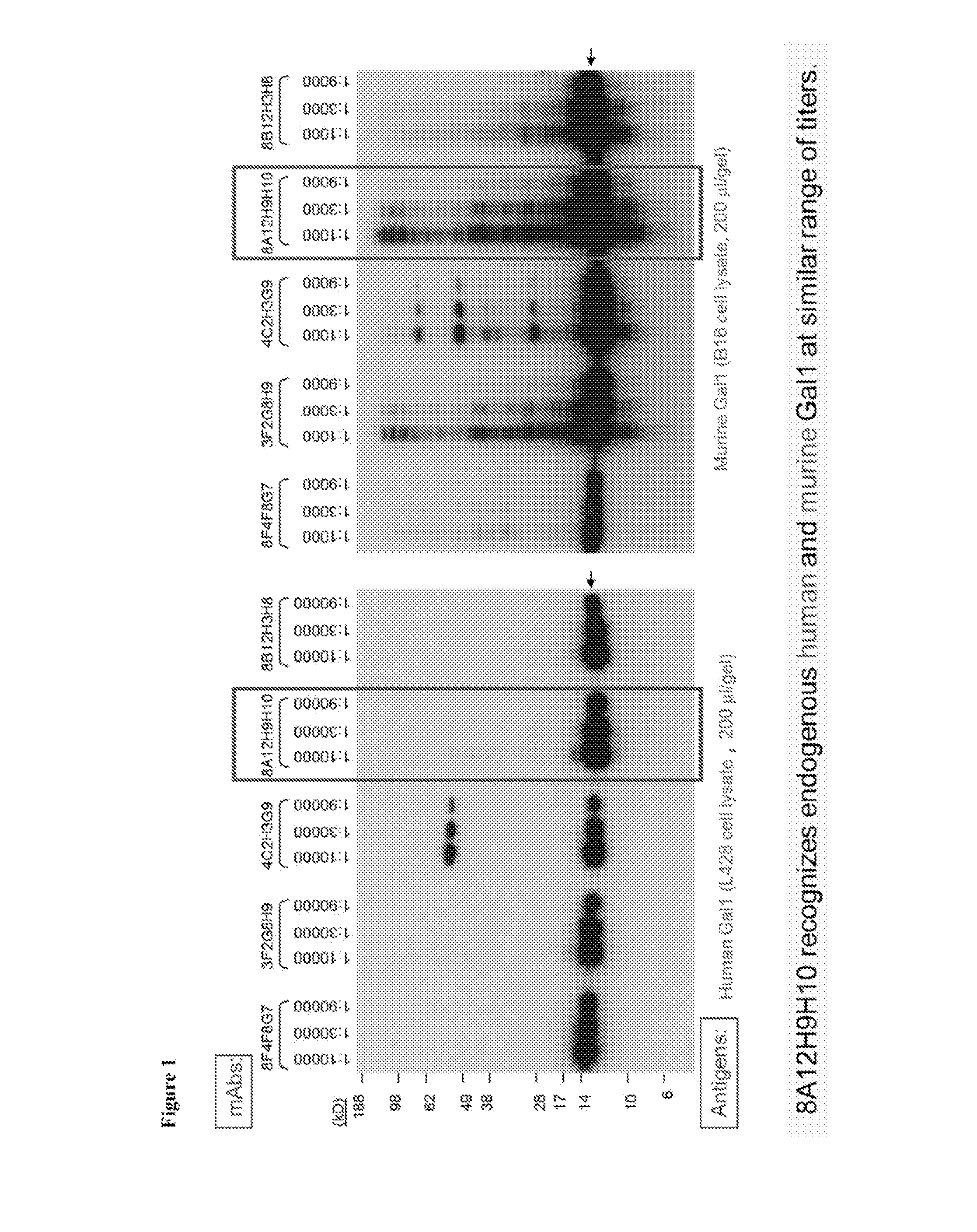
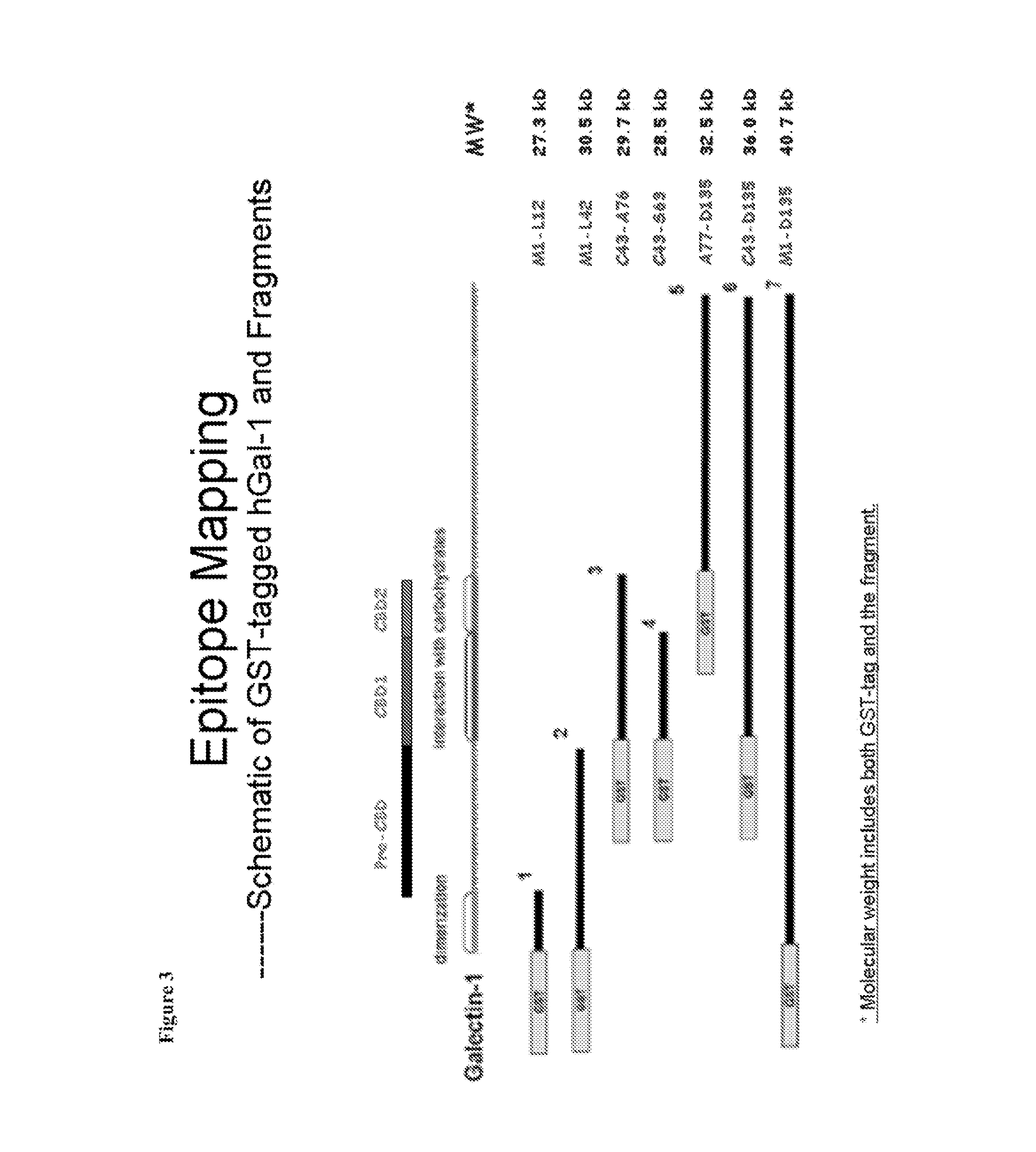
[0194]Anti-Gal1 monoclonal antibodies were generated and were determined to cross-react well with both human Gal1 and mouse Gal1 (FIG. 1). Epitope mapping indicated that the 8A12.H9.H10 (i.e., the 8A12) anti-Gal1 monoclonal antibody recognized a domain distal to the previously described carbohydrate-binding domain (CBD) of Gal1 and encompasses a kappa light chain. Six recombinant human Gal1 fragments (covering the N-terminal, CBD, post-CBD sequences) named F1, F2, F3, F4, F5, F6 and the full length protein F7 in fusion with GST-tag were produced in E. coil (FIG. 2). The 8A12 monoclonal antibody was determined to recognize recombinant GST-F5, GST-F6 and GST-F7 by Western blot analysis, but none of GST-F1, GST-F2, GST-F3 and GST-F4, indicating that the antibody binds to the post-CBD domain.
[0195]The 8A12 antibody was sequenced, which are presented in Table 1 below, and analysis of the sequences obtained from the hybrido...
example 2
Gal1 Serum Levels in Classical Hodgkin Lymphoma (cHL)
[0196]A. Patients and Samples
[0197]For the retrospective study, frozen serum samples and clinical data from 293 newly diagnosed, previously untreated cHL patients were used from patients who were enrolled on Institutional Review Board-approved risk-adapted German Hodgkin Study Group (GHSG) multicenter clinical trials: a) HD13 (ISRCTN63474366) for early-stage disease (clinical stage [CS] IA-IIB with no risk factors, 80 patients) (Diehl et. al. (2003) Hematol. Am. Soc. Hematol. Educ. Program Rev. 2003:225-247; Sieniawski et al. (2007) J. Clin. Oncol. 25:2000-2005); b) HD14 (ISRCTN04761296) for early-stage disease with risk factors (CS I-IIA with large mediastinal mass, extranodal disease, elevated erythrocyte sedimentation rate (ESR) or ≧3 nodal areas and CS IIB with elevated ESR or ≧3 nodal areas, 89 patients) (Diehl et al. (2003) Hematol. Am. Soc. Hematol. Educ. Program Rev. 2003:225-247; von Tresckow et al. (2012) J. Clin. Oncol....
example 3
Gal1 and PD-L1 are co-expressed by EBV- and HHV8-associated Malignancies
[0207]A. Case Selection
[0208]Cases were retrieved from the surgical pathology files of Brigham and Women's Hospital, Boston, Mass.; Yale School of Medicine; New Haven Conn.; UMass Memorial Hospital, Worcester, Mass.; and from the consult files of a practitioner with institutional review board approval. Representative hematoxylin and eosin stained slides were reviewed. Whole tissue sections from EBV-positive of the elderly, EBV-positive immunodeficiency-related DLBCL, BL, ENKTCL, PBL, NPC, PEL, KS, and EBV-negative PTLD were evaluated. Seventy-seven total cases were evaluated for Gal1 expression by immunohistochemistry. Due to limited availability of tissue, a subset of these cases were evaluated for PD-L1 (59 cases), JunB (66 cases), and phosphorylated c-Jun (p-cJun; 63 cases) expression by inummohistochemistry. All cases of DLBCL, NPC, and BL were shown previously to be positive for EBV-encoded RNA (EBER) by in...
PUM
| Property | Measurement | Unit |
|---|---|---|
| Fraction | aaaaa | aaaaa |
| Digital information | aaaaa | aaaaa |
Abstract
Description
Claims
Application Information
 - R&D
- Intellectual Property
- Life Sciences
- Materials
- Tech Scout
- Unparalleled Data Quality
- Higher Quality Content
- 60% Fewer Hallucinations
Browse by: Latest US Patents, China's latest patents, Technical Efficacy Thesaurus, Application Domain, Technology Topic, Popular Technical Reports.
© 2025 PatSnap. All rights reserved.Legal|Privacy policy|Modern Slavery Act Transparency Statement|Sitemap|About US| Contact US: help@patsnap.com


