
Single-use, port deployable articulating endoscope

Active Publication Date: 2016-03-17
VIVID MEDICAL
View PDF10 Cites 32 Cited by
  • Summary
  • Abstract
  • Description
  • Claims
  • Application Information

AI Technical Summary

Benefits of technology

This patent describes an endoscope design that allows for the insertion of other endoscopic devices and tools through the same port. The endoscope is flexible and can be routed to the side of the port with thin cables. Multiple endoscopes can also be inserted, providing a multi-axis view. The images from the endoscopes can be stitched together or viewed separately by different users. The endoscope has a straight tubular configuration that can be inserted through a semi-rigid or rigid insertion tube, which opens up the insufflation membrane in the port and allows the endoscope to expand. The flexible tubular membrane seals when the insertion tube is removed, creating an airtight seal. The flat cables carrying the electrical signals and used for deployment and articulation can be routed through a thin-walled flexible tubular body, which can be bend controlled to allow for maximum flexibility and bend angle control of the endoscope.

Problems solved by technology

This both reduces the risk of infection and reduces the patient's hospital stay.
Typical endoscopic procedures require a large amount of equipment.
Space is also very limited at the proximal end of the port and tools and endoscopes with proximal cameras are bulky and heavy, often propped up and locked in position with secondary mechanisms that often physically interfere with other devices used by the surgeon, especially if multiple ports are close to one another, or in Single-Port procedures.
Another common problem that occurs with endoscopic procedures is that, because the endoscope is inserted into the body, the cavity being imaged by the endoscope is small and difficult to view.
Insufflation can be problematic because of inadequate seals between the port opening and the endoscopic device used.
In addition, the smallness of the space may cause too much contact with the endoscope, which may result in the endoscope becoming smeared with blood and liquids that obscure the view for the camera on the endoscope to capture images of the cavity.

Method used


Image

  • Single-use, port deployable articulating endoscope
  • Single-use, port deployable articulating endoscope
  • Single-use, port deployable articulating endoscope

Examples


Embodiment Construction

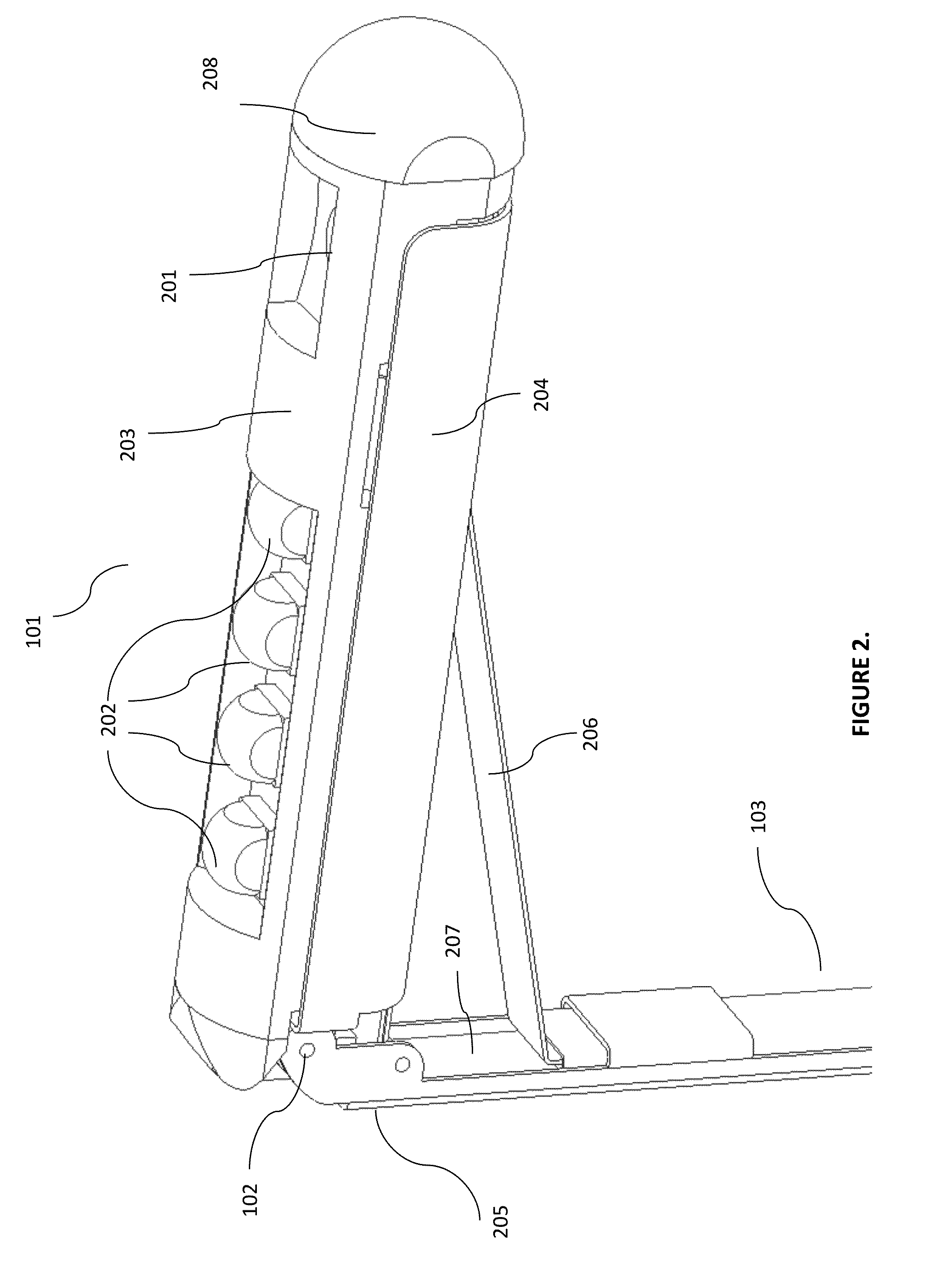
[0038]Example embodiments of the invention are directed to a deployable and / or articulating endoscope that includes a multi jointed housing, a camera, a light source at the distal section of the multi jointed endoscope, and very thin flat cables for electrical connections as well as deployment and articulation, which are routed through a secondary section of the endoscope, or incorporated within the outside body of another tool or surgical port.

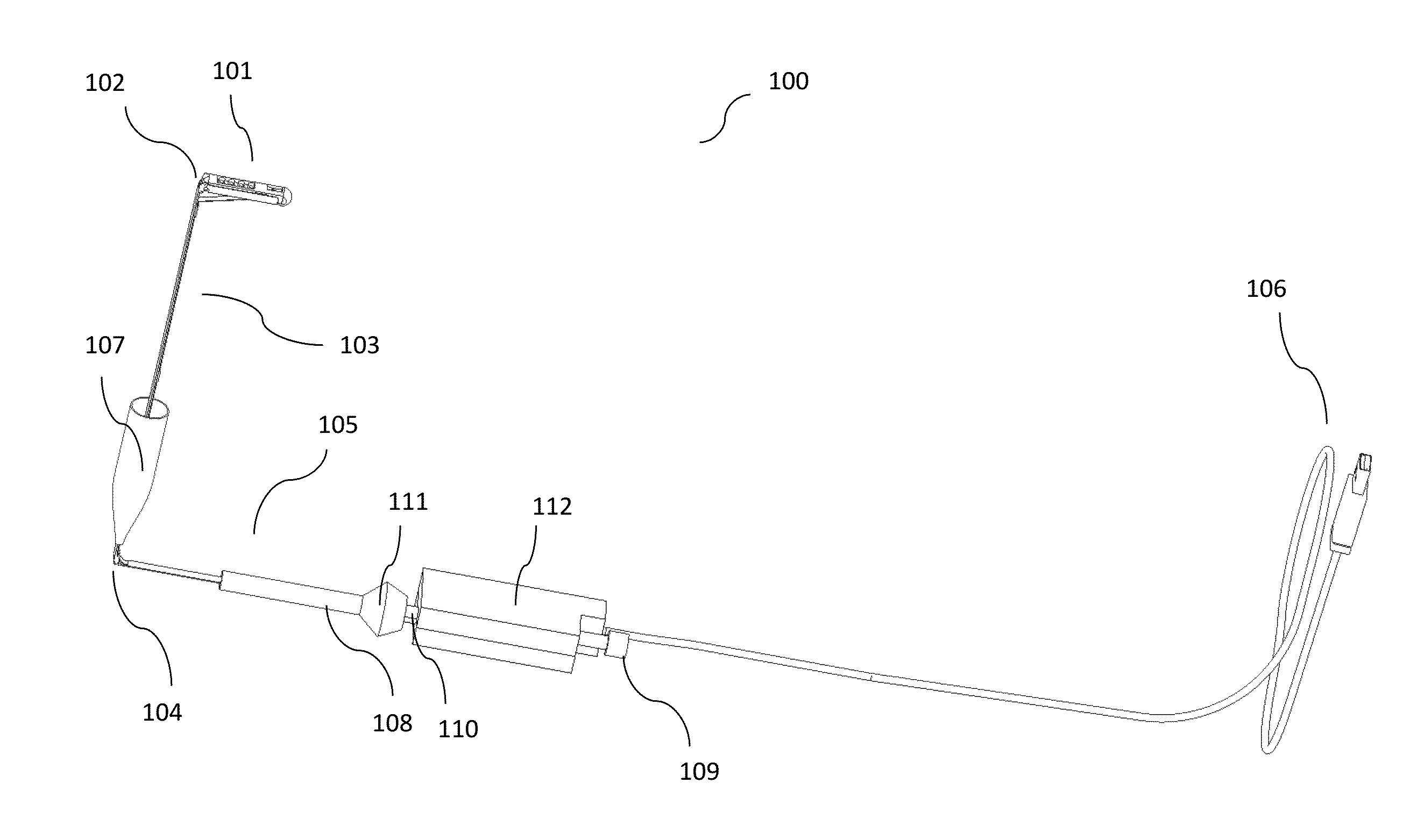
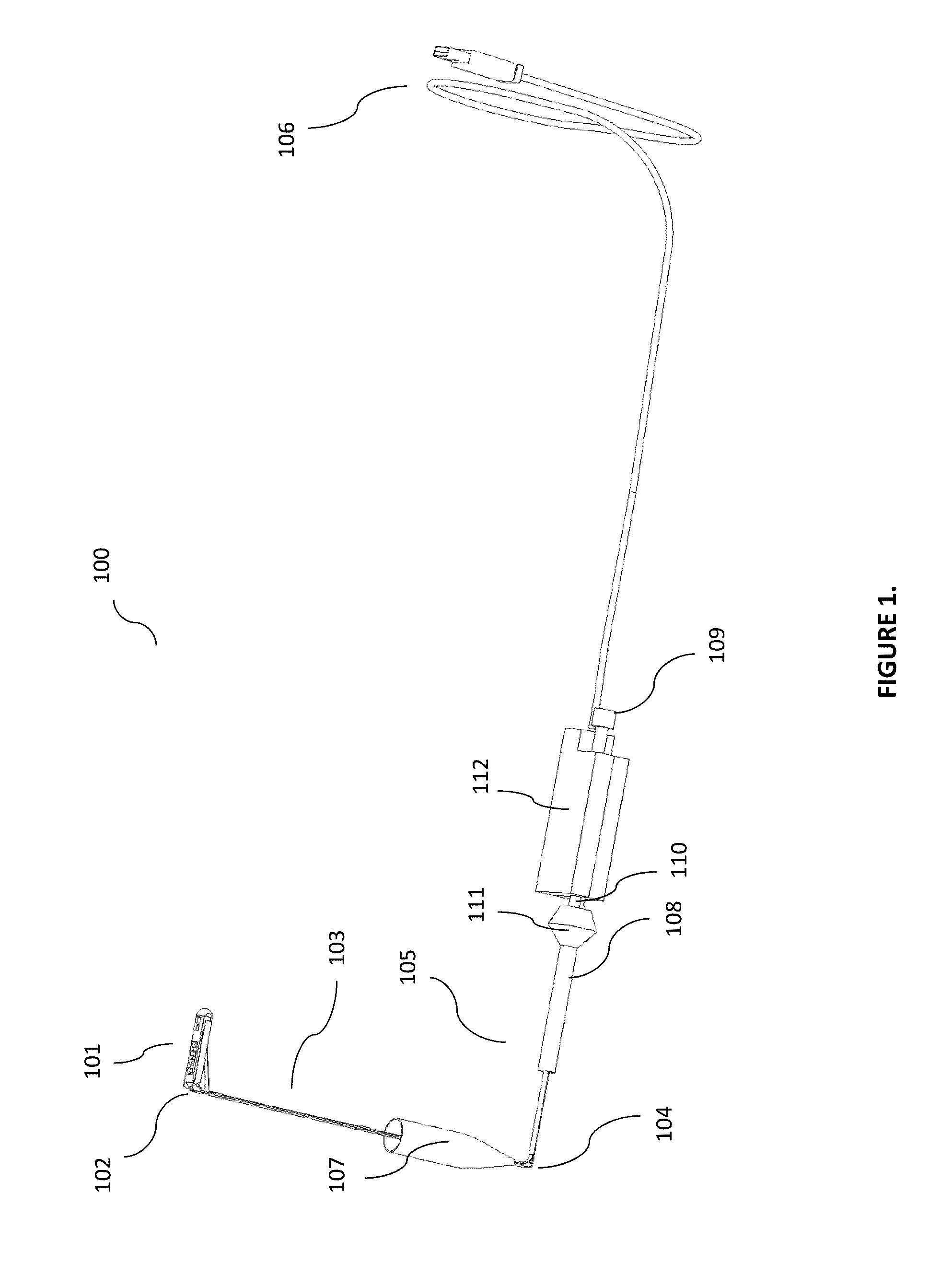
[0039]FIG. 1 illustrates an example side view of a multi jointed endoscope 100, with a distal section 101, a secondary section which may include a midsection 103, and a proximal section 105. In some embodiments, the secondary section may be coupled with the distal section 101 via a distal joint (or joints) 102; and the midsection 103 and the proximal section 105 may be coupled together via a proximal joint 104. The distal section 101 of the endoscope 100, for example, may be bent at about a 90 degree angle at the distal joint 102, directing t...


PUM


Abstract

The multi-section deployable and articulating endoscope is designed with tubing and flat cables that are small enough that the endoscope becomes a thin stick that is minimally invasive and usable with other devices in a common port. The endoscope includes very thin flat cables that are used for electrical connectivity and threaded above and below an articulation and deployment hinge for opening the endoscope passively using an adjustable tension spring, or by pulling on a first cable and closing the endoscope by pulling on a second cable, When in a tubular configuration the endoscope may be inserted into the port and an insufflation membrane inside the port forms an air seal with the endoscope, which aids in insufflation and desufflation.The endoscope includes one or more tubes for creating an air jet stream above the camera to act as a shield for keeping the camera clean.

Description

FIELD OF THE INVENTION[0001]The specification relates generally to a disposable endoscope that can be used, for example, in minimally invasive surgical (MIS) procedures, general or diagnostic medical or industrial procedures. In some embodiments, the specification relates to a fully digital endoscope that is deployed after insertion through the endoscopic port which can also be articulated at various angles to provide, for example, zero degrees and up to 90 degrees or more angled scope functionality. The endoscope body, for example, may take up only a very small fraction of space inside the port once it is deployed, allowing other devices to be concurrently used inside the same port. Some embodiments of the invention may also include a deployable and / or articulating endoscope that may include a very small profile flexibly bent and extended to the side, at the proximal end of the port, and thus transparent to the user when other devices are inserted through the same port or other por...

Claims


Application Information

Patent Timeline
IPC IPC(8): A61B1/00A61B1/005A61B1/015A61B1/045A61B1/05A61B1/06A61B1/12A61B1/313A61M13/00
CPCA61B1/00018A61B1/00103A61B1/00006A61B1/0676A61B1/00096A61B1/00114A61B1/00124A61B1/015A61B1/00091A61B1/0684A61B1/005A61B1/00045A61B1/00029A61B1/045A61B1/128A61B1/00154A61B1/3132A61M13/003A61B1/051A61B1/0615A61B1/0008A61B1/00177A61B1/0057A61B1/05
Inventor FARR, MINAKENNEY, SHANE JAMES
Owner VIVID MEDICAL
Learn More