
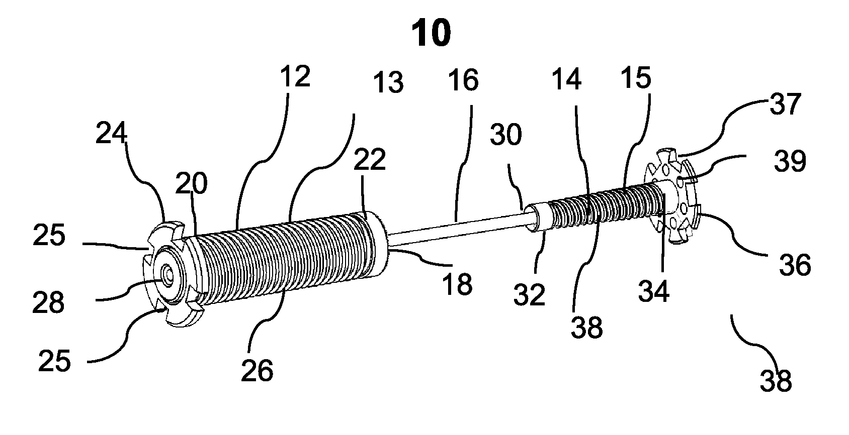
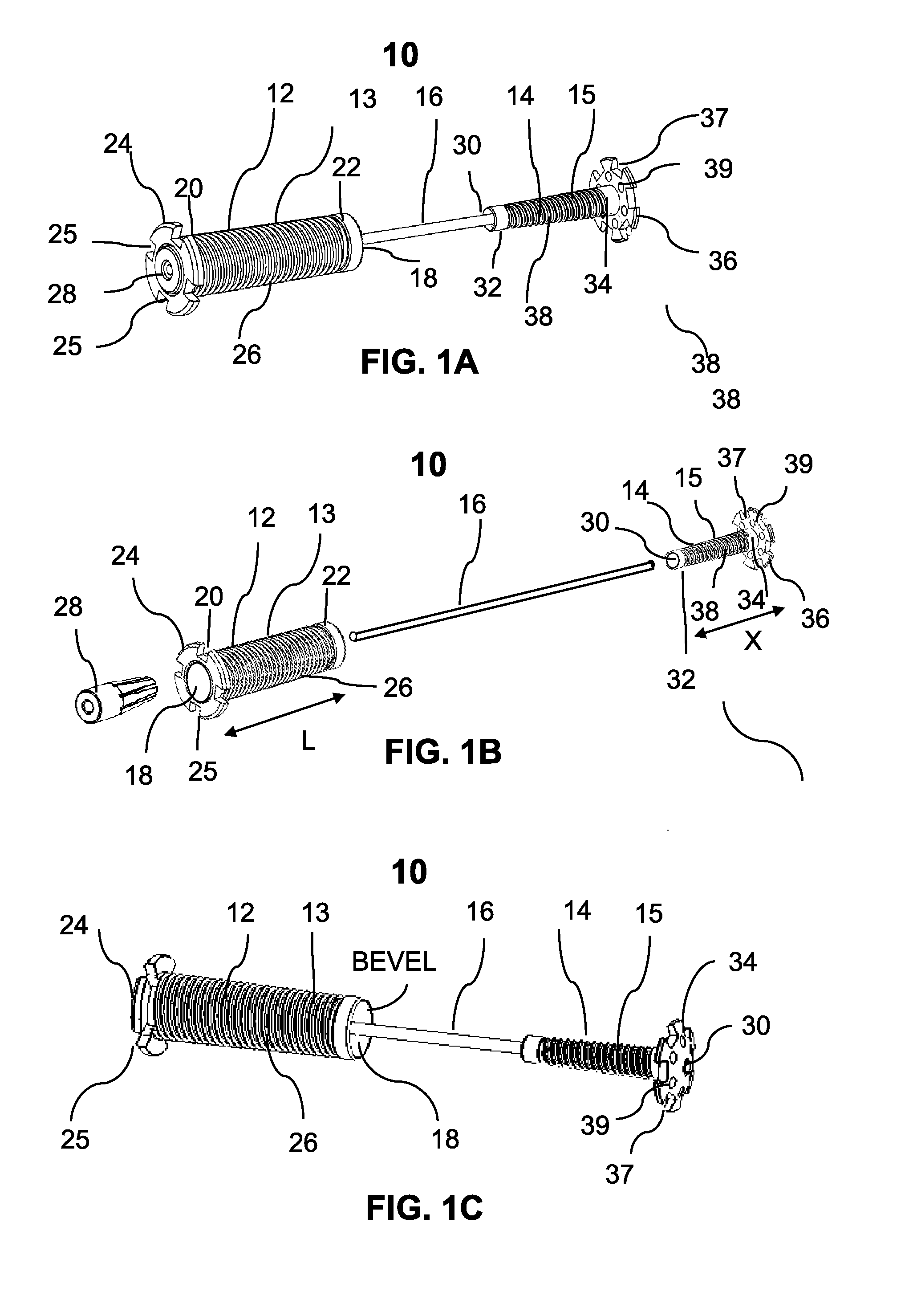
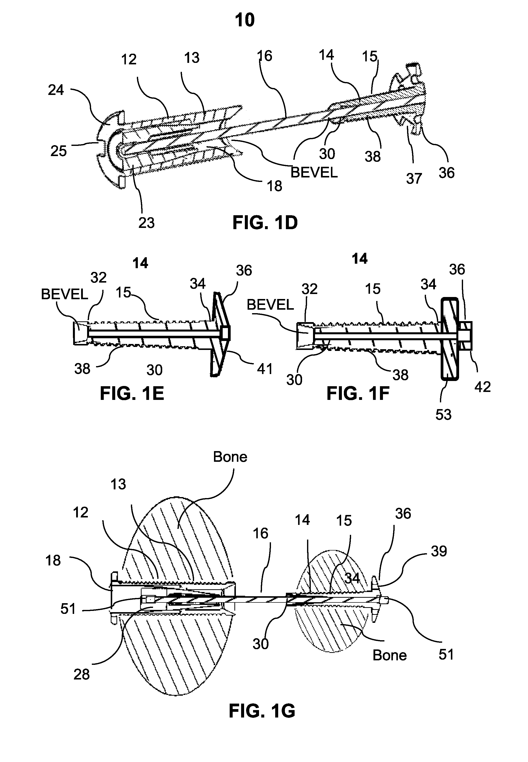
Devices and methods for bone anchoring

a bone anchoring and device technology, applied in the field of bone anchoring system, can solve the problems of limiting weight bearing activity, accompanied by pain and discomfort, and non-operative treatment may alleviate symptoms, and not correct the deformity of the big to

Inactive Publication Date: 2016-02-11
CYCLA ORTHOPEDICS
View PDF4 Cites 27 Cited by
  • Summary
  • Abstract
  • Description
  • Claims
  • Application Information

AI Technical Summary

Benefits of technology

The present invention is a bone anchoring system that can connect adjacent bones to treat bone fractures and deformities such as hallux valgus. It has overcome the shortcomings of existing configurations. The technical effects of this invention are its ability to overcome the limitations of existing bone anchoring systems and provide a more effective and reliable solution for spinal and joint fixations.

Problems solved by technology

Non-operative treatment may alleviate symptoms but does not correct the deformity of the big toe.
The corrective osteotomy of the first metatarsal is followed by a long recovery which limits weight bearing activity and in many cases is accompanied by pain and discomfort.

Method used


Image

  • Devices and methods for bone anchoring
  • Devices and methods for bone anchoring
  • Devices and methods for bone anchoring

Examples


example 1

Cadaver Study

[0136]A study was conducted in order to evaluate the transverse inter-metatarsal forces between first and second metatarsals after reduction of inter-metatarsal angle (IMA) in normal foot and in case of hallux valgus deformity.

[0137]Four fresh frozen cadaver feet (one with Hallux valgus) were used in the study. The device of the present invention was implanted in all four cadaver feet. The device was positioned between 1st and 2nd metatarsals at mid-shaft and the IMA was reduced using the dedicated wire tensioning device of the present invention. The tool includes a force indicator that shows the transverse load between the two metatarsals.

[0138]Each of the four feet was tensioned gradually reducing the IMA. Force was recorded and X-Rays were obtained (FIGS. 9a-c). Three cadaver feet were also loaded at 15° tilt under body weight and inter metatarsal force under load was recorded.

[0139]Results

[0140]Three of the cadaver feet exhibited a normal IMA (less than 10 Degrees) ...

example 2

In Vivo Human Study

[0143]A clinical study was conducted in order to evaluate the efficacy and safety of the present device in human subjects. The feet of five female patients ages 22-67 having moderate Hallux Valgus were implanted with the present device in order to realign the first metatarsal to a normal position.

[0144]The device of the present invention was implanted as described herein and the force indicator of the tensioning device was used to measure the transverse load between the two metatarsals. The force was recorded and an X-Ray was taken (FIGS. 10a-b) without loading the foot.

[0145]Results

[0146]The average pre-op Inter Metatarsal Angle (IMA) was 14.60 (STD 0.80) and the average reduction was by 8 degree to a final 6.60 degrees (STD 0.630). The device was positioned at different distal distances from the cuneiform joint of the first metatarsal at an average distance of 35.4% (STD 5.3%) of the first metatarsal length measured at base of bone (Cuneiform joint). The average...


PUM


Abstract

A device for fixation of bone tissue and methods of using same are provided. The device includes a first anchor positionable within a first bone region, a second anchor positionable within a second bone region and a wire interconnecting the first and second anchors. The wire is attached to the first anchor via an elastically deformable element having a force constant (K) of 20-80 N / mm along a longitudinal axis of the first anchor.

Description

FIELD AND BACKGROUND OF THE INVENTION[0001]The present invention relates to a bone anchoring system and methods of using same, and more particularly, to an anchor system which can be used to interconnect adjacent bones, such as metatarsal bones. Embodiments of the present invention relate to treatment of bone deformation disorders such as metatarsal bones and hallux valgus or repair of bone fractures such as Lisfranc.[0002]Deformity of skeletal bones can affect posture, locomotion and the quality of life to of active individuals. Such deformity can be caused by traumatic injury or a creeping deformity.[0003]Hallux valgus deformity is the most common forefoot disorder, with an estimated age related prevalence of 10 to 35%. Hallux valgus is characterized by outward deviation of the first metatarsal bone which leads to valgus deformity of the big toe (phalange). This deviation changes the biomechanics of the foot and may cause subluxation of the first metatarsophalangeal joint (MTP joi...

Claims


Application Information

Patent Timeline
IPC IPC(8): A61B17/68A61B17/84A61B17/86A61B17/88
CPCA61B17/683A61B17/848A61B17/8869A61B2017/681A61B17/686A61B17/864A61B17/685A61B17/8863A61B2017/565
Inventor HERZOG, RAFIVIKINSKY, OFER
Owner CYCLA ORTHOPEDICS
Learn More