Multilayer fluorescent nanoparticles and methods of making and using same
a technology of fluorescent nanoparticles and nanoparticles, which is applied in the field of multi-layer fluorescently responsive materials containing silica nanoparticles, can solve the problems of difficult large-scale analysis methodologies, difficult to apply multiplexed in-vivo or intracellular bioimaging, and high equipment set-up and reagent costs, so as to achieve maximum brightness, fluorescence brightness enhancement, and the effect of reducing energy transfer
- Summary
- Abstract
- Description
- Claims
- Application Information
AI Technical Summary
Benefits of technology
Problems solved by technology
Method used
Image
Examples
example 1
[0067]The following is an example describing synthesis and use of multilayer, FRM-containing nanoparticles of the present disclosure.
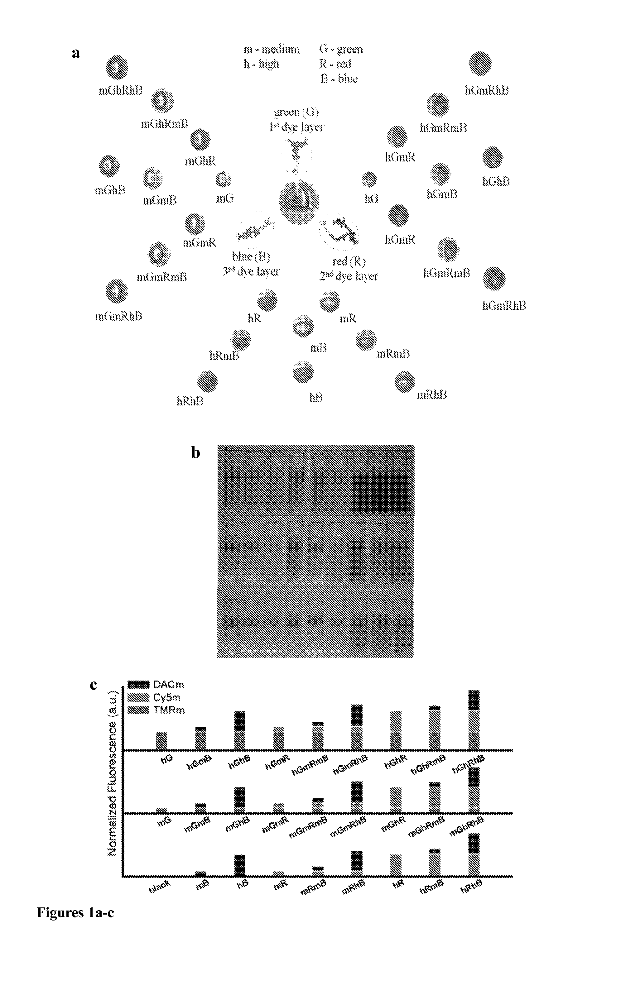
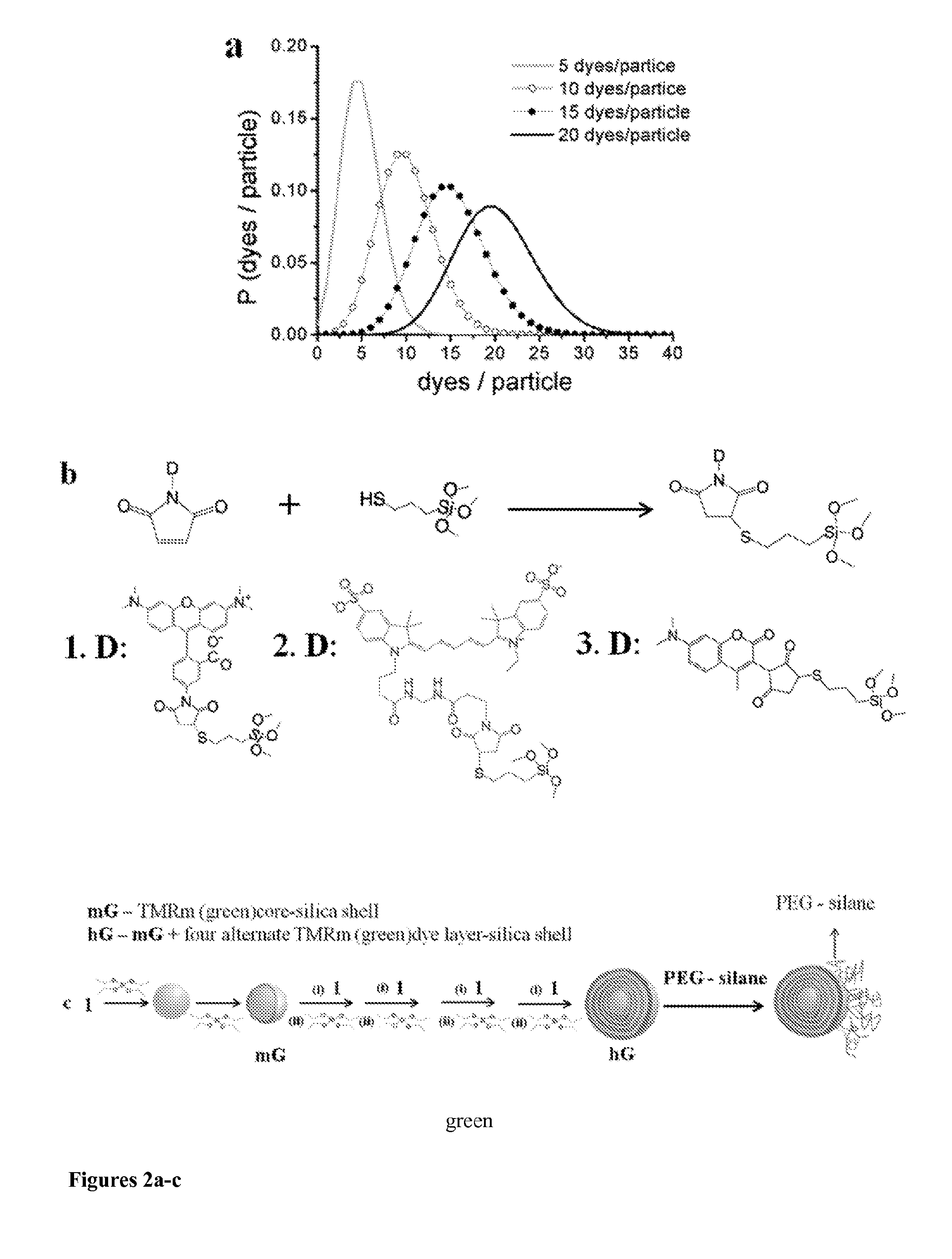
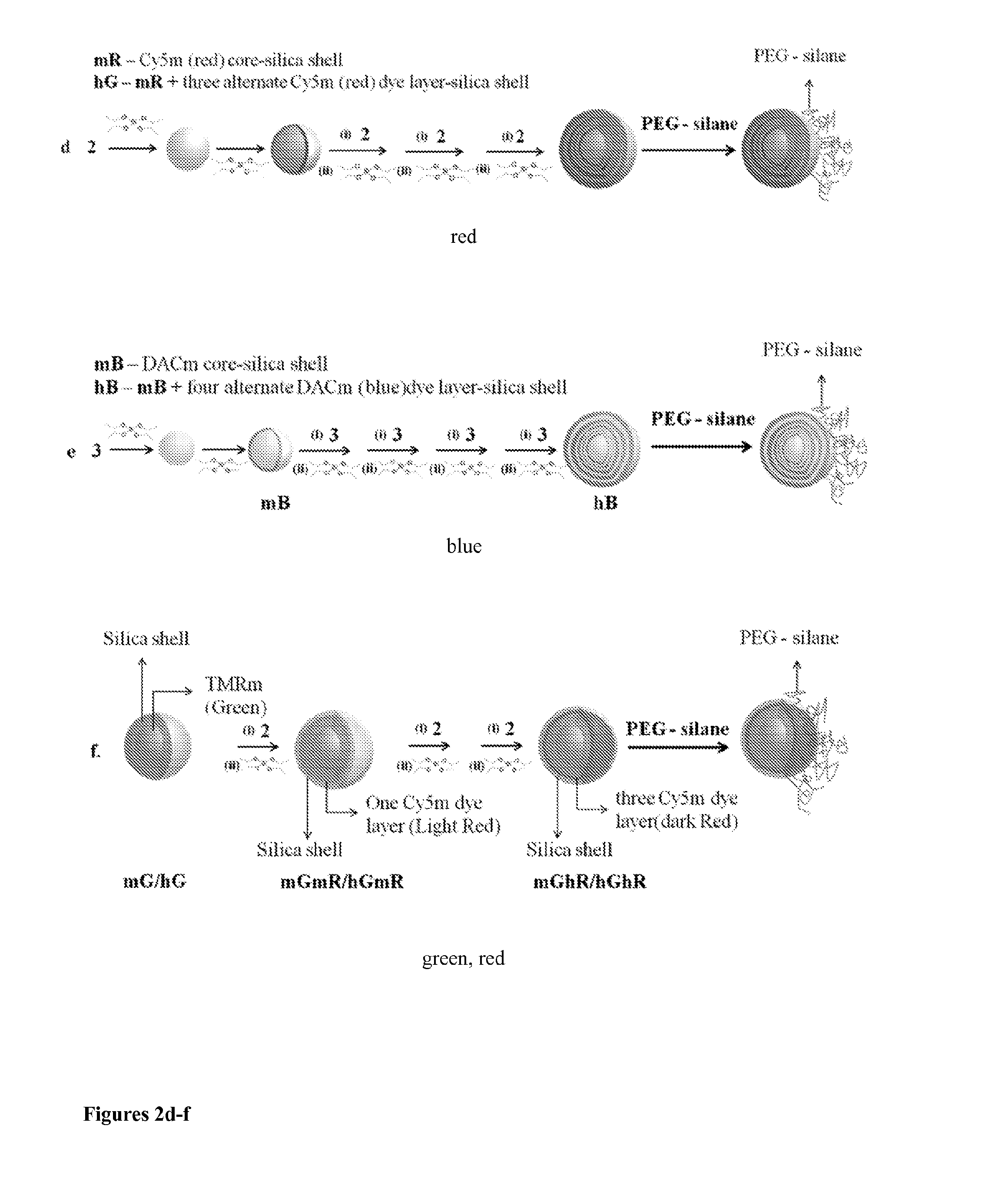
[0068]In this example, bright and optically encoded fluorescent core-shell silica nanoparticles were produced. The nanoparticles are referred to as multicolor Cornell dots or simply mcC dots and have sizes below 100 nm, which makes the nanoparticles desirable for high throughput screening and applying them to intracellular bioimaging using fluorescence multiplexing. These nanoparticles are encoded with three spectrally distinct organic fluorophores, i.e., N-(7-dimethylamino-4-methylcoumarin-3-yl)maleimide (DACm, λabs=395 nm, λem=450 nm), tetramethylrhodamine-5-maleimide (TMRm, λabs=540 nm, λem=570 nm) and Cy5-maleimide (Cy5m, λabs=640 nm, λem=670 nm), with three precisely controlled numbers of dyes, i.e. 0, 5 and 20, respectively, per particle, resulting in 26 optically distinguishable nanoparticles as shown in FIG. 1. The dyes were chosen based on com...
example 2
[0124]The following is an example describing use of the multilayer, FRM-containing nanoparticles of the present disclosure in imaging methods.
[0125]Sample Preparation. In these experiments, each of 26 Rat Basophilic Leukemia (RBL) cell samples were mechanically transfected with a single type of particle via square-wave electroporation. Cells were membrane-labeled using an Alexa Fluor® 488 and cholera toxin conjugate. This fourth spectrally distinct fluorescent dye was chosen so that the fluorescent cell membrane could be distinguished from the mc C Dots inside the cells. Single-particle samples were plated directly on glass microscope slides and fixed before imaging. Mixing single-particle cell samples before plating enabled us to create multi-particle samples having only a single type of mc C Dot per cell.
[0126]Imaging & Analysis.
[0127]Cells were imaged using a Zeiss 710 confocal scanning laser microscope, enabling simultaneous detection of the Cy5, TMR, DAC, and Alexa Fluor® 488 d...
PUM
| Property | Measurement | Unit |
|---|---|---|
| thickness | aaaaa | aaaaa |
| sizes | aaaaa | aaaaa |
| sizes | aaaaa | aaaaa |
Abstract
Description
Claims
Application Information
 - R&D
- Intellectual Property
- Life Sciences
- Materials
- Tech Scout
- Unparalleled Data Quality
- Higher Quality Content
- 60% Fewer Hallucinations
Browse by: Latest US Patents, China's latest patents, Technical Efficacy Thesaurus, Application Domain, Technology Topic, Popular Technical Reports.
© 2025 PatSnap. All rights reserved.Legal|Privacy policy|Modern Slavery Act Transparency Statement|Sitemap|About US| Contact US: help@patsnap.com


