Radiographic image detector

a detector and radiographic technology, applied in the field of radiographic image detectors, can solve the problems of image detectors that cannot detect x-ray images, serve imaging, and detection elements disposed outside the imaging area may be out of the irradiation rang

Active Publication Date: 2015-10-22
FUJIFILM CORP
View PDF1 Cites 6 Cited by
  • Summary
  • Abstract
  • Description
  • Claims
  • Application Information

AI Technical Summary

Benefits of technology

The present invention relates to a radiographic image detector that can detect the start of radiation of radioactive rays from a remote radiation source without any signal from the radiation source or other external devices, while utilizing radiated rays fully for imaging. The detector detects the start of radiation of the radioactive rays based on charge leaks from the pixels while the switching elements of the pixels are OFF. However, in this detector, the charge accumulating operation for accumulating the signal charges in the pixels is started after the start of radiation is detected. This ensures that the detector can detect the start of radiation of the radioactive rays without wasting the rays and without the need for external devices.

Problems solved by technology

However, the x-ray image detector may be equipped with such interfaces that are incompatible to interface standards (standards of cables and connectors, format of the synchronizing signal, etc.) of the control device for the x-ray source of the radiography system, for example, because of different manufacturers.
The detection method suggested in United States Patent Application Publication No. 2002 / 101527 (corresponding to Japanese Patent Laid-Open Publication No. 2002-181942), however, has a problem that the detection element disposed outside the imaging area may be out of an irradiation range by the x-rays when it is confined within the imaging area.
As a result, the image detector cannot detect any x-ray image while the test subject is exposed to the x-rays wastefully.
However, because the signal charges accumulated upon the x-ray radiation must be read out from the pixels in order to detect the start of x-ray radiation, the x-rays radiated in a first stage of a radiation time will be used merely for detecting the start of radiation and will not be served for imaging.
This is a waste of x-rays.

Method used


Image

  • Radiographic image detector
  • Radiographic image detector
  • Radiographic image detector

Examples


first embodiment

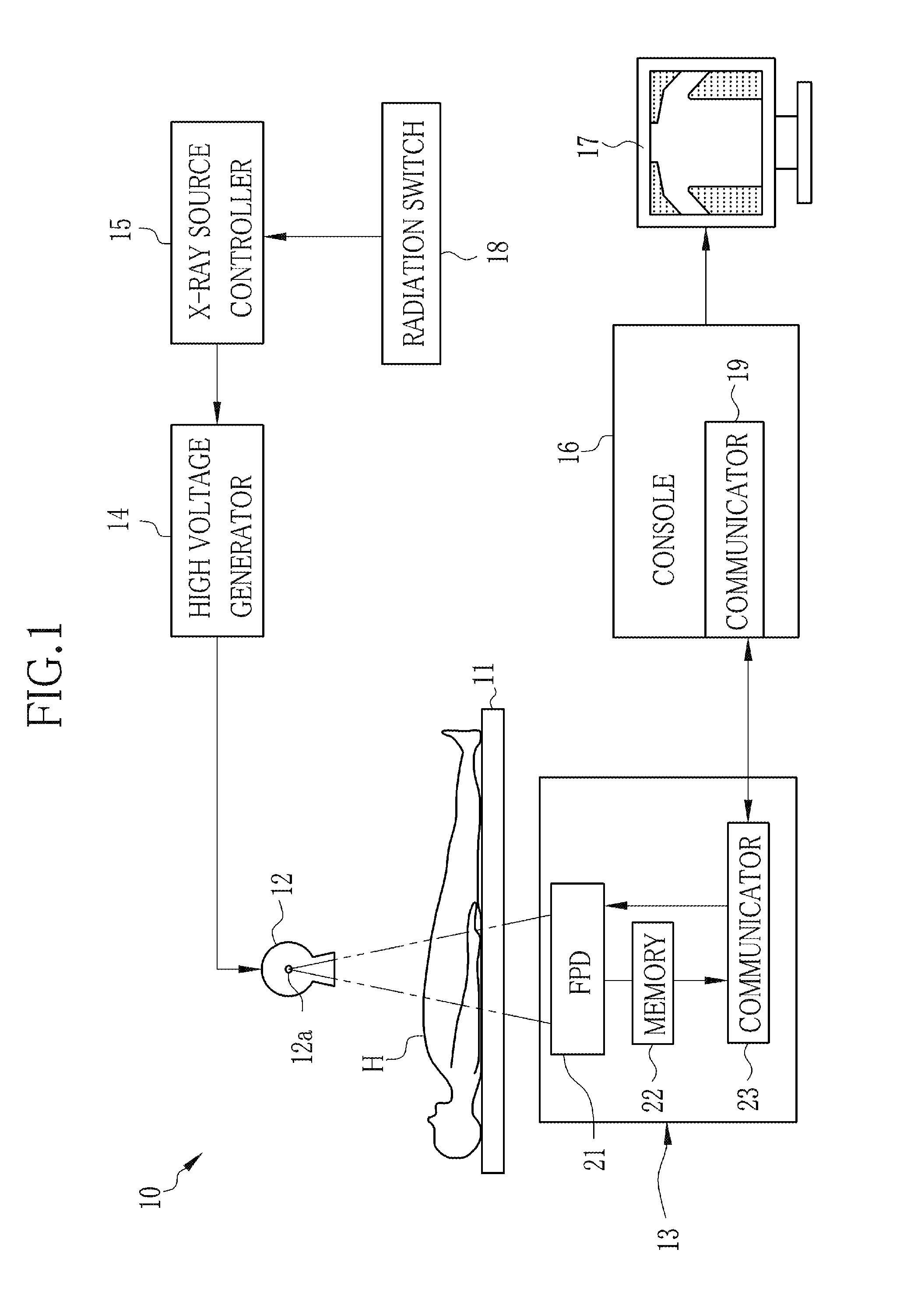
[0040]In FIG. 1, an X-ray radiography system 10 includes a radiological examination table having a table top 11 for laying a test subject H thereon, an X-ray source 12 for radiating X-rays from an X-ray focus 12a toward the subject H, and a radiographic image detector 13 for detecting an X-ray image of the test subject H from the X-rays that have penetrated through the test subject H. The radiographic image detector 13 may be formed as an electronic cassette 13 that is removably attached to the radiological examination table. The x-ray light source 12 consists of an X-ray tube and a collimator for limiting the radiation field of the X-rays from the X-ray tube.

[0041]The X-ray radiography system 10 also includes a high voltage generator 14, an X-ray source controller 15, a console 16, and a monitor 17. The x-ray source controller 15 is fed with various imaging conditions such as tube voltage, tube current and radiation time, which may be input through a not-shown operation panel or th...

second embodiment

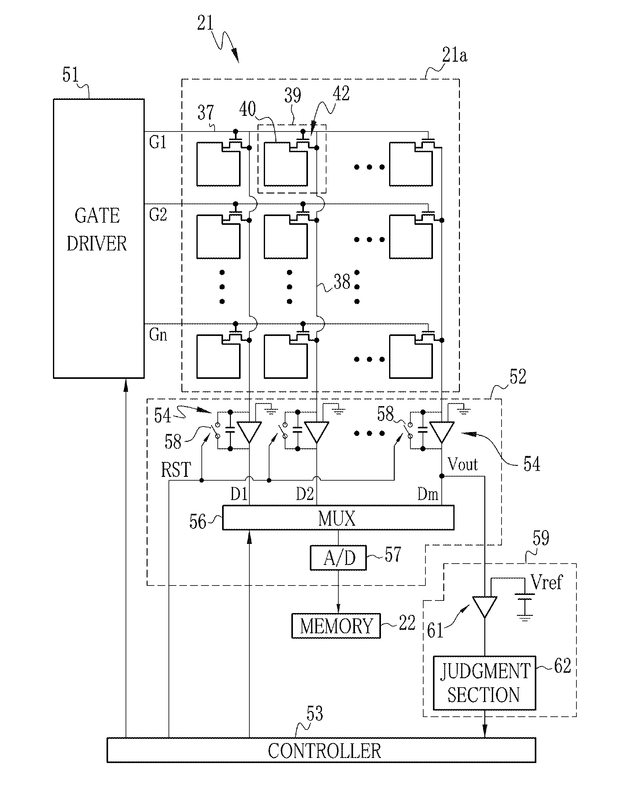
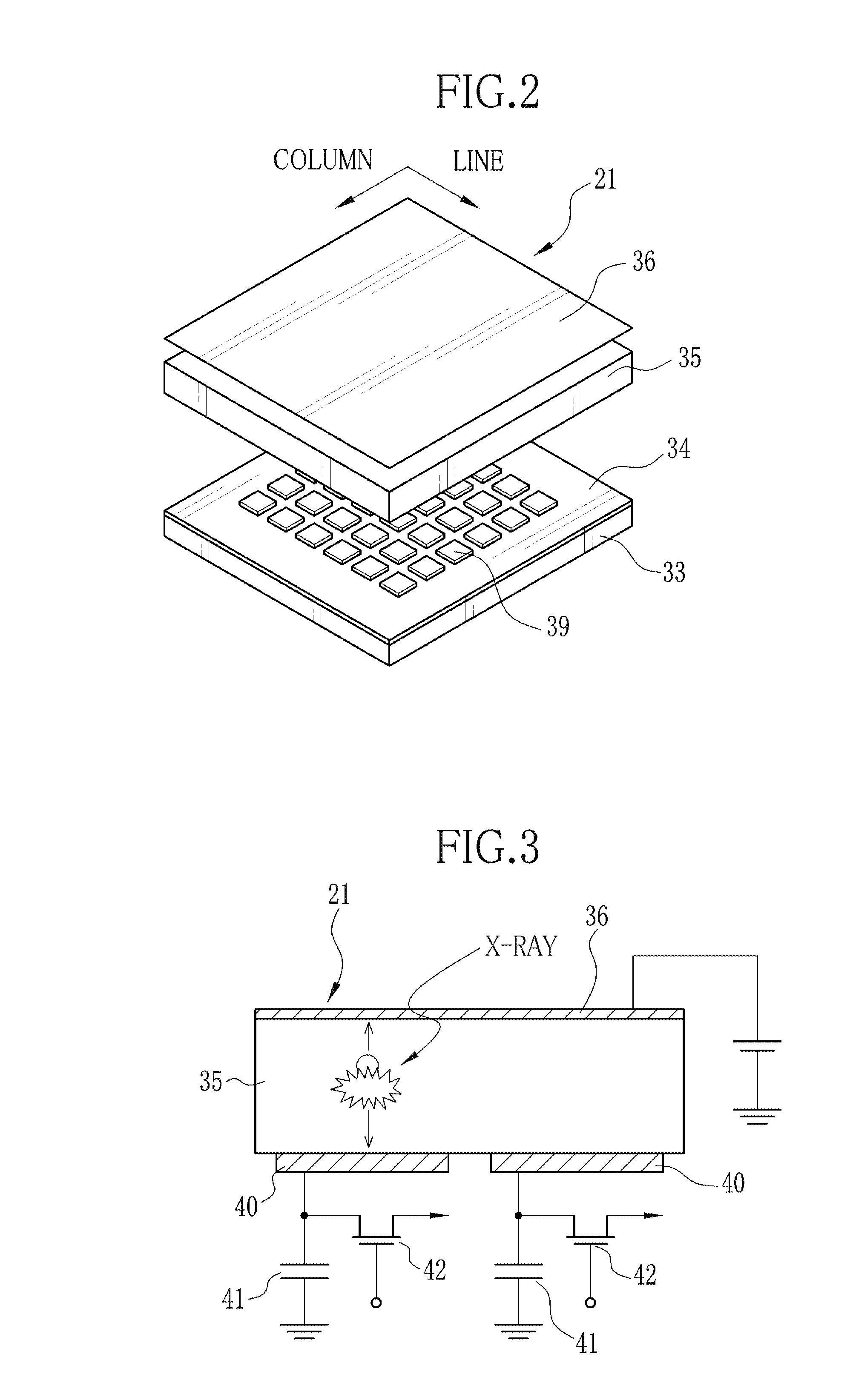
[0087]Meanwhile the console 16 processes the detected x-ray image data to eliminate such noises that result from the dark charges. Specifically, image data detected while no x-ray is radiated, hereinafter referred to as off-set image data, are previously memorized and subtracted from the detected x-ray image data, to eliminate the noises. The off-set image data may for example be such image data that represent the noises resulted from dark charges accumulated with the signal charges in the maximum radiation time. Accordingly, those dark charges accumulated from the start-of-radiation detecting operation to the start of x-ray radiation will not be reflected on the off-set image data. As a result, if the time from the start-of-radiation detecting operation to the start of x-ray radiation is too long, the noises caused by the dark charges cannot completely be eliminated using the off-set image data. To solve this problem, it is preferable to reset the pixels 39 to cancel the dark charg...

third embodiment

[0105]In the above first and second embodiments, the radiation detecting section 59 detects the start of x-ray radiation. In the following third embodiment, the end of x-ray radiation may be detected.

[0106]As shown in FIG. 10, in order to detect the end of x-ray radiation, the controller 53 inputs the reset pulses RST at constant intervals after the cassette 13 proceeds from the standby operation to the radiation detecting operation, like in the first or second embodiment, to reset the integrating amplifiers 54. When the radiation detecting section 59 detects the start of x-ray radiation (the time TA), the controller 53 however stops inputting the reset pulse RST, as indicated by arrows Pc1 and Pc2, so as not to withdraw the leak charges from the integrating amplifiers 54.

[0107]Since the reset pulse RST is not applied after the start of x-ray radiation is detected (the time TA), the output voltages Vout of the integrating amplifiers 54 continue to increase mainly because of the leak...


PUM


Abstract

A flat panel detector (FPD) includes an imaging panel having pixels arranged in a matrix, a gate driver for turning thin film transistors (TFTs) of the pixels ON and OFF, a radiation detecting section for detecting the start of x-ray radiation from the x-ray source, and a controller. The controller controls the gate driver to turn the TFTs ON periodically to reset dark charges of the pixels. Before starting a charge accumulating operation for accumulating signal charges for imaging, the controller controls the gate driver to turn the TFTs OFF so that the radiation detecting section may detect the start of x-ray radiation on the basis of charge leaks from the pixels. When the start of x-ray radiation is detected, the controller starts the charge accumulating operation while keeping the TFTs in the OFF condition. Thereafter, the TFTs are turned ON to read out the accumulated signal charges.

Description

CROSS REFERENCE TO RELATED APPLICATIONS[0001]This is a Divisional of U.S. application Ser. No. 13 / 239,741 filed Sep. 22, 2011, which claims benefit of Japanese Patent Application No. 2010-216754 filed Sep. 28, 2010. The above-noted applications are incorporated herein by reference in their entirety.BACKGROUND OF THE INVENTION[0002]1. Field of the Invention[0003]The present invention relates to a radiographic image detector that detects an image of a subject from radioactive rays penetrating through the subject.[0004]2. Description of the Related Art[0005]In the medical field, radiography using radioactive rays like x-rays has been well known as a diagnostic method. An x-ray radiography system generally consists of an x-ray source for radiating x-rays, and an x-ray image detector for detecting an x-ray image of a subject from x-rays penetrating through the subject. Among those used practice, there is an x-ray image detector that adopts a flat panel detector (FPD) having a TFT (thin f...

Claims


Application Information

Patent Timeline
Patent Type & Authority Applications(United States)
IPC IPC(8): G01T1/24H04N5/32
CPCH04N5/32G01T1/247G01T1/243H04N23/30H04N25/30
Inventor WATANABE, KEITA
Owner FUJIFILM CORP
Why Eureka
Try Eureka