
Realtime imaging and radiotherapy of microscopic disease

a radiotherapy and microscopic disease technology, applied in the field of real-time imaging and radiotherapy of microscopic disease, can solve the problems of more normal tissue at risk of collateral damage, difficult to detect and target for treatment, and difficult to achieve the effect of improving the prognosis, facilitating treatment, and reducing the risk of missing the targ

Inactive Publication Date: 2015-09-17
RGT UNIV OF CALIFORNIA
View PDF15 Cites 2 Cited by
  • Summary
  • Abstract
  • Description
  • Claims
  • Application Information

AI Technical Summary

Benefits of technology

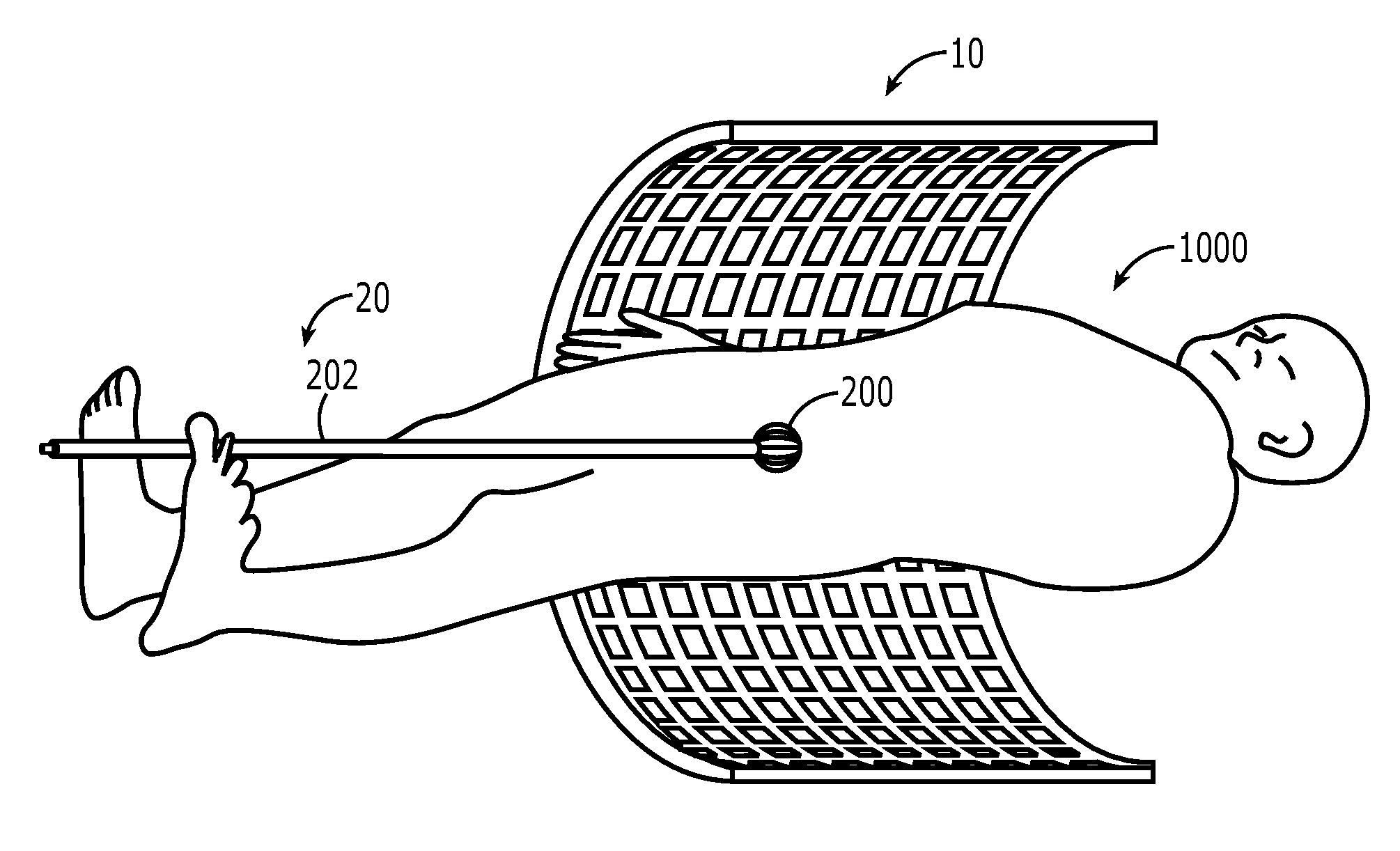
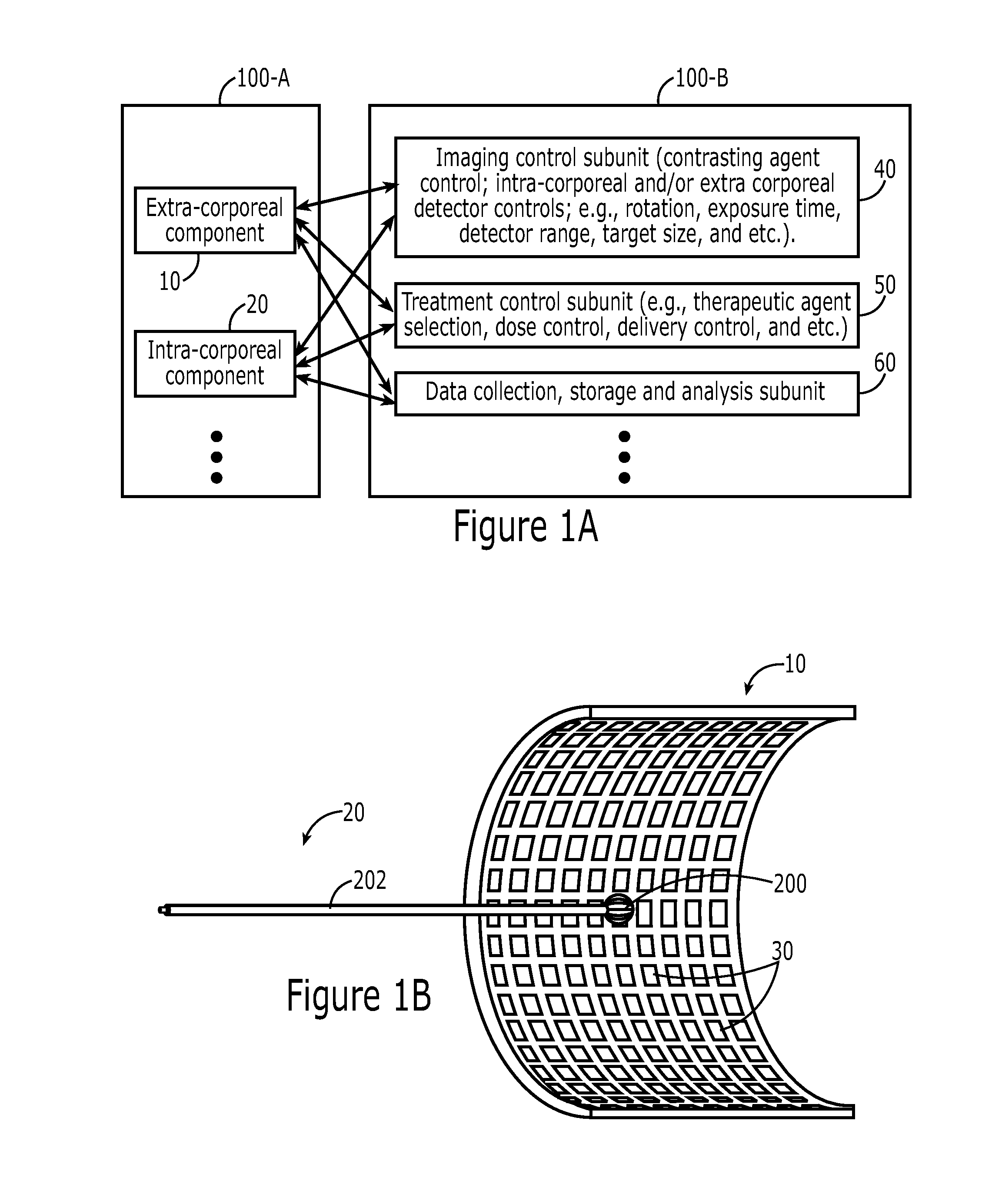
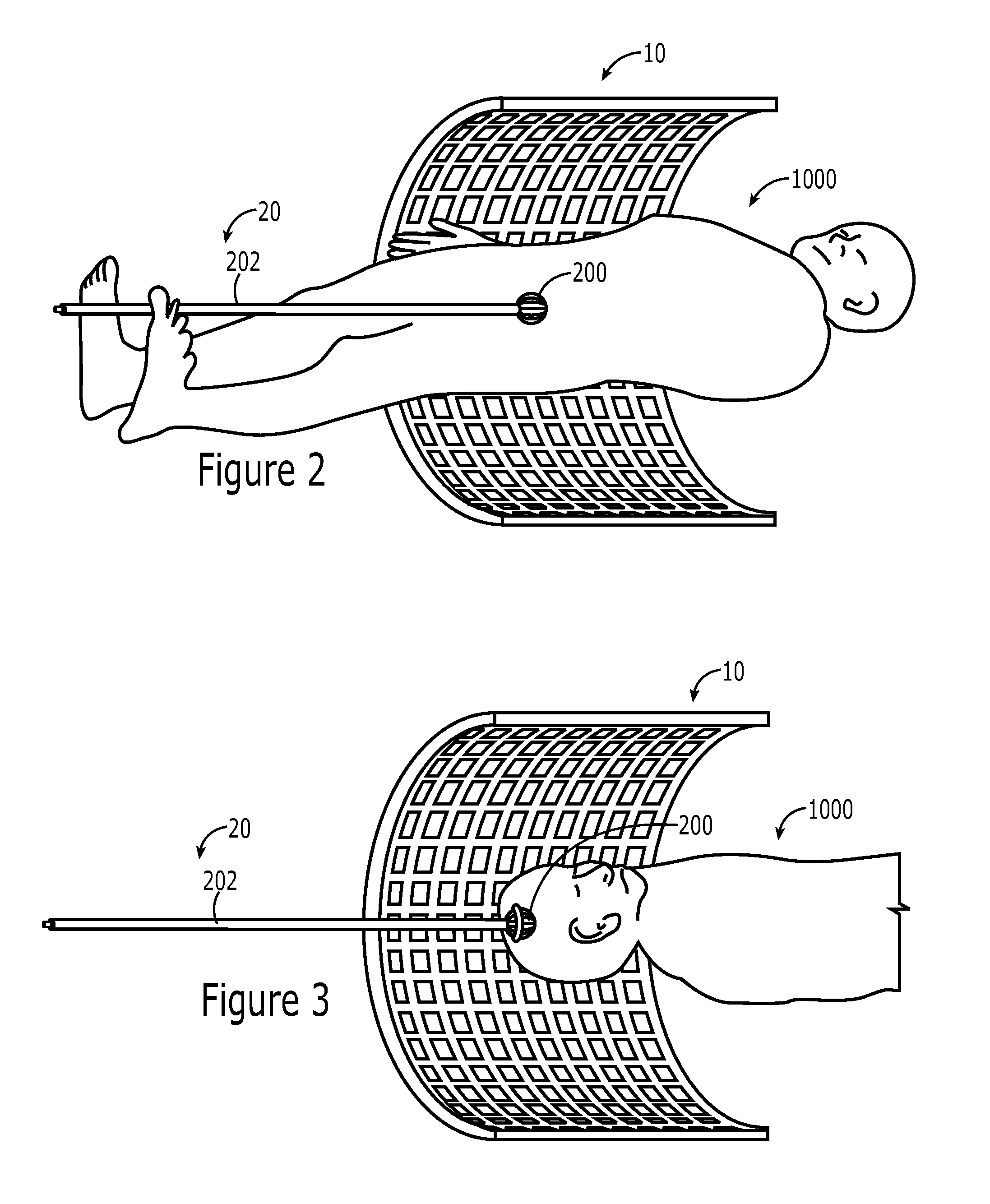
The patent text describes a system and method for detecting and treating small tumors in real-time imaging. The system includes an intra-corporeal component with a detector and a radiation source, and an extra-corporeal component with a detector that always faces the first detector. The system can be used to detect and treat tumors in various parts of the body, such as the brain, breast, or endometrium. The method involves administering a contrast agent that is taken up by tumor cells, placing the intra-corporeal component in a cavity within the body, rotating the detector and radiation source, and collecting images from different parts of the cavity to create a new image. The technical effects of this system and method include improved accuracy in detecting and targeting tumors, reduced risk of collateral damage to normal tissue, and better screening for early-stage tumors.

Problems solved by technology

On the other hand, the smaller the tumor is, the more difficult it is to detect and target for treatment.
Additionally, the smaller the tumor is, the more normal tissue is at risk from collateral damage especially if treatment accuracy is compromised by imprecise targeting.
And finally, the greater the time interval and the number of procedural steps between localization and treatment, the greater is the risk of missing the target.

Method used


Image

  • Realtime imaging and radiotherapy of microscopic disease
  • Realtime imaging and radiotherapy of microscopic disease
  • Realtime imaging and radiotherapy of microscopic disease

Examples


example 1

Miniature X-Ray Tube Characteristics

[0090]Figure A displays the photon spectrum in 5 cm of air, 1.5 cm of water, and 5 cm of water from the source operated at 50 kVp; note the substantial hardening of the beam with distance and bolus. The depth dose characteristics are illustrated in figure B for the source operated at 20 kVP and at 50 kVp with or without bolus. These data suggest that via collimation, filtration, and / or energy modulation, treatment conformation to the desired volume is possible.

example 2

Relative Biological Effectiveness (RBE) of the Source

[0091]We have measured preliminary RBEs of the x-rays from the source using U87MG human glioblastoma multiforme (GBM) and MCF7 human breast cancer cells in a standard clonogenic survival assay method. Compared to cobalt-60 irradiation for 10% survival, the RBEs are 1.3 and 1.8, respectively. (FIG. 9D) Even with a highly radioresistant GBM cell line, we show an RBE value greater than 1. The high surface dose deposition and >1 RBE of orthovoltage x-rays has always been a hindrance for EBRT of non-superficial tumors, but they become a clear advantage for the Axxent system in managing disease interstitially, intracavitarily, and intraoperatively. Sparing of normal tissues can be achieved by the rapid depth-dose fall-off (FIGS. 9B & 9C). Use of iodine for dose enhancement in the tumor and dose reduction in the normal tissue should further improve the therapeutic ratio; this study is still currently in progress.

[0092]Characterization da...

example 3

A Pilot Ambi-Cranial PET System for GBM Surgery Guidance: Characterization and Analysis

I. Introduction

[0093]Gliobastoma multiforme (GBM) is the most common and most aggressive malignant primary brain tumor in humans. GBMs are distinguished by extensive and diffuse infiltration of tumor cells into the dense network of interwoven neuronal and glial processes rendering these tumors extremely difficult to excise without large concomitant areas of normal brain.

[0094]To best identify and excise tumor during the surgical operation, many image-guided systems have been introduced, such as a stereotactic navigation system (SNS) that combines a microscope with a tracking system and the use of pre-operative MRI images to relay the 3D location of the scalpel relative to the tumor and brain structures in real-time. However, both systems suffer from brain-shift during and following removal of the gross tumor mass. Intraoperative MRI systems are costly and require MRI-compatible surgical instrument...


PUM


Abstract

Disclosed herein are methods, systems and therapeutics concerning radiotherapy of microscopic disease based on realtime imaging. The system comprises an intra-corporeal component and an extra-corporeal component. The intra-corporeal component comprises a detector / imaging subunit and a treatment subunit, where both subunits are placed in a cavity of a patient. The extra-corporeal component comprises a detector that is placed outside any cavity of the patient. Through this system, a treatment can be applied to a target tissue in a patient concurrently or within a short period time to signal detections.

Description

CROSS REFERENCE TO RELATED APPLICATION[0001]The application claims priority to U.S. provisional application No. 61 / 707,879 filed on Sep. 28, 2012 and also entitled “Realtime Imaging and Radiotherapy of Microscopic disease,” which is hereby incorporated by reference herein in its entirety.FIELD OF THE INVENTION[0002]The invention disclosed herein generally relates to methods, systems and therapeutics concerning radiotherapy of microscopic disease based on realtime imaging.BACKGROUND[0003]Devices and methods for imaging sub-millimeter-sized tumors that are embedded in tissues (e.g., at depths greater than 1-2 mm) are not available. Consequently, methods for treating such tumors are also lacking due to the inability in combining high specific and sensitive imaging with highly conformal radiation.[0004]What is needed are systems, methods and therapeutics that can overcome the deficiencies in the art.SUMMARY OF THE INVENTION[0005]In oncology, the sequence of locating the tumor and then t...

Claims


Application Information

Patent Timeline
Patent Type & Authority Applications(United States)
IPC IPC(8): A61B6/03A61N5/10A61M31/00A61B6/00
CPCA61B6/037A61B6/4057A61B6/4258A61B6/4266A61N2005/1098A61B6/4488A61B6/5217A61N5/1001A61M31/005A61B6/4452A61B6/481A61B6/50A61N5/1014A61N5/1015A61N5/1016A61N2005/1022A61N2005/1052
Inventor IWAMOTO, KEISUKE S.DAHLBOM, MAGNUSDEMARCO, JOHN J.RUAN, DAN
Owner RGT UNIV OF CALIFORNIA
Learn More