Eureka AIR delivers breakthrough ideas for toughest innovation challenges, trusted by R&D personnel around the world.

Method and apparatus for magnetic resonance imaging

a magnetic resonance imaging and apparatus technology, applied in the field of magnetic resonance imaging, can solve the problems of high noise, patient noise protection, and high noise, and achieve the effect of quiet magnetic resonance sequen

Active Publication Date: 2015-09-10
SIEMENS HEALTHCARE GMBH
View PDF15 Cites 18 Cited by
  • Summary
  • Abstract
  • Description
  • Claims
  • Application Information

AI Technical Summary

Benefits of technology

The patent text describes a method to improve the signal-to-noise ratio of magnetic resonance sequences by reducing the bandwidth of the sequences. This is achieved by increasing the echo interval between the echoes of the sequence and reducing the slew rate of the gradient pulse activation in the readout direction. The method can be executed quickly and robustly, and is thus suitable for identical repeated use. The method steps are executed by a control computer with a main memory, graphics card, or logic unit. Electronic data carriers such as a DVD, magnetic tape, or USB stick can be used to store the control information.

Problems solved by technology

During the switch-over process there is certain distortion and / or expansion of the magnetic coil, so that loud noises arise and patients need noise protection.
In particular the gradient switchings of an echo-planar imaging magnetic resonance sequence, which is typically used in examinations of the head, but also in the abdominal region of the body, have a very high slew rate, so that the execution of the magnetic resonance sequence is typically very loud.
Furthermore, strong vibrations of the magnetic resonance apparatus can occur during the execution of a magnetic resonance sequence using echo-planar imaging.
Patients with claustrophobia or who are fundamentally uncomfortable with the magnetic resonance apparatus may in addition be alarmed by loud examinations and / or unknown noises, so that an examination can only be carried out by administering tranquilizers, or is refused outright.

Method used


Image

  • Method and apparatus for magnetic resonance imaging
  • Method and apparatus for magnetic resonance imaging
  • Method and apparatus for magnetic resonance imaging

Examples


Embodiment Construction

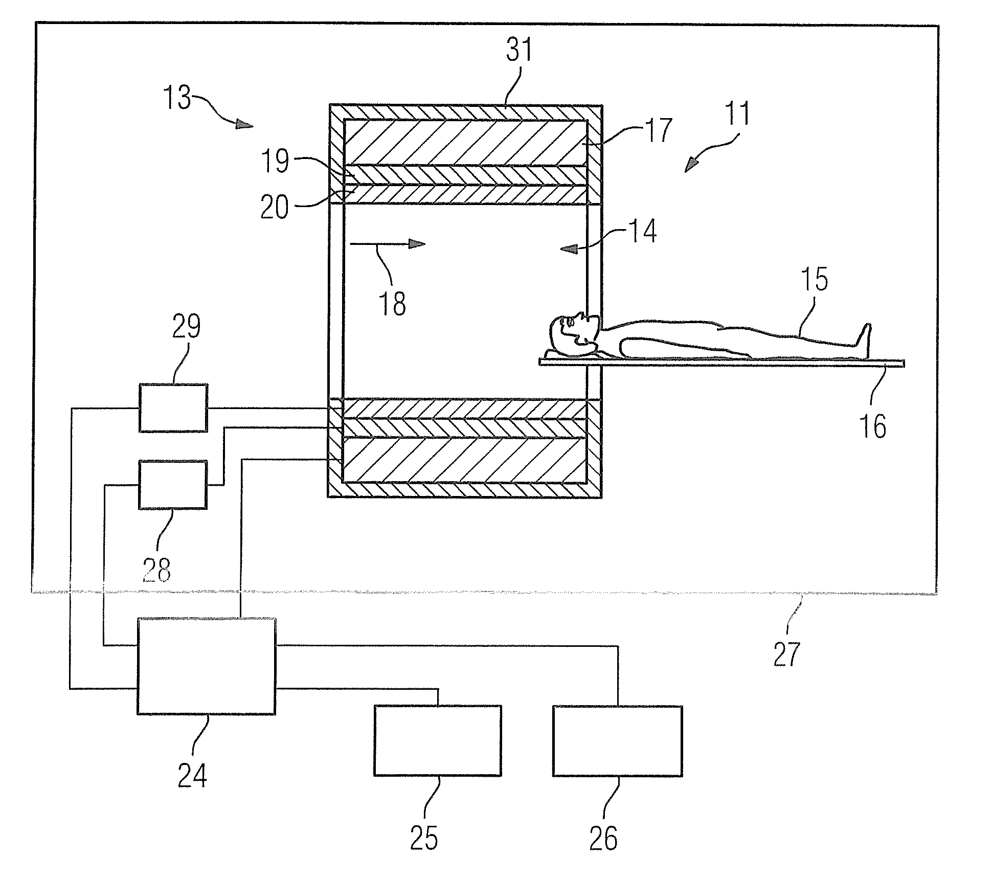
[0034]FIG. 1 schematically illustrates an inventive magnetic resonance apparatus 11. The magnetic resonance apparatus 11 has a data acquisition unit formed by a magnet unit 13, having a basic field magnet 17 for generating a strong and constant basic magnetic field 18. The magnetic resonance apparatus 11 additionally has a patient receiving zone 14 in the shape of a cylinder for receiving an examination object, in particular a patient 15, the patient receiving zone 14 being cylindrically surrounded by the magnet unit 13 in a circumferential direction. The patient 15 can be introduced into the patient receiving zone 14 by a patient positioning apparatus 16 of the magnetic resonance device 11. To this end the patient positioning apparatus 16 has a bed, which is disposed in a movable manner within the magnetic resonance device 11. The magnet unit 13 is shielded to the outside by a housing shell 31 of the magnetic resonance device.

[0035]The magnet unit 13 additionally has a gradient coi...


PUM


Abstract

In a method and apparatus for magnetic resonance imaging, a particularly quiet magnetic resonance sequence, uses echo-planar imaging with at least one gradient switching in a readout direction, wherein the at least one gradient switching in the readout direction has a slew rate that is less than a maximum slew rate defined by system specification parameters of the magnetic resonance apparatus.

Description

BACKGROUND OF THE INVENTION [0001]1. Field of the Invention[0002]The invention concerns a method for magnetic resonance imaging by operation of a magnetic resonance apparatus, as well a magnetic resonance apparatus and a non-transitory, computer-readable storage medium encoded with programming instructions to implement such a method.[0003]2. Description of the Prior Art[0004]In a magnetic resonance apparatus, also called a magnetic resonance tomography system, the body of a person to be examined, such as a patient, is normally exposed, with a basic field magnet, to a high main magnetic field, for example of 1.5 or 3 or 7 Tesla. In addition, gradient fields are activated by a gradient coil unit. Radio-frequency pulses, in particular excitation pulses, are then transmitted via a radio-frequency antenna unit using suitable antenna devices, that cause nuclear spins of particular atoms to be resonantly excited by these radio-frequency pulses are deflected by a defined flip angle relative...

Claims


Application Information

Patent Timeline
Patent Type & Authority Applications(United States)
IPC IPC(8): G01R33/561G01R33/30G01R33/48G01R33/54G01R33/385
CPCG01R33/5616G01R33/543G01R33/307G01R33/4818G01R33/3852G01R33/3854G01R33/5611
Inventor GRODZKI, DAVIDHEIDEMANN, ROBINOTT, MARTINPORTER, DAVID ANDREW
Owner SIEMENS HEALTHCARE GMBH
Why Eureka
Eureka Blog
Learn More