Eureka AIR delivers breakthrough ideas for toughest innovation challenges, trusted by R&D personnel around the world.

Eustachian tube dilation balloon with ventilation path

a technology of eustachian tubes and dilation balloons, which is applied in the direction of catheters, prostheses, therapy, etc., can solve the problems of mild hearing impairment or other ear symptoms, mucous membrane swelling, and edema, and achieve enhanced wetting of the mucosal surface and reduce edema

Active Publication Date: 2015-07-23
ACCLARENT INC
View PDF7 Cites 46 Cited by
  • Summary
  • Abstract
  • Description
  • Claims
  • Application Information

AI Technical Summary

Benefits of technology

The patent describes a medication and a device for treating a Eustachian tube. The medication aims to reduce swelling in the Eustachian tube region and is made of a surfactant that helps to wet the mucosal surface better. The device is designed to fit into the Eustachian tube and has a flexible insert with a therapeutic tool to treat the tube. The insert has a stiffness that allows it to push into the tube without getting bent or twisted. This makes the treatment more effective and easier.

Problems solved by technology

Anything that interferes with this periodic opening and closing of the Eustachian tube 26 may result in hearing impairment or other ear symptoms.
In adults, this is usually accompanied by some ear discomfort, a fullness or pressure feeling and may result in a mild hearing impairment and head noise (tinnitus).
Infection of these areas results in mucous membrane swelling which in turn may result in obstruction of the Eustachian tube 26.
In the presence of bacteria, this fluid may become infected, leading to an acute suppurative otitis media (infected or abscessed middle ear).
There may be recurrent ear pain, especially when the individual catches a cold.
The presence of fluid in the middle ear 14, however, makes it very susceptible to recurrent acute infections.
These recurrent infections may result in middle ear damage.
Finally, the fluid can become infected, which is painful and makes the patient feel ill and which may cause the patient not to be able to hear well.
However, even if antihistamines, decongestants and antibiotics are used to treat an infection or other cause of fluid build-up in the middle ear 14, these treatments will typically not immediately resolve the pain and discomfort caused by the buildup of fluid in the middle ear 14; i.e. the most immediate relief will be felt by the patient if the fluid can be removed from the Eustachian tube 26.
During the healing period, the patient can experience varying degrees of ear pressure, popping, clicking and fluctuation of hearing, occasionally with shooting pain in the ear.

Method used


Image

  • Eustachian tube dilation balloon with ventilation path
  • Eustachian tube dilation balloon with ventilation path
  • Eustachian tube dilation balloon with ventilation path

Examples


Embodiment Construction

[0174]The embodiments of the present invention are directed toward methods and systems for accessing, diagnosing and treating target tissue regions within the middle ear and the Eustachian tube.

[0175]Access

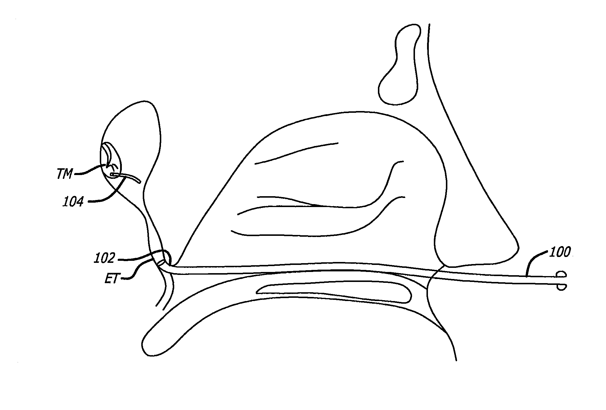
[0176]One embodiment of the present invention is directed toward using minimally invasive techniques to gain trans-Eustachian tube access to the middle ear. In one embodiment, a middle ear space may be accessed via a Eustachian tube (ET). To obtain this access to the Eustachian tube orifice, a guide catheter having a bend on its distal tip greater than about 30 degrees and less than about 90 degrees may be used. Once accessed, diagnostic or interventional devices may be introduced into the Eustachian tube. Optionally, to prevent damage to the delicate middle ear structures, a safety mechanism may be employed. In one embodiment, the safety mechanism may include a probe and / or a sensor introduced into the middle ear via the tympanic membrane as shown in FIG. 7. For example, the prob...


PUM


Abstract

An apparatus comprises a shaft, an expandable dilator, and at least one ventilation pathway. The shaft defines a longitudinal axis and comprises a distal and proximal ends with at least one shaft lumen. The expandable dilator comprises body with its own proximal and distal ends. The body is configured to transition between a contracted state and an expanded state. The body is configured to dilate a Eustachian tube of a patient in the expanded state. The at least one ventilation pathway is configured to provide ventilation from the distal end of the body to the proximal end of the body when the body is in the expanded state. In some examples, the ventilation pathway comprises a set of transversely oriented vent openings formed through the shaft. In some other examples, the ventilation pathway comprises a space defined between one or more radially outwardly protruding features of the expandable dilator.

Description

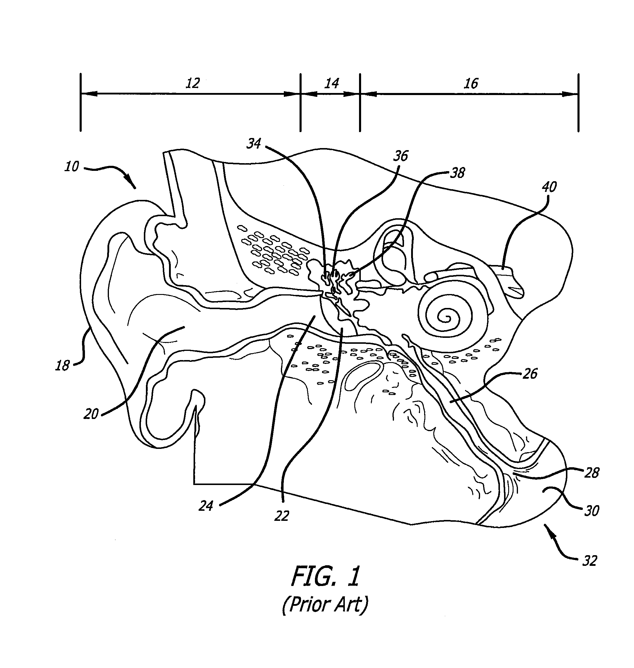
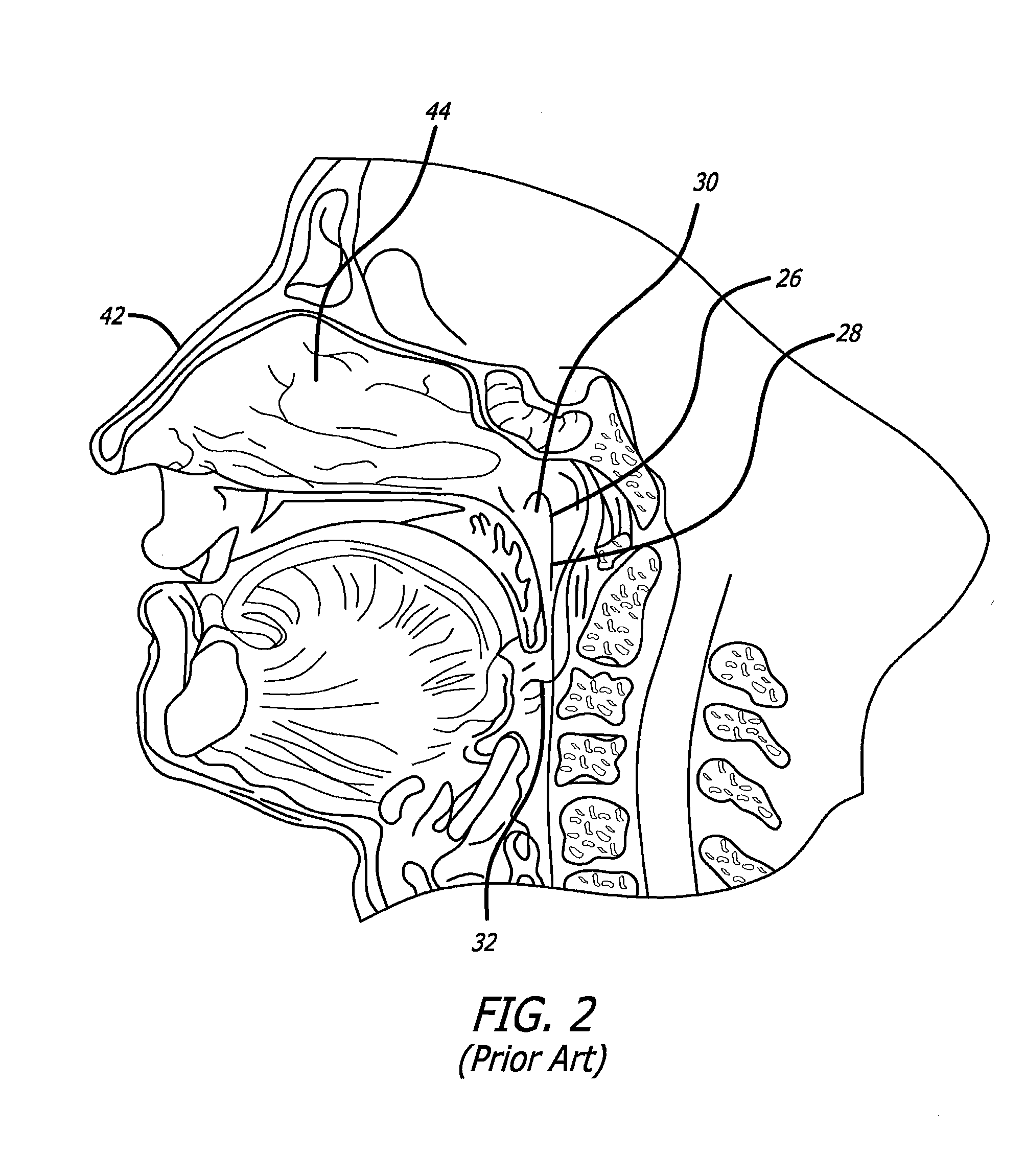
PRIORITY[0001]This application is a continuation-in-part of U.S. patent application Ser. No. 12 / 777,856, filed on May 11, 2010 (published on Oct. 28, 2010 as U.S. Pub. No. 2010 / 0274188), which is a continuation-in-part of U.S. patent application Ser. No. 12 / 649,078, filed on Dec. 29, 2009 (published on Aug. 5, 2010 as U.S. Pub. No. 2010 / 0198191), which is a continuation-in-part of U.S. patent application Ser. No. 12 / 340,226, filed on Dec. 19, 2008 (published on Jun. 25, 2009 as U.S. Pub. No. 2009 / 0163890), which claims the benefit of U.S. Provisional Pat. App. No. 61 / 015,647, filed on Dec. 20, 2007. All the disclosures of the above listed references are hereby incorporated by reference in their entirety for all purposes.BACKGROUND OF THE INVENTION[0002]The present invention is related to methods and systems for accessing, diagnosing and treating target tissue regions within the middle ear and the Eustachian tube.[0003]Referring to FIGS. 1-2, the ear 10 is divided into three parts: a...

Claims


Application Information

Patent Timeline
Patent Type & Authority Applications(United States)
IPC IPC(8): A61F11/00A61B1/07A61B17/00A61B5/00A61B17/12A61M25/09A61B1/233
CPCA61F11/002A61M25/09A61B1/07A61B1/233A61B5/6852A61B17/12104A61M2025/09183A61B17/00234A61F2250/0067A61M2025/09066A61B2017/00787A61B2017/00296A61B17/12131A61B1/227A61B8/12A61N7/02A61B5/036A61B5/411A61B5/415A61M25/0032A61M25/0041A61B18/20A61M25/0054A61M25/0068A61M25/007A61M25/01A61M25/0102A61M25/0105A61M25/0147A61M25/10A61M29/02A61M31/00A61M31/002A61N1/30A61M2025/0008A61M2025/0166A61M2025/09008A61M2025/105A61M2025/1075A61B2018/00327A61B2018/144A61M2210/0681A61F11/20A61F11/202
Inventor CAMPBELL, PETER F.HARFE, DANIEL T.HA, HUNG V.MUNI, KETAN P.NGUYEN, ANDREWLAM, SIVETTECHANG, JOHN Y.GOLDFARB, ERIC
Owner ACCLARENT INC
Why Eureka
Eureka Blog
Learn More