Apparatus and method for displaying ultrasound image

an ultrasound image and apparatus technology, applied in the field of methods and apparatus for displaying ultrasound images, can solve the problems of difficult to find out the overall stenosis degree of the blood vessel, difficult to recognize fiber plagues, and expensive and invasive imaging methods of coronary angiography

Inactive Publication Date: 2015-06-25
SAMSUNG MEDISON
View PDF6 Cites 5 Cited by
  • Summary
  • Abstract
  • Description
  • Claims
  • Application Information

AI Technical Summary

Benefits of technology

The invention provides a way to accurately measure the narrowing of a blood vessel by measuring the speed of blood flow and creating an ultrasound image that shows the degree of narrowing. This is helpful in identifying the location and degree of stenosis in a blood vessel.

Problems solved by technology

However, the coronary angiography is an expensive and invasive imaging method.
However, it is difficult to find out the overall stenosis degrees of the blood vessel based on the image of one cross-section of the blood vessel and diagnosis accuracy may vary from one user to another.
Furthermore, it is easy to recognize fat plagues in the B mode image, but it is difficult to recognize fiber plagues.

Method used


Image

  • Apparatus and method for displaying ultrasound image
  • Apparatus and method for displaying ultrasound image
  • Apparatus and method for displaying ultrasound image

Examples


Embodiment Construction

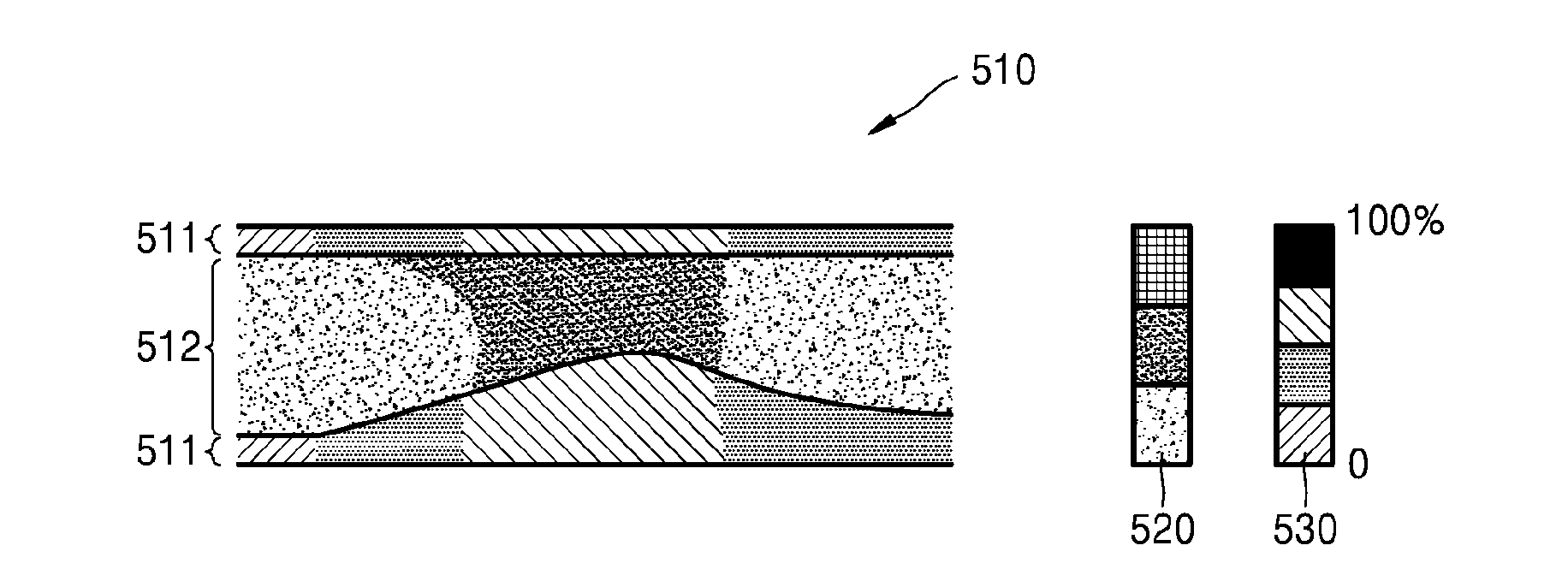
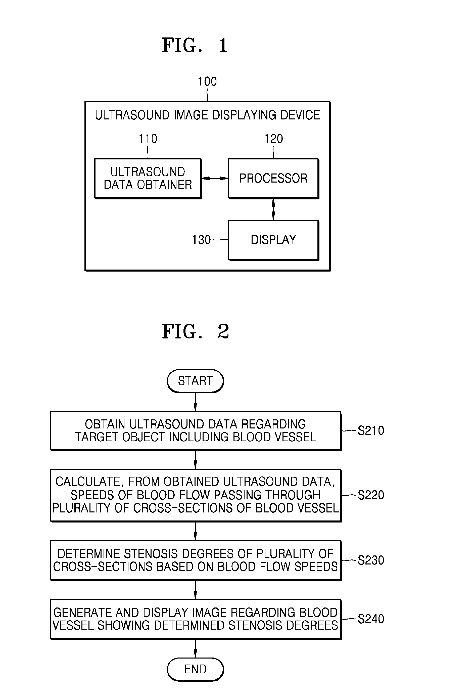
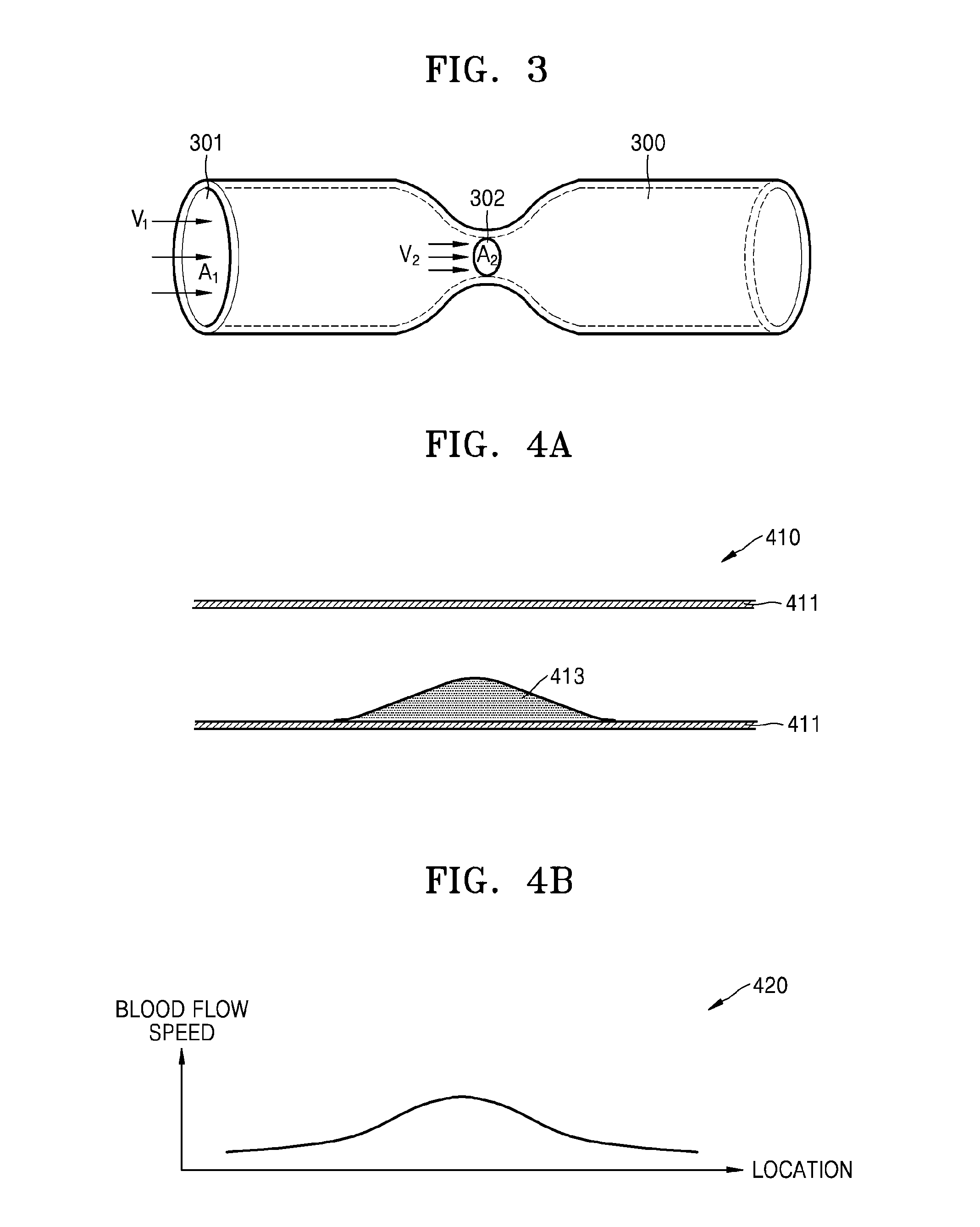
[0041]Reference will now be made in detail to embodiments, examples of which are illustrated in the accompanying drawings. In this regard, the present embodiments may have different forms and should not be construed as being limited to the descriptions set forth herein. Accordingly, the embodiments are merely described below, by referring to the figures, to explain aspects of the present description. As used herein, the term “and / or” includes any and all combinations of one or more of the associated listed items. In the description of the present invention, certain detailed explanations of related art are omitted when it is deemed that they may unnecessarily obscure the essence of the invention. Like reference numerals refer to like elements throughout

[0042]It will be understood that when an element or layer is referred to as being “connected” to another element or layer, the element or layer can be “directly connected” to another element or layer or “electrically connected” across ...


PUM


Abstract

A method of displaying an ultrasound image, the method including obtaining ultrasound data regarding a target object including a blood vessel; obtaining, from the ultrasound data, speeds of blood flows passing through a plurality of cross-sections of the blood vessel; determining stenosis degrees of the plurality of cross-sections based on the blood flow speeds; and generating and displaying an ultrasound image of the blood vessel showing the determined stenosis degrees.

Description

RELATED APPLICATIONS[0001]This application claims the benefit of Korean Patent Application No. 10-2013-0158675, filed on Dec. 18, 2013, in the Korean Intellectual Property Office, the disclosure of which is incorporated herein in its entirety by reference.BACKGROUND[0002]1. Field[0003]One or more embodiments of the present invention relate to a method and an apparatus for displaying an ultrasound image, and more particularly, to a method and an apparatus for determining a stricture of a blood vessel based on the speed of blood flow in the blood vessel and generating and displaying an ultrasound image showing the determined stricture of the blood vessel.[0004]2. Description of the Related Art[0005]An ultrasonography system is non-invasive and non-destructive, and thus, it is widely used in various medical fields for obtaining information regarding the interior of a target object. A medical practitioner may use the ultrasonography system to acquire high resolution images of tissues in...

Claims


Application Information

Patent Timeline
IPC IPC(8): A61B8/00A61B8/14A61B8/08A61B8/06
CPCA61B8/463A61B8/06A61B8/523A61B8/5207A61B8/5223A61B8/14A61B8/0891A61B8/488A61B8/5246G06T7/00
Inventor LEE, JIN-YONG
Owner SAMSUNG MEDISON
Why Eureka
Try Eureka