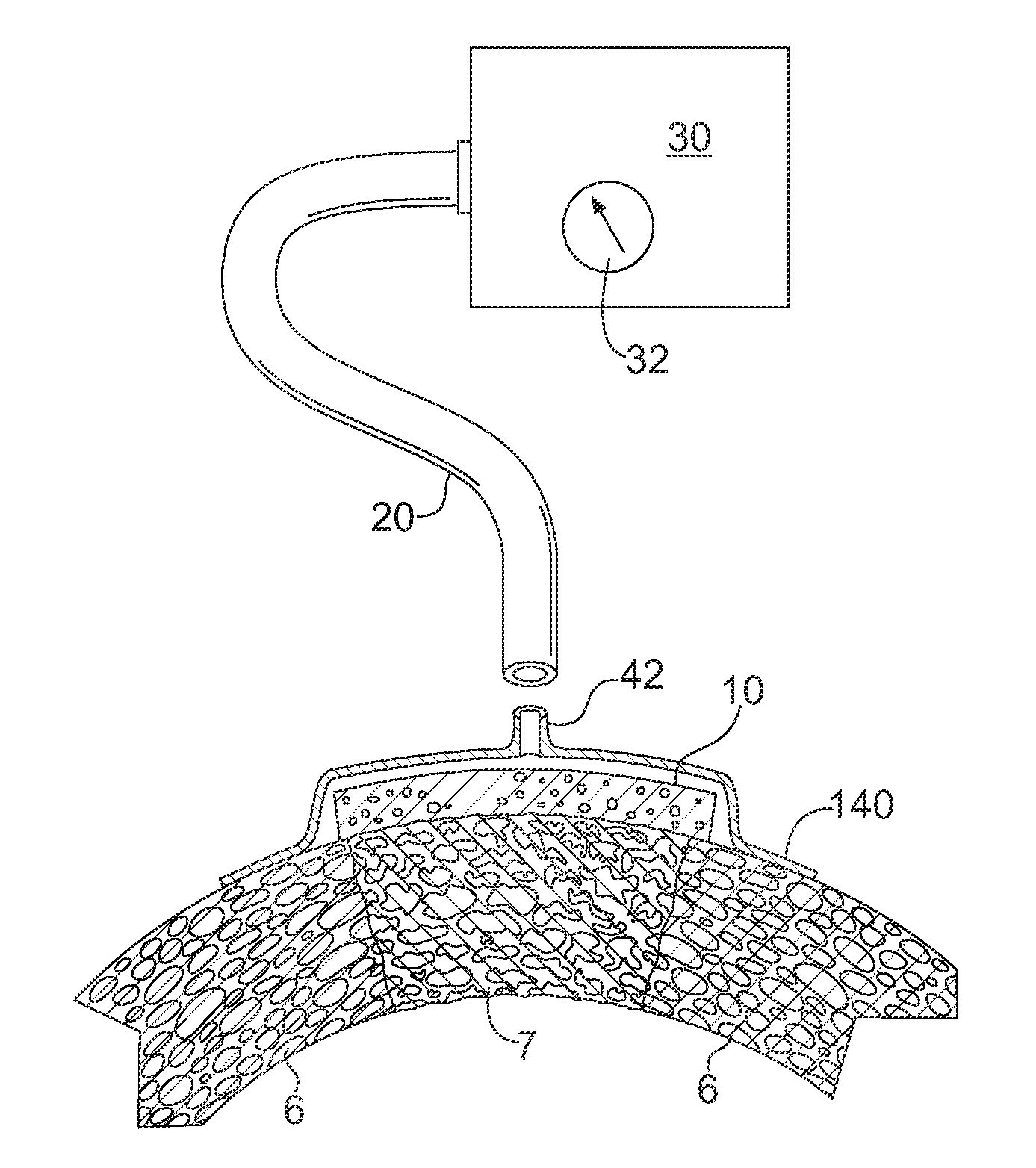
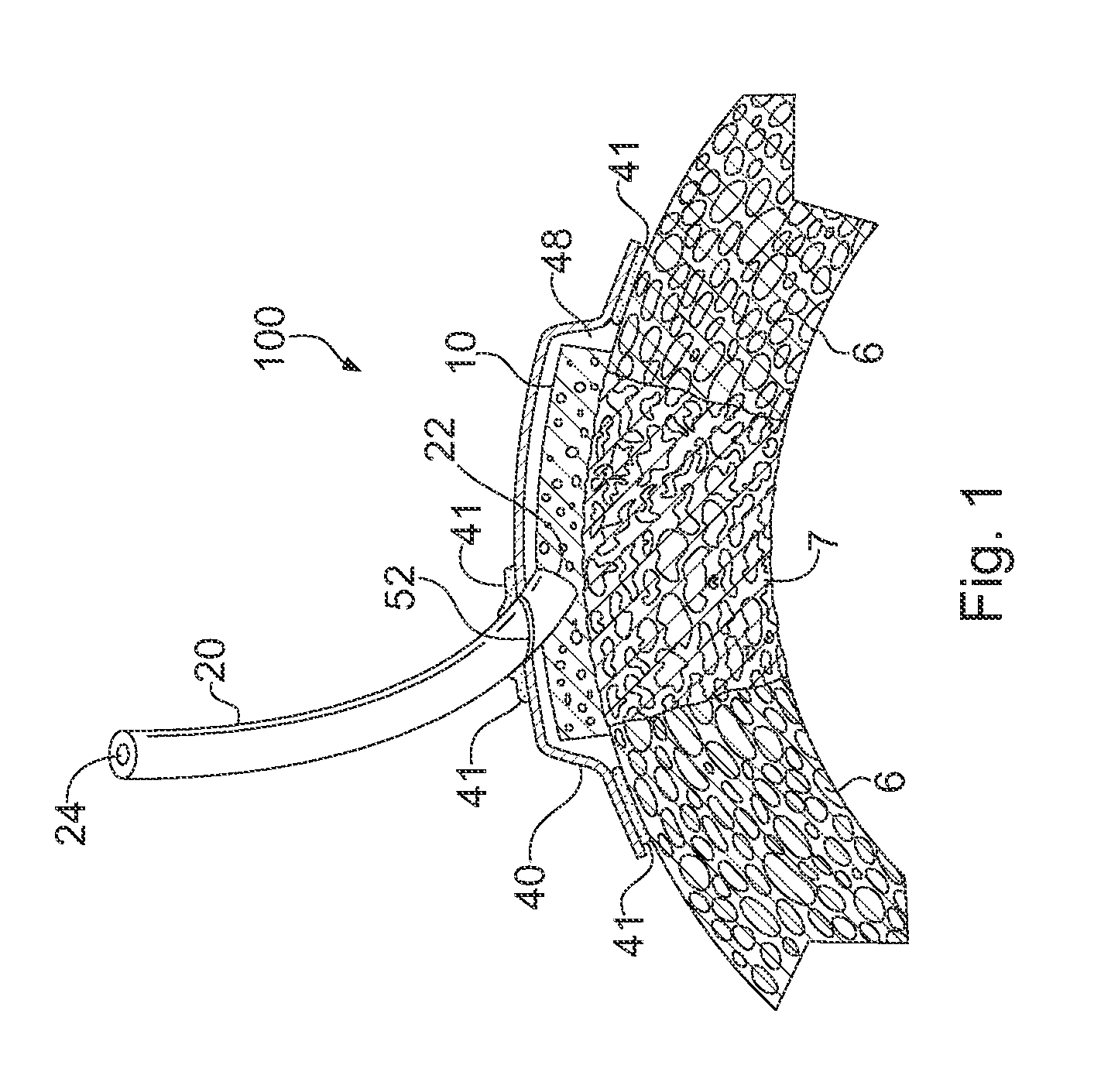
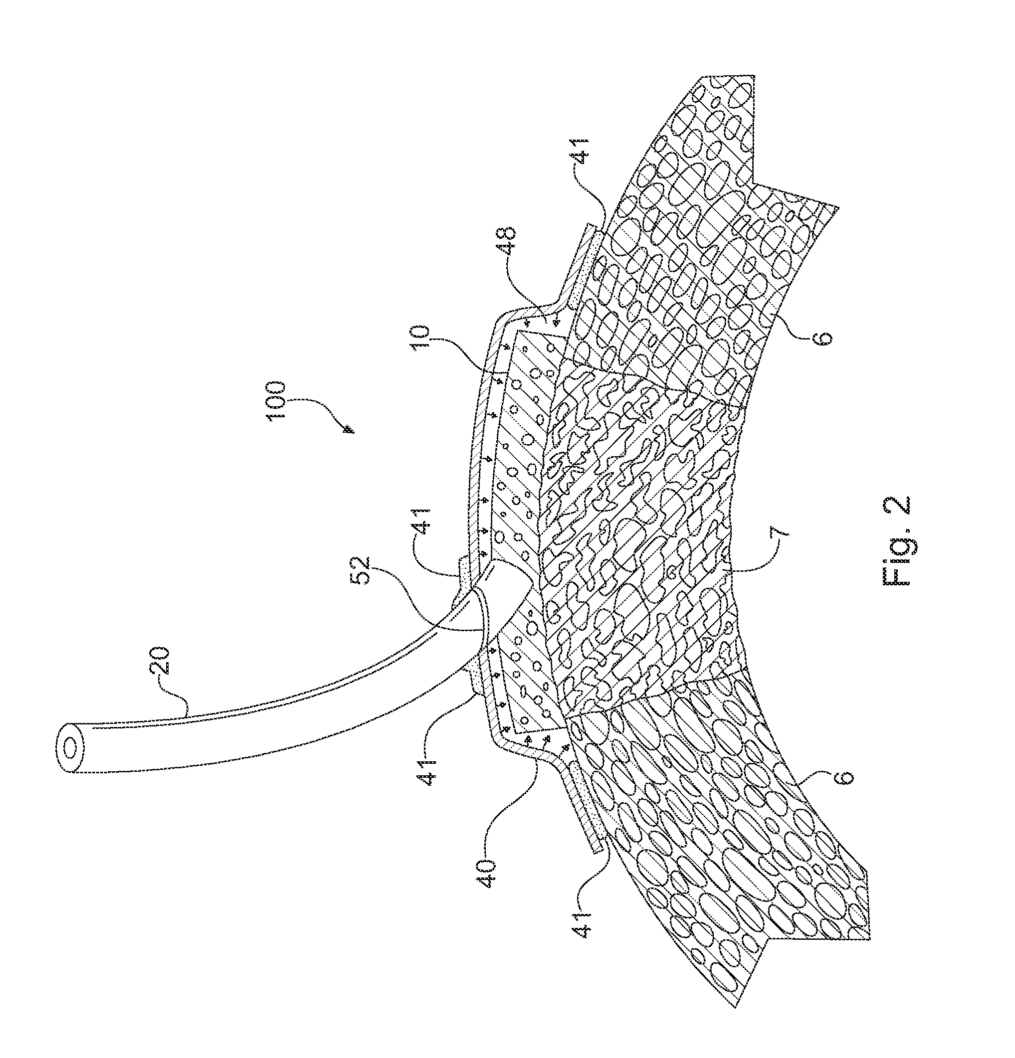
Apparatus and Method for Cardiac Tissue Modulation by Topical Application of Vacuum to Minimize Cell Death and Damage
a technology of cardiac tissue and vacuum, applied in the field of cardiac tissue modulation apparatus and vacuum topical application, can solve the problems of finite life cycle of cells and expected fatigue, and achieve the effects of minimizing the progression of pathologic processes, minimizing the disruption of physiological cardiac integrity, and minimizing the interference with cardiac blood flow
- Summary
- Abstract
- Description
- Claims
- Application Information
AI Technical Summary
Benefits of technology
Problems solved by technology
Method used
Image

Examples
example 1
[0058]The porcine heart has anatomy similar to that of humans with the main vasculature consisting of the right and left coronary arteries. The left main coronary artery splits into the circumflex coronary artery and the left anterior descending (LAD) coronary artery. The LAD runs down along the anterior septum and perfuses the anterior portion of the left ventricle with diagonal branches. For these studies, a porcine model of ischemia-reperfusion was used that included the temporary ligation of 2-3 diagonal branches of the LAD in order to create an ischemic area on the anterior portion of the heart. These coronary arteries were occluded for 75 minutes and then reperfused for 3 hours to allow for ischemia / reperfusion injury to develop. The negative pressure therapy was applied only during the reperfusion phase of the experiments to simulate a clinically relevant treatment window.
[0059]To begin the study, the animals were sedated and transported to the operating room. The first 13 an...
example 2
[0067]Another experiment was conducted using 50 mm Hg vacuum for treatment for comparison to original control animals from Example 1 above. The surgical technique in this experiment was similar to that used for those of Example 1. These animals were sedated and prepped for surgery. The heart was exposed through a midline sternotomy. Branches of the left anterior descending artery were ligated for 75 minutes. A polyvinyl alcohol vacuum dressing was placed over the ischemic area and an AlloDerm® cover was placed over the vacuum dressing and sealed into place with a combination of sutures and fibrin glue. Negative pressure of 50 mm Hg was applied for 3 hours. At the end of this time the heart was stained for area of risk, removed and then counter stained for area of necrosis. The infarct size results for these five, 50 mm Hg negative pressure therapy animals were significantly smaller (P<0.001) than for the control animals. The infarct size for the 50 mm Hg treated animals was smaller ...
example 3
[0071]A subsequent study was performed to examine resorbable vacuum dressings and overlay covers. One animal was sedated, prepared for surgery as described, and the heart exposed through a mid-line sternotomy. Branches of the LAD were ligated for 90 minutes. The dressing was prepared by freeze drying. A solution of chitosan (1.33% weight / volume in 2% acetic acid, 20 ml total volume) was poured into an appropriately sized mold. The solution was frozen for 2 hours at −70° C., then transferred to the lyophylizer for 24 hours. The dressing was cross-linked by 2.5% glutaraldehyde vapor for 12 hours to provide a porous material. The overlay cover was an electrospun mixture of Type I collagen and poly 1,8-octanediol citrate (POC) (80%:20% weight / weight). The solution concentration was 15% dissolved in hexafluoro-20proponal (HFIP) with a total volume of 9.5 ml. The solution was ejected from a syringe through an 18 gauge needle at a flow rate of 3 ml / hour. The voltage was 25 KV with a workin...
PUM
 Abstract
Description
Claims
Application Information
 - R&D
- Intellectual Property
- Life Sciences
- Materials
- Tech Scout
- Unparalleled Data Quality
- Higher Quality Content
- 60% Fewer Hallucinations
Browse by: Latest US Patents, China's latest patents, Technical Efficacy Thesaurus, Application Domain, Technology Topic, Popular Technical Reports.
© 2025 PatSnap. All rights reserved.Legal|Privacy policy|Modern Slavery Act Transparency Statement|Sitemap|About US| Contact US: help@patsnap.com