
Cell binding peptides for diagnosis and detection

a technology of cell binding and detection, applied in the direction of peptides, peptide/protein ingredients, instruments, etc., can solve the problems of cytology and slide-based diagnostic testing being confounded, putting a significant financial burden on both the patient and the healthcare facility, and requiring expensive methods

Active Publication Date: 2015-05-07
AFFINERGY INC
View PDF1 Cites 1 Cited by
  • Summary
  • Abstract
  • Description
  • Claims
  • Application Information

AI Technical Summary

Benefits of technology

The patent text describes a cell binding peptide that can be used to capture cells from a sample for cytological evaluation. The peptide includes a specific sequence of amino acids that is attached to a support. The peptide can be used in a device or method to capture cells from the sample. The technical effect of this invention is a more efficient and effective tool for capturing cells for cytological evaluation.

Problems solved by technology
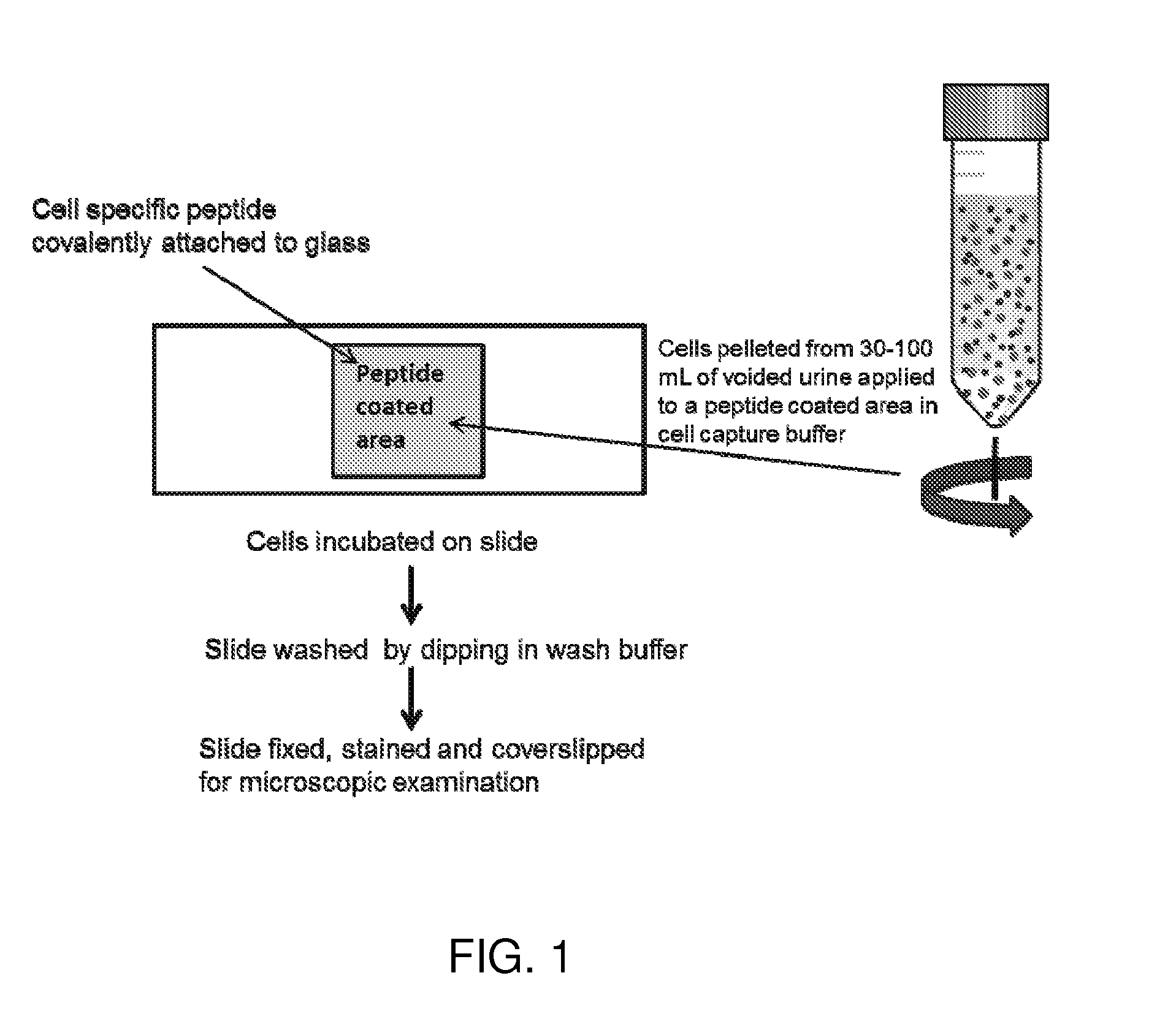
However, hematuria is one of the most common presenting symptoms with bladder cancer, and both cytology and slide-based diagnostic testing can be confounded by the presence of red blood cells and inflammatory cells.
Cytology slides prepared from specimens containing a high concentration of obscuring blood cells often lead to non-diagnostic results that necessitate repeat testing, unnecessary cystoscopy, and in some cases, exploratory biopsies.
In all cases, these unnecessary procedures place a significant financial burden on both the patient and the healthcare facility.
Although methods are available for processing specimens to concentrate urothelial cells and remove obscuring cell types, these methods are expensive, time-consuming, disruptive to cell phenotype, or all of the above.
Hemorrhaging is common during the procedure, however, leading to an aspirate that is diluted by blood.
Subsequent slide preparations are often suboptimal, resulting in nondiagnostic outcomes that necessitate additional testing or diagnostic surgery.
In fact, quality is so unpredictable that a pathologist must often be present in the operating room to certify that the slides are adequate, thus adding substantial cost and complexity to the procedure.
Furthermore, even with an optimal slide preparation, cytology yields indeterminate results up to 30% of the time.
Immunocytochemistry and fluorescence in-situ hybridization are useful tools for diagnosing indeterminate cases, but such tests are not usually carried out on existing smears due to a paucity of follicular cells and an abundance of red blood cells.
As a result, additional slides must be prepared using expensive and time-consuming techniques.

Method used


Image

  • Cell binding peptides for diagnosis and detection
  • Cell binding peptides for diagnosis and detection
  • Cell binding peptides for diagnosis and detection

Examples


example 1

Generation of Synthetic Binding Peptides

[0063]Peptide Synthesis:

[0064]Binding peptide sequences were synthesized using standard Solid-Phase peptide synthesis techniques on a SYMPHONY Peptide Synthesizer (Protein Technologies, Inc., Tucson, Ariz.) using standard Fmoc / t-Bu chemistry with the following coupling reagents at a 1:1:1:2 ratio—Amino acids; O-(benzotriazol-1-yl)-N,N,N′,N′-tetramethyluronium hexafluoroborate (HBTU); 1-hydroxybenzotriazole (HOBt); and N-Methylmorpholine (NMM). A20% (v / v) solution of piperidine in DMF was utilized for Fmoc removal. Peptides were synthesized on Fmoc-NH-Rink-Ahx-MBHA resin or on Fmoc-PAL-Peg-PS resin yielding peptides with C-terminal amides.

[0065]Amino acids with orthogonal side-chain protecting groups were coupled in 5-fold excess in the synthesis cycles, and all residues were doubly or triply coupled for 1 h. The coupling reactions were monitored by Kaiser ninhydrin test or chloranil test. After all amino acid residues were coupled and followin...

example 2

Peptide Cell Binding

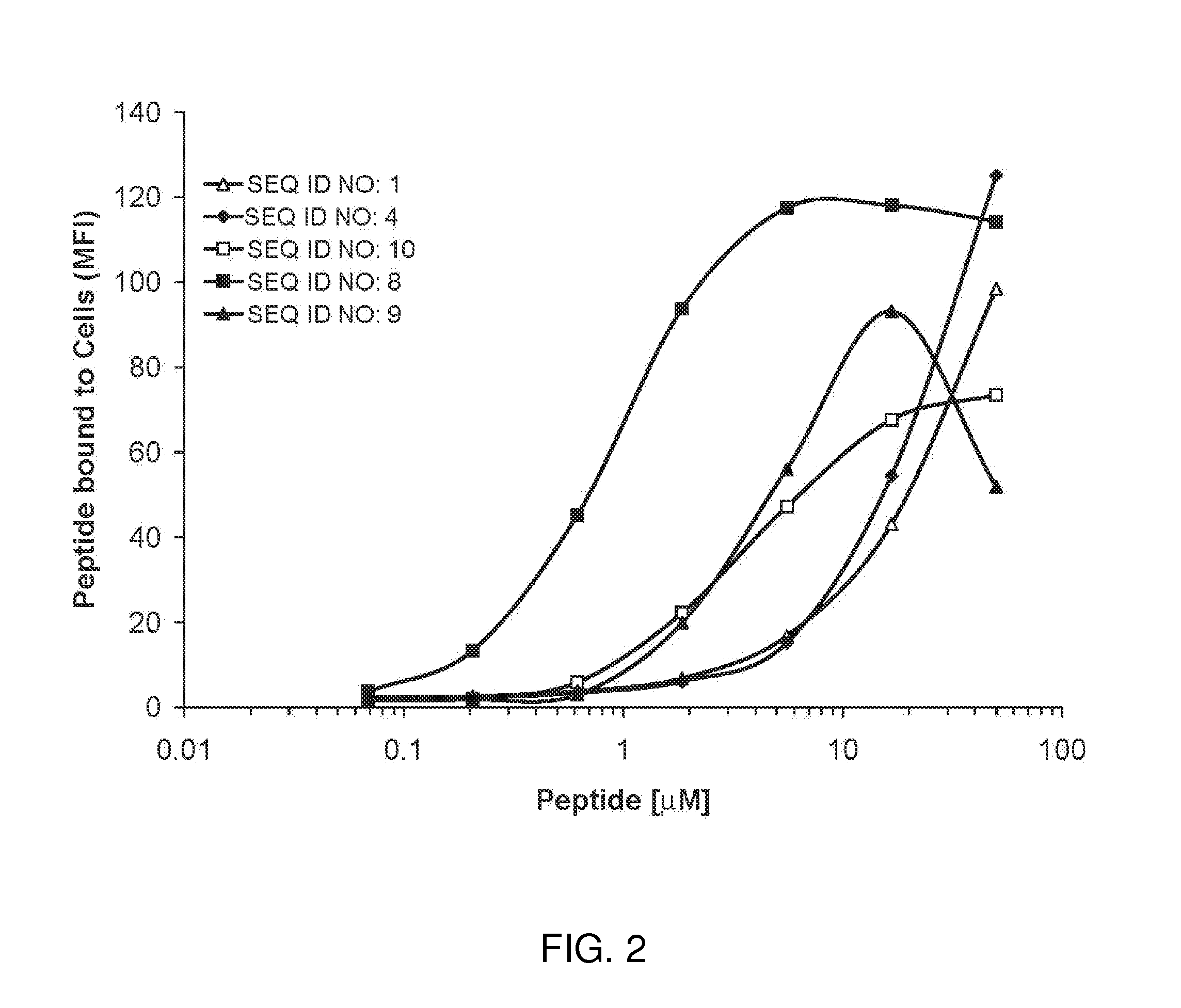
[0066]Synthetic biotinylated cell binding peptides were examined for their ability to bind to a bladder cancer cell line, J82 (ATCC, HTB1). The cell binding peptide was synthesized with a PEG-10 spacer and a carboxyl terminal lysine-biotin. J82 cells were grown to confluency on tissue-culture treated polystyrene. Cells were washed with PBS and released from the support with 0.05% trypsin. Trypsin was removed by centrifuging cells at 500×g and resuspending in PBS supplemented with 0.5% BSA and 2 mM EDTA to a concentration of 68,000 cells / ml. Aliquots of cells (50 μL) were separately incubated in 100 μl PBS / 0.5% BSA / 2 mM EDTA containing a final concentration of 0, 0.07, 0.21, 0.62, 1.85, 5.55, 16.7, and 50 μM peptide for 60 min at 4° C. Cells were then washed thrice in 1.8 ml PBS / 0.5% BSA / 2 mM EDTA with 500×g centrifugation for 5 min between washes. Approximately 50 μl of wash buffer remained with the cells after the final wash. Fluorescently-tagged neutravidin (Ne...

example 3

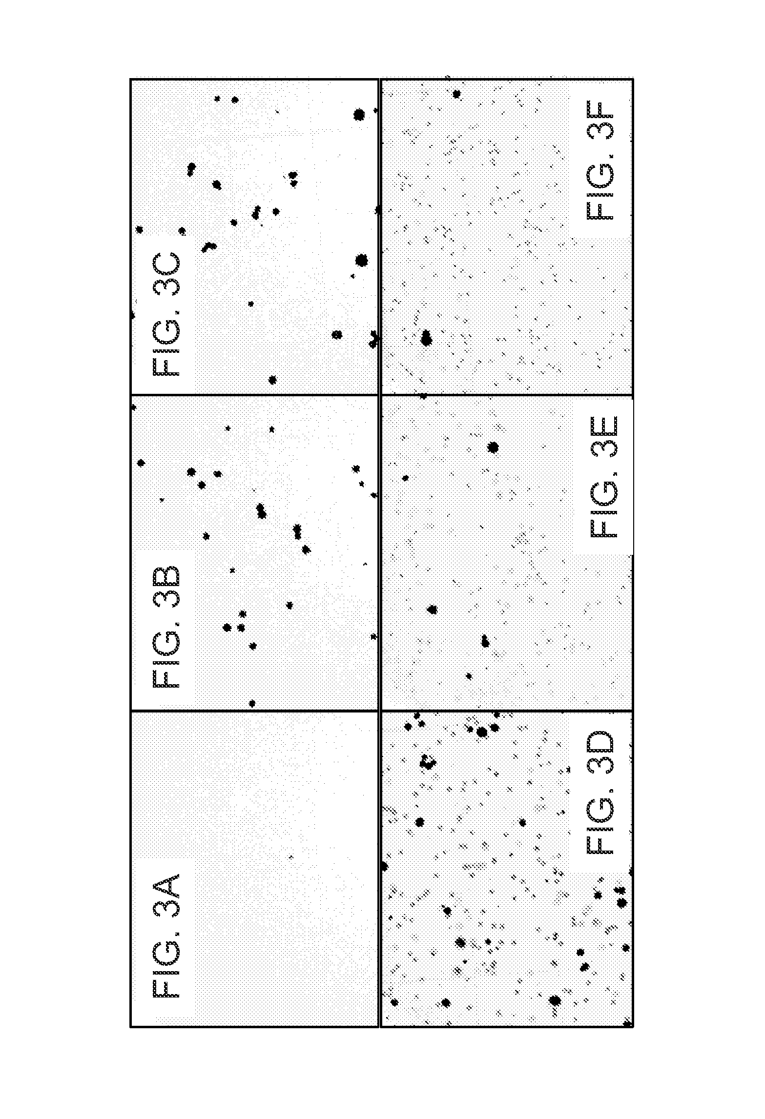
Capture of Cultured J82 Tumor Cells from Urine with Cell Binding Peptide Attached to a Support

[0067]To demonstrate the ability of cell binding peptides to recover tumor cells from urine, biotinylated peptides were immobilized on Streptavidin coated magnetic beads and used to separate tumor cells from white blood cells.

[0068]A green fluorescent leukocyte preparation was prepared as follows. Red blood cells in 2 ml anti-coagulated human blood were lysed by the addition of 30 ml of a hypotonic medium and incubation for 10 min at room temperature. Intact cells and debris were recovered by centrifugation at 300×g. Pelleted material was resuspended with PBS / 0.5% BSA / 2 mM EDTA and filtered through a 100 micron nylon filter. Leukocytes (3×10⁶) were labeled with 5 μM Cell Tracker Green CMFDA (LIFE TECHNOLOGIES / MOLECULAR PROBES) in EMEM growth medium without serum for 45 min. Excess dye was removed by centrifugation at 500×g for 5 min and washing with PBS / 0.5% BSA / 2 mM EDTA and cells were exp...


PUM

PropertyMeasurementUnit
RHaaaaaaaaaa
pHaaaaaaaaaa
fluorescence in-aaaaaaaaaa

Abstract

Cell binding peptides are provided for binding to cells including urothelial and thyroid follicular cells. The peptides are useful for detection and diagnosis of cancer including bladder and thyroid cancer. A device and method for using the device for capturing cells is provided, the device includes a support having attached cell binding peptide. The support can be a slide and the device can be used for detection and diagnosis of cancer including bladder and thyroid cancer. A kit is provided with instructions for capturing cells and a support with attached cell binding peptide for detection and diagnosis of bladder and thyroid cancer.

Description

CROSS-REFERENCE TO RELATED APPLICATIONS[0001]This application claims the benefit of U.S. Provisional Application No. 61 / 895,889 filed Oct. 25, 2013, which is hereby incorporated in its entirety by reference herein.STATEMENT OF GOVERNMENT SUPPORT[0002]This invention was made with U.S. Government support under the National Institutes of Health grant No.'s 2R44AR054229, 2R44GM077753, and 1R43DE022487. The U.S. Government has certain rights in the invention.TECHNICAL FIELD[0003]The present disclosure relates to the use of cell binding peptides for diagnosis and detection. More specifically, the present disclosure relates to the use of cell binding peptides for the detection and diagnosis of cancer.BACKGROUND[0004]Urine cytology is a non-invasive diagnostic method for detecting and monitoring bladder cancer, based on the collection of voided urine to examine exfoliated epithelial cells on glass slides. Follow-up tests, such as immunocytochemistry (ICC) and fluorescence in-situ hybridizat...

Claims


Application Information

Patent Timeline
Patent Type & Authority Applications(United States)
IPC IPC(8): G01N33/569G01N33/543G01N33/552C07K7/08
CPCG01N33/56966C07K7/08G01N33/54353G01N33/552C07K17/14
Inventor CULP, WILLIAM DAVIDDARBY, MARTYN KERRYJUZUMIENE, DALIA ISOLDAKRAJEWSKA, MAGDALENALYGINA, NATALIAMORRISON, JUHUANAIR, SHRIKUMAR AMBUJAKSHANSIESSER, WILLIAM BOURCHIERWRONSKA, DANUTA
Owner AFFINERGY INC
Learn More